. 2012 Sep;72(3):455-63.
doi: 10.1002/ana.23614.

Glucocerebrosidase deficiency in substantia nigra of parkinson disease brains

Affiliations
Free PMC article

Glucocerebrosidase deficiency in substantia nigra of parkinson disease brains

Matthew E Gegg et al. Ann Neurol. 2012 Sep.
Free PMC article

Abstract

Objective: Mutations in the glucocerebrosidase gene (GBA) represent a significant risk factor for developing Parkinson disease (PD). We investigated the enzymatic activity of glucocerebrosidase (GCase) in PD brains carrying heterozygote GBA mutations (PD+GBA) and sporadic PD brains.

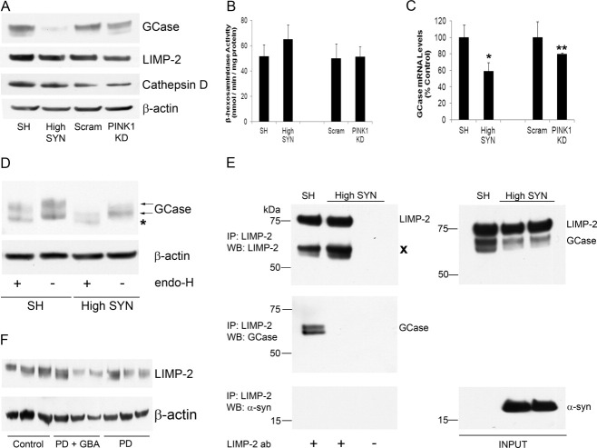
Methods: GCase activity was measured using a fluorescent assay in cerebellum, frontal cortex, putamen, amygdala, and substantia nigra of PD+GBA (n = 9-14) and sporadic PD brains (n = 12-14). Protein expression of GCase and other lysosomal proteins was determined by western blotting. The relation between GCase, α-synuclein, and mitochondria function was also investigated in vitro.

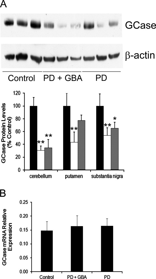
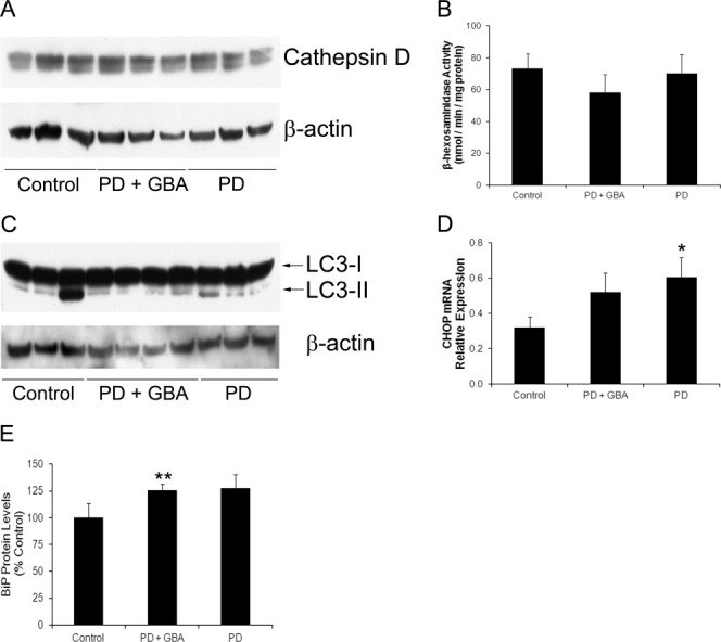
Results: A significant decrease in GCase activity was observed in all PD+GBA brain areas except the frontal cortex. The greatest deficiency was in the substantia nigra (58% decrease; p < 0.01). GCase activity was also significantly decreased in the substantia nigra (33% decrease; p < 0.05) and cerebellum (24% decrease; p < 0.05) of sporadic PD brains. GCase protein expression was lower in PD+GBA and PD brains, whereas increased C/EBP homologous protein and binding immunoglobulin protein levels indicated that the unfolded protein response was activated. Elevated α-synuclein levels or PTEN-induced putative kinase 1 deficiency in cultured cells had a significant effect on GCase protein levels.

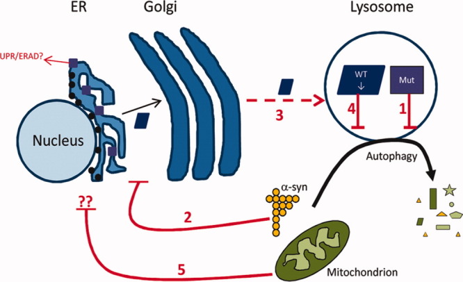
Interpretation: GCase deficiency in PD brains with GBA mutations is a combination of decreased catalytic activity and reduced protein levels. This is most pronounced in the substantia nigra. Biochemical changes involved in PD pathogenesis affect wild-type GCase protein expression in vitro, and these could be contributing factors to the GCase deficiency observed in sporadic PD brains.


Figures

FIGURE 1
FIGURE 1
Glucocerebrosidase enzyme (GCase) deficiency in Parkinson disease (PD) brains carrying GBA mutations and sporadic PD. GCase activity was significantly decreased in the cerebellum (CBM, n = 14), putamen (PUT, n = 12), amygdala (AMYG, n = 12), and SN (SN, n = 9), but not the frontal cortex (FCX, n = 14) of PD brains with GBA mutations (PD+GBA, white bars), when compared to controls (n = 6–10, black bars). GCase activity was also significantly decreased in the CBM (n = 14) and SN (n = 14) of sporadic PD brains (gray bars), but not the FCX (n = 14), PUT (n = 14), or AMYG (n = 12). *p < 0.05 versus control, **p < 0.01 versus control.
FIGURE 2
FIGURE 2
Glucocerebrosidase enzyme (GCase) protein expression was decreased in Parkinson disease (PD) brains carrying GBA mutations and sporadic PD brains. (A) Western blotting for GCase protein levels in cerebellum, putamen, and substantia nigra (example blot shown). GCase protein was significantly decreased in the cerebellum of both PD+GBA (white bars) and sporadic PD brains (gray bars, PD+GBA, 30.4 ± 7.0%, n = 14; PD, 34.6 ± 13.1%, n = 6, of control brain optical density [black bars], 100 ± 13%, n = 9). GCase was significantly decreased in the putamen of PD+GBA brains but not sporadic PD brains (PD+GBA, 43.4 ± 16.0%, n = 7; PD, 77.2 ± 8.8%, n = 6, of control brain optical density, 100 ± 20%, n = 7). GCase was significantly decreased in the substantia nigra of PD+GBA brains and sporadic PD brains (PD+GBA, 53.9 ± 12.0%, n = 9; PD, 65.1 ± 9.3%, n = 11, of control brain optical density, 100 ± 19%, n = 7). (B) RNA was extracted from the putamen of control (n = 6), PD+GBA (n = 10), and sporadic PD brains (n = 6), and GCase mRNA relative expression was measured with quantitative real time polymerase chain reaction. No changes in mRNA were detected. *p < 0.05 versus control, **p < 0.01 versus control.
FIGURE 3
FIGURE 3
Glucocerebrosidase enzyme deficiency was not due to decreased lysosomal content, but C/EBP homologous protein (CHOP) and binding immunoglobulin protein (BiP) levels were increased. (A) Western blots for cathepsin D in the substantia nigra (SN) of control brains, PD brains carrying GBA mutations (PD+GBA), and sporadic PD brains (PD) was unaffected. (B) The activity of the lysosomal enzyme β-hexosaminidase in the SN of control (n = 7), PD+GBA (n = 9), and PD (n = 14) was unaffected. (C) Measurement of LC3-II levels in the putamen did not differ significantly between the groups (control, 0.624 ± 0.329, n = 6; PD+GBA, 1.122 ± 0.596, n=7; PD, 0.816 ± 0.213, n=6). (D) CHOP mRNA levels were increased in the putamen of both PD+GBA (n = 10) and PD brains (n = 6) compared to control brain (n = 6). (E) Protein levels of the chaperone BiP were found to be increased by western blotting in the putamen of both PD+GBA (n = 8) and PD brains (n = 10) compared to control brain (n = 8). *p < 0.05 versus control, **p < 0.01 versus control.
FIGURE 4
FIGURE 4
Glucocerebrosidase enzyme (GCase) protein levels were decreased in SH-SY5Y cells overexpressing α-synuclein (α-syn) or with PTEN-induced putative kinase 1 (PINK1) deficiency. (A) Western blotting for GCase in α-synuclein overexpressing cells (High SYN) or PINK1 knockdown (KD) cells showed a significant decrease in protein levels (High SYN, 13.4 ± 7.1%, n = 3, p < 0.001; PINK1 KD, 65.3 ± 9.3%, n = 3, p < 0.05, of respective control optical density [SH-SY5Y cells, 100 ± 16%, n = 3; scram short hairpin RNA cells, 100 ± 9%, n = 3]). Cathepsin D and lysosomal integral membrane protein (LIMP)-2 protein levels were unaffected in either cell type, compared to their respective controls. (B) The activity of the lysosomal enzyme β-hexosaminidase was unaffected in High SYN or PINK1 KD cells (n = 6), when compared to respective control cell lines. (C) Quantitative real time polymerase chain reaction indicated that steady-state GCase mRNA levels were decreased in cells with exogenous α-synuclein or PINK1 KD (High SYN, 58.9 ± 10.1%, n = 3; PINK1 KD, 79.9 ± 1.3%, n = 3 of respective control GCase mRNA levels, n = 3). (D) SH or High SYN cell lysates (20μg) were treated with or without endoglycosidase-H (endo-H) and GCase protein species analyzed by Western blot. The 2 normal species of GCase detected in SH cells are indicated by arrows. An additional lower molecular weight band was observed in High SYN but not SH cells following endo-H treatment (asterisk). (E) LIMP-2 was immunoprecipitated (Ip) from lysates of SH or High SYN cells to a similar extent (left panel). No LIMP-2 was immunoprecipitated from lysates incubated without antibody (ab). Cross denotes heavy chain of antibody used for immunoprecipitation. Two bands corresponding to GCase were pulled down in SH cells, but not High SYN cells (left panel). α-Synuclein was not pulled down in SH or High SYN cells (left panel). The right panel shows the expression of GCase, LIMP-2, and α-synuclein in the initial lysates (INPUT). WB, western blotting (F) Western blotting for LIMP2 in the substantia nigra from control, PD+GBA, or sporadic PD brains showed that there was no difference in expression between the groups (PD+GBA, 92.0 ± 17.2%, n = 8; PD, 123.3 ± 10.7%, n = 8, of control optical density, n = 7). *p < 0.05 versus SH, **p < 0.01 versus scram.
FIGURE 5
FIGURE 5
Scheme of glucocerebrosidase enzyme (GCase) deficiency in PD pathogenesis. Mutant (Mut) GCase (purple squares) disrupts the degradation of α-synuclein and organelles such as mitochondria by the autophagy–lysosomal pathway (1). Some GCase mutants may also become trapped in the endoplasmic reticulum (ER) and activate the unfolded protein response (UPR)/endoplasmic reticulum-associated degradation (ERAD). Increased levels of α-synuclein (α-syn) impair the trafficking of wild-type (WT) GCase (blue rectangle) via the ER/Golgi apparatus (2), resulting in less WT GCase being delivered to the lysosomes by lysosomal integral membrane protein-2 (3). This will exacerbate any lysosomal/autophagic dysfunction (4). Dysfunctional mitochondria can also affect WT GCase protein levels by an unidentified mechanism (5).

References

    1. Grabowski GA. Phenotype, diagnosis, and treatment of Gaucher's disease. Lancet. 2008;372:1263–1271. - PubMed
    1. Schapira AHV. Mitochondria in the aetiology and pathogenesis of Parkinson's disease. Lancet Neurol. 2008;7:97–109. - PubMed
    1. Neudorfer O, Giladi N, Elstein D, et al. Occurrence of Parkinson's syndrome in type I Gaucher disease. QJM. 1996;89:691–694. - PubMed
    1. Aharon-Peretz J, Rosenbaum H, Gershoni-Baruch R. Mutations in the glucocerebrosidase gene and Parkinson's disease in Ashkenazi Jews. N Engl J Med. 2004;351:1972–1977. - PubMed
    1. Wong K, Sidransky E, Verma A, et al. Neuropathology provides clues to the pathophysiology of Gaucher disease. Mol Genet Metab. 2004;82:192–207. - PubMed

Publication types

MeSH terms