. 2012 Oct 3;32(40):13906-16.
doi: 10.1523/JNEUROSCI.2108-12.2012.

Microtubule plus-end tracking protein CLASP2 regulates neuronal polarity and synaptic function

Affiliations

Microtubule plus-end tracking protein CLASP2 regulates neuronal polarity and synaptic function

Uwe Beffert et al. J Neurosci. .

Abstract

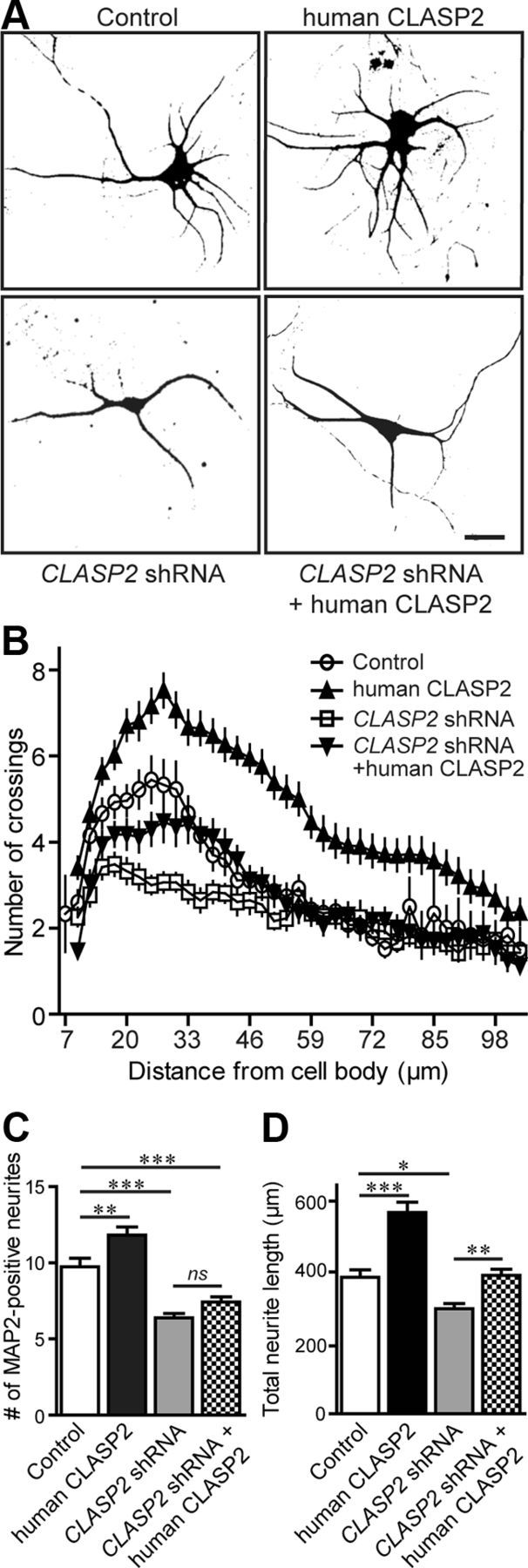
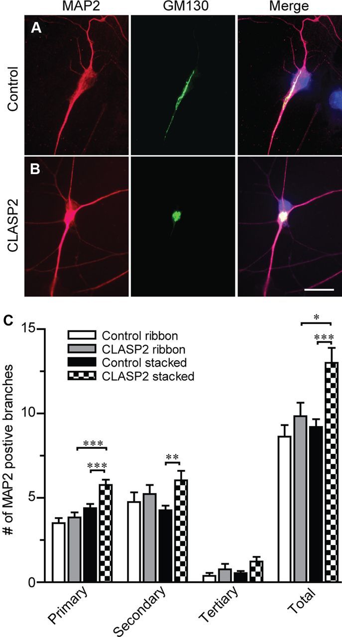
Microtubule organization and dynamics are essential during axon and dendrite formation and maintenance in neurons. However, little is known about the regulation of microtubule dynamics during synaptic development and function in mammalian neurons. Here, we present evidence that the microtubule plus-end tracking protein CLASP2 (cytoplasmic linker associated protein 2) is a key regulator of axon and dendrite outgrowth that leads to functional alterations in synaptic activity and formation. We found that CLASP2 protein levels steadily increase throughout neuronal development in the mouse brain and are specifically enriched at the growth cones of extending neurites. The short-hairpin RNA-mediated knockdown of CLASP2 in primary mouse neurons decreased axon and dendritic length, whereas overexpression of human CLASP2 caused the formation of multiple axons, enhanced dendritic branching, and Golgi condensation, implicating CLASP2 in neuronal morphogenesis. In addition, the CLASP2-induced morphological changes led to significant functional alterations in synaptic transmission. CLASP2 overexpression produced a large increase in spontaneous miniature event frequency that was specific to excitatory neurotransmitter release. The changes in presynaptic activity produced by CLASP2 overexpression were accompanied by increases in presynaptic terminal circumference, total synapse number, and a selective increase in presynaptic proteins that are involved in neurotransmitter release. Also, we found a smaller increase in miniature event amplitude that was accompanied by an increase in postsynaptic surface expression of GluA1 receptor localization. Together, these results provide evidence for involvement of the microtubule plus-end tracking protein CLASP2 in cytoskeleton-related mechanisms underlying neuronal polarity and interplay between microtubule stabilization and synapse formation and activity.


Figures

Figure 1.
Figure 1.
Localization and expression of CLASP2 in the developing brain. A, Representative images of endogenous CLASP2 enriched in the growing tip of a growth cone of mouse hippocampal neurons at 4–5 DIV double labeled with anti-CLASP2 (green) and anti-SMI-312 (red) antibodies. Representative images of hippocampal neuron (B) and a glial cell at 4–5 DIV double labeled with anti-CLASP2 (green) and anti-α-tubulin (red) antibody (C). Scale bars: A, 2.5 μm; B, 16 μm; C, 15 μm. D, Protein expression levels of CLASP2, MAP2, SMI-312, and β-tubulin during postnatal development examined by immunoblotting of total mouse brain lysate at the indicated ages (Adult = 3 months). E, Expression levels of CLASP2 was examined by immunoblotting of total lysate from 7–14 DIV cortical neurons infected with control, human CLASP2, or CLASP2 shRNA lentivirus.
Figure 2.
Figure 2.
CLASP2 increases axon length and induces multiple axon formation. A, Hippocampal neurons infected at 1 DIV with control, human CLASP2, CLASP2 shRNA, or CLASP2 shRNA coexpressing human CLASP2 were fixed at 2 DIV and stained with SMI-312 (axonal marker). Scale bar, 20 μm. B, Quantitative analysis showed that human CLASP2 significantly increased axon length at 2 DIV (n = 49 neurons for each group; *p < 0.05, **p < 0.001). C, Hippocampal neurons infected with human CLASP2 were fixed at 4 DIV and stained with SMI-132 showed multiple axons compared with control and CLASP2 shRNA (n = 62 neurons for each group; ***p < 0.0001). Neuronal polarity phenotypes were categorized into two groups: single axon (white bar) and multiple axons (black bar).
Figure 3.
Figure 3.
CLASP2 regulates dendritic growth and branching. A, Hippocampal neurons infected at 1 DIV with control, human CLASP2, CLASP2 shRNA, or CLASP2 shRNA coexpressing human CLASP2 were fixed at 5 DIV and stained with MAP2 (dendritic marker). Scale bar, 20 μm. B, Sholl analysis based on the number of dendritic crossings distributed over the distance from the cell body (n = 17 neurons for each group). CLASP2 significantly increased the number of MAP2-positive neurites (C) (n = 32 for each group; **p < 0.01, ***p < 0.001; ns, not significant) and the total length of all neurites (D) (n = 19 for each group; *p < 0.05, **p < 0.01, ***p < 0.001).
Figure 4.
Figure 4.
CLASP2 regulates Golgi morphology in neurons. A, Representative images of anti-CLASP2 (green) and anti-GM130 (red) antibodies showing colocalization of endogenous CLASP2 with Golgi complex in a hippocampal neuron at 5 DIV. B, Neurons treated at 1 DIV with control, human CLASP2, or CLASP2 shRNA lentivirus were stained for GM130 (red), MAP2 (cyan), and DAPI (blue) to assess ribbon, stacked, and fragmented Golgi morphology in hippocampal neurons at 5 DIV. Scale bars: A, 1.5 μm; B, 10 μm. C, CLASP2 led to a significant increase in the percentage of neurons with a stacked Golgi phenotype compared with those with a ribbon-shaped Golgi (n = 100 for each group; ***p < 0.0001). D, The Golgi is highly fragmented in CLASP2 knockdown neurons (n = 100 for each group; ***p < 0.0001).
Figure 5.
Figure 5.
CLASP2 increases neuritic branching in neurons with stacked but not ribbon Golgi phenotypes. A, B, Primary hippocampal neurons infected at 1 DIV with control or CLASP2 were fixed at 4 DIV and immunostained for MAP2 (red) and GM130 (green). Scale bar, 23 μm. C, All primary, secondary, tertiary, and total number of MAP2-positive branches were counted and separated by treatment and Golgi phenotype (n > 13 for each group; *p = 0.029, **p = 0.004, ***p < 0.0009).
Figure 6.
Figure 6.
CLASP2 increases spontaneous synaptic activity at excitatory but not inhibitory synapses. A, Sample traces showing mEPSC of control, human CLASP2, CLASP2 shRNA, or CLASP2 shRNA coexpressing human CLASP2 at 14–17 DIV. B, Bar graph of mEPSC frequency revealed a significant 4.6-fold increase in miniature frequency in neurons infected with human CLASP2 compared with control (n ≥ 14 for each group; *p < 0.05, ***p < 0.0001). C, Bar graph of mEPSC amplitude demonstrating a significant increase in mEPSC amplitude in neurons infected with human CLASP2 (n ≥ 14 for each group; *p < 0.05). D, Sample traces showing mIPSCs of control and overexpressed human CLASP2. E, No changes in mIPSC frequency were detected in neurons infected with human CLASP2 compared with control (n ≥ 10 for each group). F, Bar graph of mIPSC amplitude showed no changes in miniature amplitude in neurons infected with human CLASP2 (n ≥ 10 for each group).
Figure 7.
Figure 7.
CLASP2 regulates synaptic structure and synaptic protein levels. A, Ultrastructural analysis by electron microscopy of synaptic structure in hippocampal neuronal cultures infected with control and overexpressed human CLASP2 at 14 DIV. Representative images of asymmetric excitatory synapses of control and CLASP2 infected neurons. Scale bar, 500 nm. B, Hippocampal neuronal processes infected with control, human CLASP2, CLASP2 shRNA, or CLASP2 shRNA coexpressing human CLASP2 at 14 DIV stained with synapsin (Syn; green) and PSD95 (red). Scale bar, 4 μm. C, D, CLASP2 increases overall synapse number and area of colocalization of synapsin and PSD95 in neuronal processes at 14 DIV (*p < 0.05, ***p < 0.0001). E, Western blotting of CLASP2, various synaptic markers, and β-tubulin in response to control, human CLASP2, CLASP2 shRNA, or CLASP2 shRNA coexpressing human CLASP2 (rescue) in cortical neurons. F, We detected a significant increase of surface (S) compared with total (T) endogenous GluA1 receptor subunit in cultured cortical neurons that overexpressed CLASP2 at 14 DIV using a biotinylation assay (left, immunoblot; right, quantification). Total GluA1 levels were determined from input lysates, whereas surface GluA1 was determined after elution from NeutrAvidin precipitation (*p = 0.04).
Figure 8.
Figure 8.
CLASP2 is required for PI3K-induced axon elongation. A, Hippocampal neurons infected at 1 DIV with control, human CLASP2, or treated with PI3K inhibitor LY 294002 or PI3K inhibitor LY 294002 coexpressing human CLASP2 were fixed at 2 DIV and stained with SMI-312 (axonal marker). Scale bar, 20 μm. B, Quantitative analysis showed that overexpressed human CLASP2 significantly increased axon length at 2 DIV and rescued the axonal growth defect in neurons treated with LY 294002 (n = 47 neurons for each group; **p < 0.01, ***p < 0.001).

References

    1. Akhmanova A, Hoogenraad CC. Microtubule plus-end-tracking proteins: mechanisms and functions. Curr Opin Cell Biol. 2005;17:47–54. - PubMed
    1. Akhmanova A, Steinmetz MO. Tracking the ends: a dynamic protein network controls the fate of microtubule tips. Nat Rev Mol Cell Biol. 2008;9:309–322. - PubMed
    1. Akhmanova A, Hoogenraad CC, Drabek K, Stepanova T, Dortland B, Verkerk T, Vermeulen W, Burgering BM, De Zeeuw CI, Grosveld F, Galjart N. Clasps are CLIP-115 and -170 associating proteins involved in the regional regulation of microtubule dynamics in motile fibroblasts. Cell. 2001;104:923–935. - PubMed
    1. Akhmanova A, Mausset-Bonnefont AL, van Cappellen W, Keijzer N, Hoogenraad CC, Stepanova T, Drabek K, van der Wees J, Mommaas M, Onderwater J, van der Meulen H, Tanenbaum ME, Medema RH, Hoogerbrugge J, Vreeburg J, Uringa EJ, Grootegoed JA, Grosveld F, Galjart N. The microtubule plus-end-tracking protein CLIP-170 associates with the spermatid manchette and is essential for spermatogenesis. Genes Dev. 2005;19:2501–2515. - PMC - PubMed
    1. Al-Bassam J, Kim H, Brouhard G, van Oijen A, Harrison SC, Chang F. CLASP promotes microtubule rescue by recruiting tubulin dimers to the microtubule. Dev Cell. 2010;19:245–258. - PMC - PubMed

Publication types

MeSH terms

LinkOut - more resources