. 2012 Nov 23;287(48):40119-30.
doi: 10.1074/jbc.M112.418889. Epub 2012 Oct 3.

Tissue specificity of a human mitochondrial disease: differentiation-enhanced mis-splicing of the Fe-S scaffold gene ISCU renders patient cells more sensitive to oxidative stress in ISCU myopathy

Affiliations

Tissue specificity of a human mitochondrial disease: differentiation-enhanced mis-splicing of the Fe-S scaffold gene ISCU renders patient cells more sensitive to oxidative stress in ISCU myopathy

Daniel R Crooks et al. J Biol Chem. .

Abstract

Background: ISCU myopathy is a disease caused by muscle-specific deficiency of the Fe-S cluster scaffold protein ISCU.

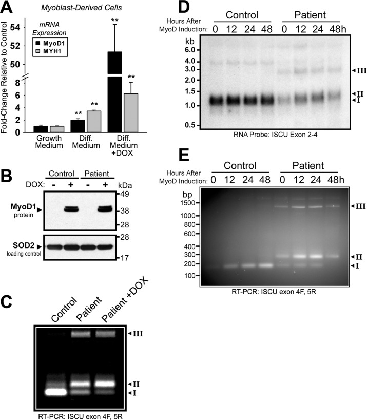
Results: MyoD expression enhanced ISCU mRNA mis-splicing, and oxidative stress exacerbated ISCU depletion in patient cells.

Conclusion: ISCU protein deficiency in patients results from muscle-specific mis-splicing as well as oxidative stress.

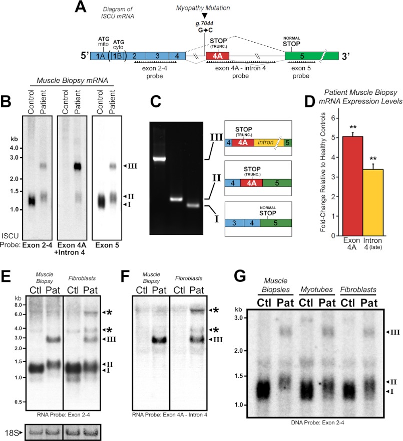
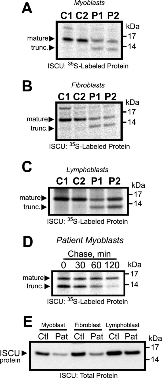
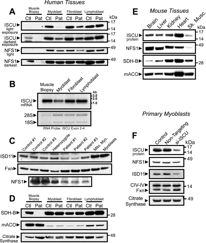
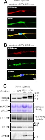
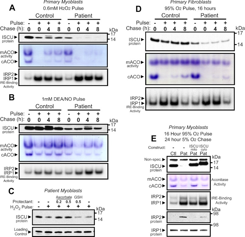
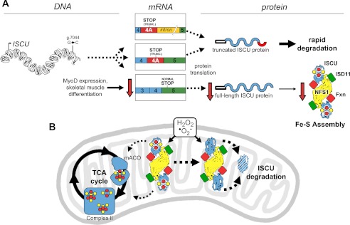
Significance: Oxidative stress negatively influences the mammalian Fe-S cluster assembly machinery by destabilization of ISCU. Iron-sulfur (Fe-S) cluster cofactors are formed on the scaffold protein ISCU. ISCU myopathy is a disease caused by an intronic mutation that leads to abnormally spliced ISCU mRNA. We found that two predominant mis-spliced ISCU mRNAs produce a truncated and short-lived ISCU protein product in multiple patient cell types. Expression of the muscle-specific transcription factor MyoD further diminished normal splicing of ISCU mRNA in patient myoblasts, demonstrating that the process of muscle differentiation enhances the loss of normal ISCU mRNA splicing. ISCU protein was nearly undetectable in patient skeletal muscle, but was higher in patient myoblasts, fibroblasts, and lymphoblasts. We next treated patient cells with pro-oxidants to mimic the oxidative stress associated with muscle activity. Brief hydrogen peroxide treatment or incubation in an enriched oxygen atmosphere led to a marked further reduction of ISCU protein levels, which could be prevented by pretreatment with the antioxidant ascorbate. Thus, we conclude that skeletal muscle differentiation of patient cells causes a higher degree of abnormal ISCU splicing and that oxidative stress resulting from skeletal muscle work destabilizes the small amounts of normal ISCU protein generated in patient skeletal muscles.


Figures

FIGURE 1.
FIGURE 1.
Altered ISCU mRNA expression patterns in ISCU myopathy patients. A, a schematic representation of ISCU mRNA showing the single intronic point mutation within the fourth intron, canonical, and aberrant splicing patterns and regions of Northern blot probe complementarity. mito, mitochondrial; cyto, cytosolic; TRUNC., truncated. B, hybridization of ISCU-specific 32P-labeled DNA probes with control and patient vastus lateralis biopsy RNA demonstrated normal (I) and additional patient-specific (II and III) ISCU mRNA bands. C, RT-PCR and sequencing using primers flanking ISCU exons 4 and 5 confirmed the identity of the three major ISCU bands (I–III), with transcript II containing 100 bp and transcript III containing 1173 bp of intronic sequence. D, qRT-PCR analysis of three ISCU myopathy patients and seven control biopsies using primer sets for ISCU exon 4A and downstream intron 4 sequences. E and F, Northern blot analysis of ISCU mRNA expression in control (Ctl) and patient (Pat) muscle biopsies and fibroblasts using high specific activity RNA probes revealed several larger ISCU-specific mRNA bands in ISCU myopathy patient fibroblasts. Methylene blue-stained 18 S ribosomal RNA served as a loading control. G, Northern blot analysis of control and patient muscle biopsies alongside myotubes and fibroblasts demonstrates a decreased residual level of normally spliced ISCU mRNA in the patient muscle biopsy.
FIGURE 2.
FIGURE 2.
MyoD-induced differentiation in patient myoblasts leads to decreased amounts of the normally spliced ISCU mRNA isoform. A, pooled clones of control and patient primary myoblasts stably harboring the episomal plasmid pEBTetD-MyoD (see “Experimental Procedures”) were incubated in growth medium or differentiation medium (Diff. Medium) in the presence or absence of 1 μg/ml doxycycline (DOX) for 48 h. MyoD1 and MYH1 mRNA expression was measured by qRT-PCR. Two-tailed t tests were used to evaluate statistical significance (**, p < 0.01) B, inducible MyoD protein expression in control and patient myoblasts was verified by Western blot. C, the pattern of ISCU mRNA expression in patient myoblasts in the presence or absence of MyoD overexpression was assessed by qualitative RT-PCR using primers flanking ISCU exons 4 and 5. The control is undifferentiated myoblast mRNA from a healthy individual. PCR bands I–III correspond to the bands identified in Fig. 1C. D, the time course of ISCU mRNA expression in differentiating myoblasts was followed by Northern blot using a 32P-labeled ISCU RNA probe. E, RT-PCR using primers flanking ISCU exons 4 and 5 was performed on cDNA generated from the same RNA as in panel D.
FIGURE 3.
FIGURE 3.
An unstable truncated patient-specific ISCU protein is produced in ISCU myopathy patient myoblasts. A–C, metabolic labeling of control (C1 and C2) and patient (P1 and P2) primary cells with [35S]cysteine and [35S]methionine for 30 min followed by immunoprecipitation and SDS-PAGE revealed the presence of a truncated (trunc.) mitochondrial ISCU band as well as decreased synthesis of the normal-sized mitochondrial ISCU in patient myoblasts (A), fibroblasts (B), and lymphoblasts (C). D, a pulse-chase experiment was performed to follow the fate of the normal-sized and truncated ISCU protein products over time (see “Experimental Procedures”). E, Western blots using an antibody against ISCU demonstrated decreased total ISCU protein levels in patient (Pat) primary cells. Ctl, control.
FIGURE 4.
FIGURE 4.
Steady-state ISCU and NFS1 protein levels are inherently low in muscle tissue. Protein levels in lysates from control (Ctl) and patient (Pat) vastus lateralis biopsies and primary cells were assessed by Western blots. A, Fe-S assembly proteins ISCU and ISCS. Short and long film exposures are shown for completeness. B, ISCU mRNA levels were assessed by Northern blot in a healthy control vastus lateralis biopsy and in primary cells derived from a healthy individual. Methylene blue-stained 18 S and 28 S ribosomal RNA bands served as loading control. C, Western blots assessed levels of Fe-S assembly cofactors ISD11 (LYRM4), Fxn, and NFS1 in control and patient muscle biopsies. D, Fe-S recipient proteins succinate dehydrogenase subunit B and mitochondrial aconitase (mACO). All proteins above were probed on the same filter, and citrate synthase served as a loading control for mitochondrial protein. E, expression of the Fe-S assembly proteins ISCU and NFS1, as well as the Fe-S recipient proteins succinate dehydrogenase subunit B and mitochondrial aconitase in multiple mouse tissues was evaluated by Western blotting. F, siRNA-mediated knockdown of ISCU protein in control patient myoblasts led to decreased NFS1 and ISD11 protein levels, whereas FXN levels increased. Citrate synthase and mitochondrial complex IV, subunit IV (CIV-IV) proteins served as loading controls.
FIGURE 5.
FIGURE 5.
Expression of mitochondrial ISCU restores Fe-S protein activities and iron homeostasis in ISCU myopathy patient-derived myoblasts. A and B, primary patient myoblasts were stably transfected with a plasmid encoding Myc-tagged cytosolic ISCU (ISCU1) or mitochondrial ISCU (ISCU2). Proper subcellular localization was verified by confocal microscopy, with double-immunostaining with anti-Myc and SOD1 (cytosol) or TOM20 (mitochondrion); nuclei were stained with DAPI. C, ISCU, IRP2, and ferritin protein levels were assessed by Western blot, and mitochondrial aconitase (mACO) and cytosolic aconitase (cACO) activity levels and IRP-IRE binding activity were assessed as described under “Experimental Procedures.”
FIGURE 6.
FIGURE 6.
Impaired synthesis of ISCU protein renders patient myoblasts and fibroblasts more sensitive to oxidative stress. A, control and patient myoblasts were pulsed with H2O2 for 1 h followed by a chase in normal culture medium for 4 or 8 h. ISCU protein levels, mitochondrial aconitase (mACO) and cytosolic aconitase (cACO) activity levels, and IRP-IRE binding activity were measured. B, control and patient myoblasts were also tested for their ability to recover from a 1-h pulse with 1 mm DEA/NO. C, preincubation of primary patient myoblasts with sodium ascorbate (vitamin C) prevented H2O2-mediated depletion of ISCU protein. Preincubation with reduced glutathione (GSH) had no effect on ISCU depletion. D, control and patient primary fibroblasts were tested for their ability to recover aconitase activity and IRP-IRE binding activity in a 5% O2 atmosphere following a 16-h challenge in a 95% O2/5% CO2 atmosphere. E, control (Ctl) and patient (Pat) myoblasts expressing either mitochondrial or cytosolic ISCU were grown in a 95% O2 atmosphere for 16 h followed by recovery at 6% O2 for 24 h.
FIGURE 7.
FIGURE 7.
The impact of aberrant ISCU mRNA expression and oxidative stress on ISCU protein synthesis and degradation in ISCU myopathy patient muscle mitochondria. A, an intronic point mutation (g.7044 G→C) leads to decreased expression of the normally spliced ISCU mRNA and the appearance of two predominant patient-specific ISCU mRNA transcripts in all tested patient tissues, both of which encode a truncated (TRUNC.) ISCU protein that is rapidly degraded after synthesis. MyoD expression and maturation of skeletal muscle myofibers lead to further decreases in the expression of normally spliced ISCU mRNA, eventually leading to pathologically low ISCU protein levels in mature skeletal muscles. B, generation of reactive oxygen species such as hydrogen peroxide and superoxide can exacerbate Fe-S cluster deficiency in enzymes and respiratory chain complexes of patient skeletal muscles by causing further depletion of ISCU protein, which cannot be rapidly resynthesized due to the ISCU mRNA splicing defect. TCA cycle, tricarboxylic acid cycle; mACO, mitochondrial aconitase.

Similar articles

Cited by

References

    1. Larsson L. E., Linderholm H., Mueller R., Ringqvist T., Soernaes R. (1964) Hereditary metabolic myopathy with paroxysmal myoglobinuria due to abnormal glycolysis. J. Neurol. Neurosurg. Psychiatry 27, 361–380 - PMC - PubMed
    1. Drugge U., Holmberg M., Holmgren G., Almay B. G., Linderholm H. (1995) Hereditary myopathy with lactic acidosis, succinate dehydrogenase, and aconitase deficiency in northern Sweden: a genealogical study. J. Med. Genet. 32, 344–347 - PMC - PubMed
    1. Linderholm H., Müller R., Ringqvist T., Sörnäs R. (1969) Hereditary abnormal muscle metabolism with hyperkinetic circulation during exercise. Acta Med. Scand. 185, 153–166 - PubMed
    1. Kollberg G., Melberg A., Holme E., Oldfors A. (2011) Transient restoration of succinate dehydrogenase activity after rhabdomyolysis in iron-sulphur cluster deficiency myopathy. Neuromuscul. Disord. 21, 115–120 - PubMed
    1. Haller R. G., Henriksson K. G., Jorfeldt L., Hultman E., Wibom R., Sahlin K., Areskog N. H., Gunder M., Ayyad K., Blomqvist C. G., et al. (1991) Deficiency of skeletal muscle succinate dehydrogenase and aconitase. Pathophysiology of exercise in a novel human muscle oxidative defect. J. Clin. Invest. 88, 1197–1206 - PMC - PubMed

Publication types

MeSH terms