. 2012 Oct 16;109(42):16858-63.
doi: 10.1073/pnas.1209751109. Epub 2012 Oct 3.

A fundamental protein property, thermodynamic stability, revealed solely from large-scale measurements of protein function

Affiliations

A fundamental protein property, thermodynamic stability, revealed solely from large-scale measurements of protein function

Carlos L Araya et al. Proc Natl Acad Sci U S A. .

Abstract

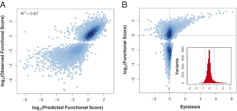
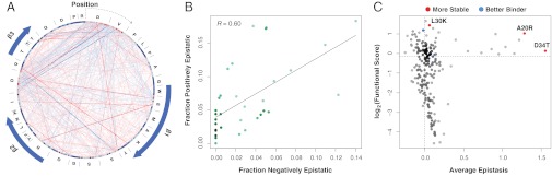
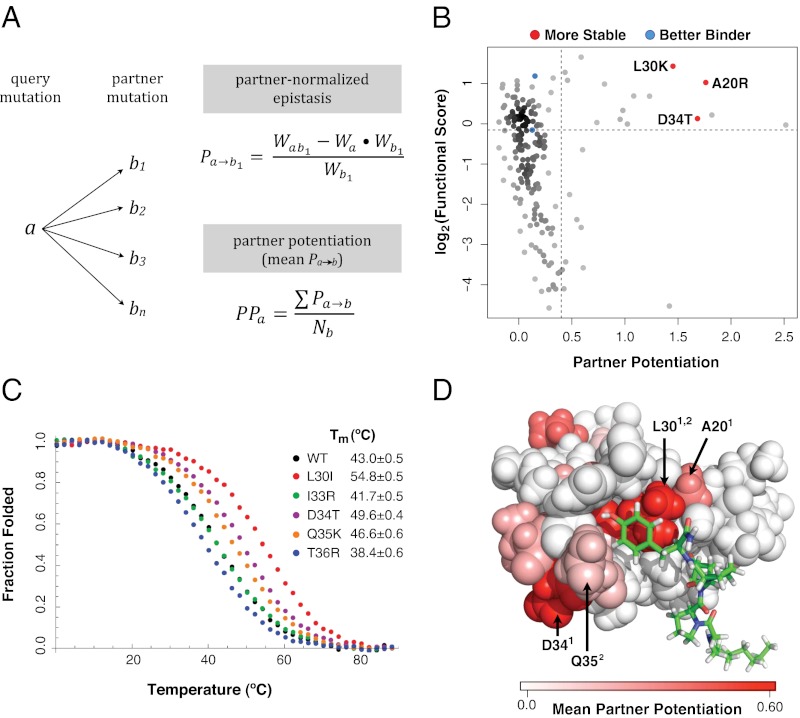
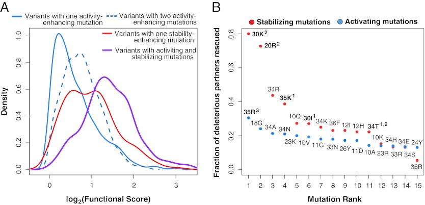
The ability of a protein to carry out a given function results from fundamental physicochemical properties that include the protein's structure, mechanism of action, and thermodynamic stability. Traditional approaches to study these properties have typically required the direct measurement of the property of interest, oftentimes a laborious undertaking. Although protein properties can be probed by mutagenesis, this approach has been limited by its low throughput. Recent technological developments have enabled the rapid quantification of a protein's function, such as binding to a ligand, for numerous variants of that protein. Here, we measure the ability of 47,000 variants of a WW domain to bind to a peptide ligand and use these functional measurements to identify stabilizing mutations without directly assaying stability. Our approach is rooted in the well-established concept that protein function is closely related to stability. Protein function is generally reduced by destabilizing mutations, but this decrease can be rescued by stabilizing mutations. Based on this observation, we introduce partner potentiation, a metric that uses this rescue ability to identify stabilizing mutations, and identify 15 candidate stabilizing mutations in the WW domain. We tested six candidates by thermal denaturation and found two highly stabilizing mutations, one more stabilizing than any previously known mutation. Thus, physicochemical properties such as stability are latent within these large-scale protein functional data and can be revealed by systematic analysis. This approach should allow other protein properties to be discovered.


Conflict of interest statement

The authors declare no conflict of interest.

Figures

Fig. 1.
Fig. 1.
Relationship between function and epistasis in a massive collection of double mutants. (A) The functional score of 5,010 doubly mutated variants was predicted from the functional scores of the component singly mutated variants using the product model. Predicted functional score is plotted against observed functional score and the two are highly correlated (Pearson’s R2 = 0.67). For each doubly mutated variant, the linear models used to generate the functional score had an R2 ≥ 0.75. (B) Epistasis scores calculated using the product model for the 5,010 variants are plotted against the functional score of the doubly mutated variant. The distribution of epistasis scores is shown in the Inset. Dashed lines are placed at ±1 SD from the mean.
Fig. 2.
Fig. 2.
Epistasis alone does not reliably identify stabilizing mutations. (A) A network view of epistatic interactions between mutations is shown. Individual mutations are presented as nodes in the graph and colored by functional scores (red corresponds to mutations with higher functional scores than WT, and blue corresponds to mutations with lower functional scores than WT). Mutations are arranged first by position and second by alphabet along the circumference of the graph in clockwise order from the 12:00 coordinate (zoom in to see individual mutations). The WT sequence is shown around the outside of the graph. Positive and negative epistatic interactions between mutations are shown as gradient red and blue edges, with width and shading proportional to the magnitude of the interaction. The position of the β-strands in the WW domain is indicated by the blue arrows. For clarity, only epistatic interactions at least 1 SD from the mean are shown. (B) For each position in the domain, the fraction of epistasis scores that are negative is plotted on the x axis, and the fraction of epistasis scores that are positive is plotted on the y axis. The fractions of positive and negative epistasis scores are correlated among positions (R = 0.60, P = 8.8 × 10⁻⁵). (C) The average epistasis score of each of the 192 single mutants found in 10 or more double mutants is plotted against the single-mutant functional score of each mutation. The known stabilizing (A20R, L30K, and D34T) and activity-enhancing (K21R and Q35R) mutations are highlighted in red and blue, respectively.
Fig. 3.
Fig. 3.
Partner potentiation reveals stabilizing mutations. (A) Partner potentiation is calculated for a query mutation a that forms doubly mutated variants with mutations b1, b2, b3 … bn. Partner-normalized epistasis scores are calculated for a by dividing the epistasis score of each double-mutant combination that a is found in by the functional score of the partner mutation (an example calculation for b1 is shown). The partner potentiation of a is calculated as the mean of the normalized epistasis scores (in this case, the scores for b1 … bn). (B) Partner potentiation is plotted for each single mutation against its functional score. The known stabilizing (A20R, L30K, and D34T) and activity-enhancing (K21R and Q35R) mutations are highlighted in red and blue, respectively. Mutations with a partner potentiation score greater than 0.4 and a functional score greater than 0.9 were considered to be stabilizing. (C) Stabilizing mutations were validated by thermal denaturation. The WT hYAP65 WW domain, the known stabilizing mutant D34T, and four candidate stabilizing mutants (L30I, I33R, Q35K, and T36R) are shown in black, purple, red, green, orange, and blue, respectively. (D) Positions in the hYAP65 NMR structure (Protein Data Bank ID code 1k9q) with stabilizing variants as judged by partner potentiation are shown, colored by the magnitude of the mean partner potentiation score using the PyMol software. Stabilizing mutations are distributed throughout the WW domain. Positions of previously known (superscript 1) and previously unknown (superscript 2) validated stabilizing mutations (20, 30, 34, 35) are highlighted.
Fig. 4.
Fig. 4.
Stabilizing mutations combine with activating mutations to drive large functional gains. (A) Mutations were classified as either stabilizing (functional score > 0.9 and partner potentiation > 0.4) or activating (functional score > 1 and partner potentiation ≤ 0.4). Double mutants with functional scores greater than WT were grouped into those mutants harboring both activating and stabilizing mutations (purple; n = 170), a single stabilizing mutation but no activating mutations (red; n = 156), a single activating mutation but no stabilizing mutation (solid blue; n = 1071), or two activating mutations (dashed blue; n = 852). The functional score distributions for each class are presented. Curves for variants with greater functional scores than WT containing paired stabilizing mutations are not drawn because of their low numbers (n = 4). (B) For each stabilizing (red) and activating mutation (blue), the fraction of deleterious single mutations rescued (i.e., found in double mutants with greater functional scores than WT) was calculated. Analysis was restricted to deleterious mutations that both stabilizing and activating mutations were paired with in double mutants in our library. Experimentally validated beneficial mutations are highlighted in bold; 1 denotes stabilizing mutations validated in this study, 2 denotes previously identified stabilizing mutations, and 3 denotes previously identified activating mutations.

References

    1. Anfinsen CB. Principles that govern the folding of protein chains. Science. 1973;181:223–230. - PubMed
    1. Fowler DM, et al. High-resolution mapping of protein sequence-function relationships. Nat Methods. 2010;7:741–746. - PMC - PubMed
    1. Hietpas RT, Jensen JD, Bolon DN. Experimental illumination of a fitness landscape. Proc Natl Acad Sci USA. 2011;108:7896–7901. - PMC - PubMed
    1. Araya CL, Fowler DM. Deep mutational scanning: Assessing protein function on a massive scale. Trends Biotechnol. 2011;29:435–442. - PMC - PubMed
    1. Brange J, et al. Monomeric insulins obtained by protein engineering and their medical implications. Nature. 1988;333:679–682. - PubMed

Publication types

MeSH terms

LinkOut - more resources