

Very distal apical prostate tumours: identification on multiparametric MRI at 3 Tesla
- PMID: 23035719
- PMCID: PMC6301064
- DOI: 10.1111/j.1464-410X.2012.11503.x
Very distal apical prostate tumours: identification on multiparametric MRI at 3 Tesla
Abstract
What's known on the subject? and What does the study add? MRI has been shown to improve prostate cancer detection rates. Pinto et al. (J Urol 2011; 86: 1281-5) reported their outcomes on 101 patients with low, moderate or high suspicion lesions on multiparametric MRI that were subsequently targeted via an MRI/ultrasound fusion biopsy platform. The prostate cancer detection rates were 27%, 66% and 89% respectively. Sciarra et al. (Clin Cancer Res 2010; 16: 1875-83) performed a prospective trial in 180 patients with prior negative biopsy and persistent PSA elevation. Patients were randomized to either MRI targeted biopsy followed by random 12-core TRUS biopsy vs random TRUS guided biopsy alone. Prostate cancer detection in the MRI targeted group was 45.5% vs 24.4% in the random group. Although MRI has been shown to improve prostate cancer detection rates, there has not previously been any work looking at the ability of MRI to detect prostate cancer localized to the very distal apex of the prostate. This is an important topic in that it might lead clinicians to counsel their patients in treatment decisions if it is felt that a treatment might not treat this section of the prostate well, e.g. high intensity focused ultrasound therapy that might spare the distal apex.
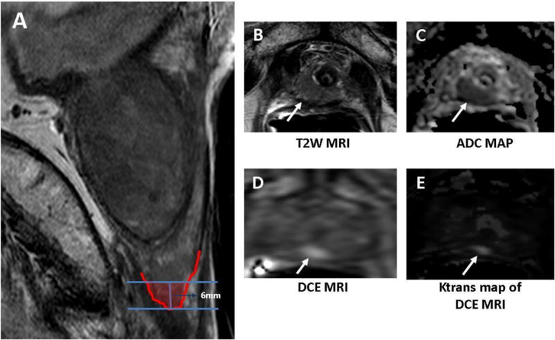
Objective: • To describe an undescribed 'very distal' apical prostate cancer on multiparametric MRI (mpMRI) since apical prostate cancer can be difficult to detect in transrectal ultrasound guided biopsy and might therefore be missed in treatment decisions such as high intensity focused ultrasound or surgical therapy.
Patients and methods: • From January 2011 to December 2012 a total of 210 consecutive patients underwent 3 T mpMRI with endorectal coil followed by our previously described MRI/ultrasound image fused and directed TRUS biopsies. • Patients also underwent 12-core TRUS sextant biopsies. • The inclusion criteria required at least one distal apical prostate lesion visualized on mpMRI and targeted for biopsy.
Results: • A total of 38 men (median age 62 years, median PSA 7.68 ng/dL) were identified as having distal apical prostate cancer on mpMRI. • Thirteen patients (34%) had a prior diagnosis of cancer and were on active surveillance protocols while 25 (66%) did not. Of those patients, 21 (55%) had undergone a median of two prior negative biopsies. • Twenty-two patients (58%) were positive on biopsy for prostate cancer. On breakdown of patients who were positive, 17 (77%) were positive on TRUS random biopsies and 21 (95%) were positive on MRI targeted biopsies with the majority of patients having multifocal disease. • At the distal apical lesions of interest, 80% were positive on MRI targeted biopsy. In addition 33% of these patients were upgraded based on MRI targeted biopsy at the distal lesion.
Conclusions: • Very distal apical prostate cancer can be accurately detected and sampled with mpMRI and subsequent MRI/ultrasound fusion biopsy. • This may aid clinicians and patients in decision making for therapeutic modalities.
© 2012 BJU INTERNATIONAL. NO CLAIM TO ORIGINAL US GOVERNMENT WORKS.
Figures
Comment in
-
Re: Very distal apical prostate tumours: identification on multiparametric MRI at 3 Tesla.J Urol. 2013 Oct;190(4):1248. doi: 10.1016/j.juro.2013.06.069. Epub 2013 Jun 26. J Urol. 2013. PMID: 24029313 No abstract available.
References
-
- Siegel R, Naishadham D, Jemal A. Cancer statistics, 2012. CA Cancer J Clin. 2012;62:10. - PubMed
-
- Ishii J, Ohori M, Scardino P, Tsuboi T, Slawin K, Wheeler T. Significance of the craniocaudal distribution of cancer in radical prostatectomy specimens. Int J Urol. 2007;14:817. - PubMed
-
- Dimmen M, Vlatkovic L, Hole KH, Nesland JM, Brennhovd B, Axcrona K. Transperineal prostate biopsy detects significant cancer in patients with elevated prostate-specific antigen (PSA) levels and previous negative transrectal biopsies. BJU Int. 2011 - PubMed
-
- Lee MC, Moussa AS, Zaytoun O, Yu C, Jones JS. Using a Saturation Biopsy Scheme Increases Cancer Detection During Repeat Biopsy in Men With High-grade Prostatic Intra-epithelial Neoplasia. Urology. 2011;78:1115. - PubMed
-
- Smith JA, Jr, Chan RC, Chang SS, Herrell SD, Clark PE, Baumgartner R, et al. A comparison of the incidence and location of positive surgical margins in robotic assisted laparoscopic radical prostatectomy and open retropubic radical prostatectomy. J Urol. 2007;178:2385. - PubMed
Publication types
MeSH terms
Grants and funding
LinkOut - more resources
Full Text Sources
Medical
Research Materials
Miscellaneous
