

PAM50 breast cancer subtyping by RT-qPCR and concordance with standard clinical molecular markers
- PMID: 23035882
- PMCID: PMC3487945
- DOI: 10.1186/1755-8794-5-44
PAM50 breast cancer subtyping by RT-qPCR and concordance with standard clinical molecular markers
Abstract
Background: Many methodologies have been used in research to identify the "intrinsic" subtypes of breast cancer commonly known as Luminal A, Luminal B, HER2-Enriched (HER2-E) and Basal-like. The PAM50 gene set is often used for gene expression-based subtyping; however, surrogate subtyping using panels of immunohistochemical (IHC) markers are still widely used clinically. Discrepancies between these methods may lead to different treatment decisions.
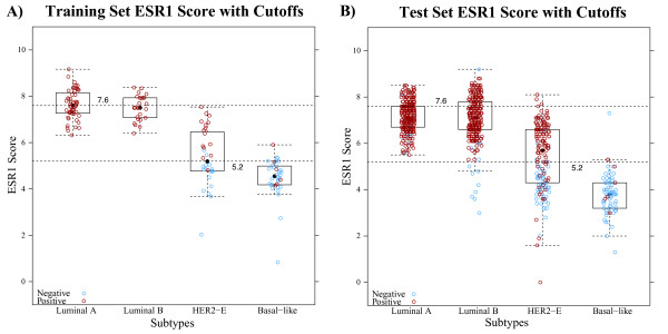
Methods: We used the PAM50 RT-qPCR assay to expression profile 814 tumors from the GEICAM/9906 phase III clinical trial that enrolled women with locally advanced primary invasive breast cancer. All samples were scored at a single site by IHC for estrogen receptor (ER), progesterone receptor (PR), and Her2/neu (HER2) protein expression. Equivocal HER2 cases were confirmed by chromogenic in situ hybridization (CISH). Single gene scores by IHC/CISH were compared with RT-qPCR continuous gene expression values and "intrinsic" subtype assignment by the PAM50. High, medium, and low expression for ESR1, PGR, ERBB2, and proliferation were selected using quartile cut-points from the continuous RT-qPCR data across the PAM50 subtype assignments.
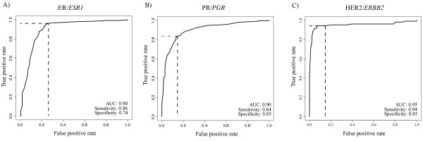
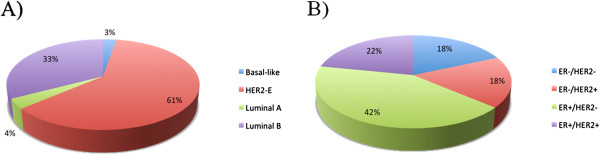
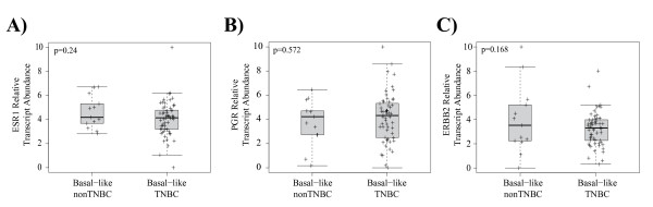
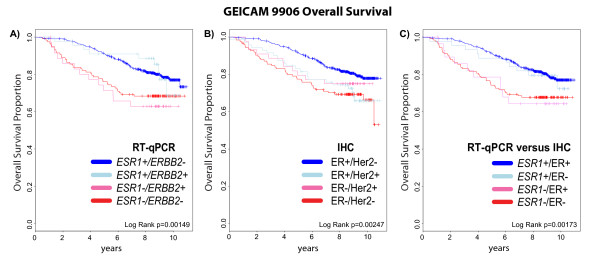
Results: ESR1, PGR, and ERBB2 gene expression had high agreement with established binary IHC cut-points (area under the curve (AUC) ≥ 0.9). Estrogen receptor positivity by IHC was strongly associated with Luminal (A and B) subtypes (92%), but only 75% of ER negative tumors were classified into the HER2-E and Basal-like subtypes. Luminal A tumors more frequently expressed PR than Luminal B (94% vs 74%) and Luminal A tumors were less likely to have high proliferation (11% vs 77%). Seventy-seven percent (30/39) of ER-/HER2+ tumors by IHC were classified as the HER2-E subtype. Triple negative tumors were mainly comprised of Basal-like (57%) and HER2-E (30%) subtypes. Single gene scoring for ESR1, PGR, and ERBB2 was more prognostic than the corresponding IHC markers as shown in a multivariate analysis.
Conclusions: The standard immunohistochemical panel for breast cancer (ER, PR, and HER2) does not adequately identify the PAM50 gene expression subtypes. Although there is high agreement between biomarker scoring by protein immunohistochemistry and gene expression, the gene expression determinations for ESR1 and ERBB2 status was more prognostic.
Figures
References
-
- Sorlie T, Perou CM, Tibshirani R, Aas T, Geisler S, Johnsen H, Hastie T, Eisen MB, van de Rijn M, Jeffrey SS. et al. Gene expression patterns of breast carcinomas distinguish tumor subclasses with clinical implications. Proc Natl Acad Sci U S A. 2001;98(19):10869–10874. doi: 10.1073/pnas.191367098. - DOI - PMC - PubMed
-
- Nielsen TO, Parker JS, Leung S, Voduc D, Ebbert M, Vickery T, Davies SR, Snider J, Stijleman IJ, Reed J. et al. A comparison of PAM50 intrinsic subtyping with immunohistochemistry and clinical prognostic factors in tamoxifen-treated estrogen receptor-positive breast cancer. Clin Cancer Res. 2010;16(21):5222–5232. doi: 10.1158/1078-0432.CCR-10-1282. - DOI - PMC - PubMed
Publication types
MeSH terms
Substances
Grants and funding
LinkOut - more resources
Full Text Sources
Other Literature Sources
Medical
Research Materials
Miscellaneous
