. 2012 Oct 4;16(5):R182.
doi: 10.1186/cc11666.

Angiopoietin-1 variant reduces LPS-induced microvascular dysfunction in a murine model of sepsis

Angiopoietin-1 variant reduces LPS-induced microvascular dysfunction in a murine model of sepsis

Alessio Alfieri et al. Crit Care. .

Abstract

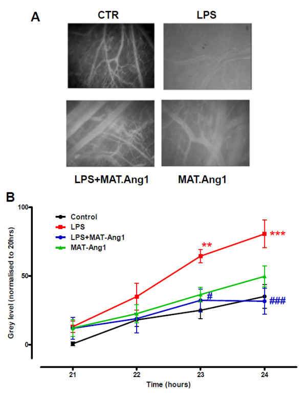
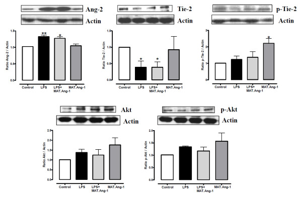
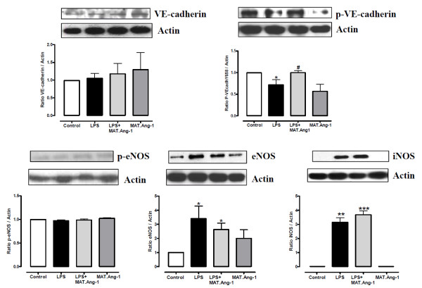
Introduction: Severe sepsis is characterised by intravascular or extravascular infection with microbial agents, systemic inflammation and microcirculatory dysfunction, leading to tissue damage, organ failure and death. The growth factor angiopoietin (Ang-1) has therapeutic potential but recombinant Ang-1 tends to aggregate and has a short half-life in vivo. This study aimed to investigate the acute effects of the more stable Ang-1 variant matrilin-1-angiopoietin-1 (MAT.Ang-1) on the function of the microcirculation in an experimental model of sepsis, and whether any protection by MAT-Ang-1 was associated with modulation of inflammatory cytokines, angiogenic factors or the endothelial nitric oxide synthase (eNOS)-Akt and vascular endothelial (VE)-cadherin pathways.

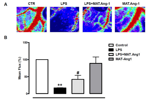
Methods: Aluminium window chambers were implanted into the dorsal skinfold of male C3H/HeN mice (7 to 10 weeks old) to expose the striated muscle microcirculation. Endotoxemia was induced by intraperitoneal injection of lipopolysaccharide (LPS, 1 mg/kg at 0 and 19 hours). MAT.Ang-1 was administered intravenously 20 hours after the onset of sepsis. Microcirculatory function was evaluated by intravital microscopy and Doppler fluximetry.

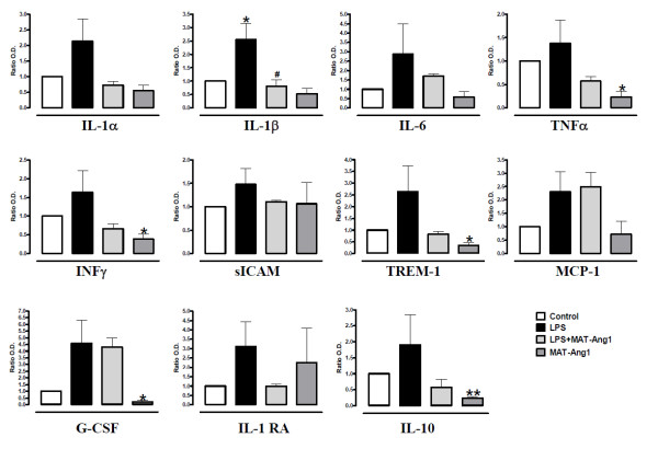
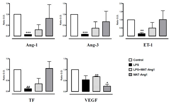
Results: Endotoxemia resulted in macromolecular leak, which was ameliorated by MAT.Ang-1 post-treatment. LPS induced a dramatic reduction in tissue perfusion, which was improved by MAT.Ang-1. Proteome profiler array analysis of skeletal muscle also demonstrated increased inflammatory and reduced angiogenic factors during endotoxemia. MAT.Ang-1 post-treatment reduced the level of IL-1β but did not significantly induce the expression of angiogenic factors. MAT.Ang-1 alone did not induce leak or increase angiogenic factors but did reduce vascular endothelial growth factor expression in controls.

Conclusion: Administration of MAT.Ang-1 after the onset of sepsis protects the microcirculation from endotoxemia-induced vascular dysfunction through reducing inflammation but without pro-angiogenic actions, thus representing a novel, potential pharmacotherapeutic agent for the treatment of sepsis.


Figures

Figure 1
Figure 1
MAT.Ang-1 reduces lipopolysaccharide-induced microvascular leak. (A) Representative images at 24 hours of murine skeletal muscle microvasculature from control (CTR), lipopolysaccharide (LPS)-treated, LPS + matrilin-1-angiopoietin-1 (MAT-Ang1)-treated and MAT-Ang1-treated groups. (B) Data expressed as mean ± standard error of the mean for change in grey level indicating macromolecular leak from 20 hours (n = 6). **P < 0.01 and ***P < 0.001 vs. control. #P < 0.05 and ###P < 0.001 vs. LPS.
Figure 2
Figure 2
MAT.Ang-1 ameliorates microvascular blood flow in sepsis. (A) Representative images from Doppler scanning at 24 hours of control (CTR), lipopolysaccharide (LPS), LPS + matrilin-1-angiopoietin-1 (MAT-Ang1) and MAT-Ang1 groups. Colours correspond to a perfusion range from 0 (black; lowest flux) to 1,000 (red; highest flux). (B) Data expressed as mean ± standard error of the mean of flux calculated as a percentage of control (n = 5). **P < 0.01 vs. control. #P < 0.05 vs. LPS.
Figure 3
Figure 3
MAT.Ang-1 reduces the inflammatory response in sepsis. Cytokine, chemokine and growth factor levels detected at 24 hours in skeletal muscle from control, lipopolysaccharide (LPS)-treated, LPS + matrilin-1-angiopoietin-1 (MAT-Ang1)-treated and MAT.Ang1-treated groups. Data expressed as mean ± standard error of the mean of fold-change from control (n = 3 different experiments performed in duplicate). *P < 0.05, **P < 0.01 vs. control. #P < 0.05 vs. LPS. G-CSF, granulocyte colony-stimulating factor; IL-1RA, IL-1 receptor antagonist; MCP-1, monocyte chemotactic protein-1; sICAM, soluble intracellular adhesion molecule-1; TREM-1, triggering receptor expressed on myeloid cells-1.
Figure 4
Figure 4
Angiogenic factors are not induced by MAT.Ang-1. Angiogenic markers detected at 24 hours in skeletal muscle from control, lipopolysaccharide (LPS), LPS + matrilin-1-angiopoietin-1 (MAT-Ang1) and MAT.Ang1 groups. Data are mean ± standard error of the mean of fold-change from control (n = 3 different experiments performed in duplicate). *P < 0.05, **P < 0.01 and ***P < 0.001 vs. control. Ang, angiopoietin; ET-1, endothelin-1; TF, tissue factor; VEGF, vascular endothelial growth factor.
Figure 5
Figure 5
Western blot analysis of angiopoietin-2, Tie-2 receptor and Akt. Detection of angiopoietin (Ang)-2, Tie-2, phospho-Tie-2, Akt and phospho-Akt by immunoblotting at 24 hours in skeletal muscle from control, lipopolysaccharide (LPS)-treated, LPS + matrilin-1-angiopoietin-1 (MAT-Ang1)-treated and MAT.Ang1-treated groups. Blots shown are representative of four animals per experimental group. Data on graphs expressed as mean ± standard error of the mean of fold-change from control (n = 4). *P < 0.05 and **P < 0.01 vs. control.
Figure 6
Figure 6
Western blot analysis of vascular endothelial-cadherin, endothelial nitric oxide synthase and inducible nitric oxide synthase. Detection of vascular endothelial (VE)-cadherin, phospho-VE-cadherin, phospho-endothelial nitric oxide synthase (phospho-eNOS), eNOS and inducible nitric oxide synthase (iNOS) by western blot analysis at 24 hours in skeletal muscle from control, lipopolysaccharide (LPS)-treated, LPS + matrilin-1-angiopoietin-1 (MAT-Ang1)-treated and MAT.Ang1-treated groups. Blots shown are representative of four animals per experimental group. Data on graphs expressed as mean ± standard error of the mean of fold-change from control (n = 4). *P < 0.05, **P < 0.01, ***P <0.001 vs. control. #P < 0.05 vs. LPS.

Comment in

Similar articles

Cited by

References

    1. Hawiger J, Musser JM. How to approach gene wars in sepsis? Crit Care. 2011;16:1007. doi: 10.1186/cc10482. - DOI - PMC - PubMed
    1. Spronk PE, Zandstra DF, Ince C. Bench-to-bedside review: sepsis is a disease of the microcirculation. Crit Care. 2004;16:462–468. doi: 10.1186/cc2894. - DOI - PMC - PubMed
    1. Ince C. The microcirculation is the motor of sepsis. Crit Care. 2005;16(Suppl 4):S13–S19. doi: 10.1186/cc3753. - DOI - PMC - PubMed
    1. Jones AE, Puskarich MA. Sepsis-induced tissue hypoperfusion. Crit Care Clin. 2009;16:769–779, ix. doi: 10.1016/j.ccc.2009.06.003. - DOI - PubMed
    1. Angus DC, Linde-Zwirble WT, Lidicker J, Clermont G, Carcillo J, Pinsky MR. Epidemiology of severe sepsis in the United States: analysis of incidence, outcome, and associated costs of care. Crit Care Med. 2001;16:1303–1310. doi: 10.1097/00003246-200107000-00002. - DOI - PubMed

Publication types