

Remodeling of axo-spinous synapses in the pathophysiology and treatment of depression
- PMID: 23036622
- PMCID: PMC3566360
- DOI: 10.1016/j.neuroscience.2012.09.057
Remodeling of axo-spinous synapses in the pathophysiology and treatment of depression
Abstract
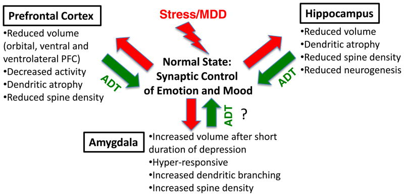
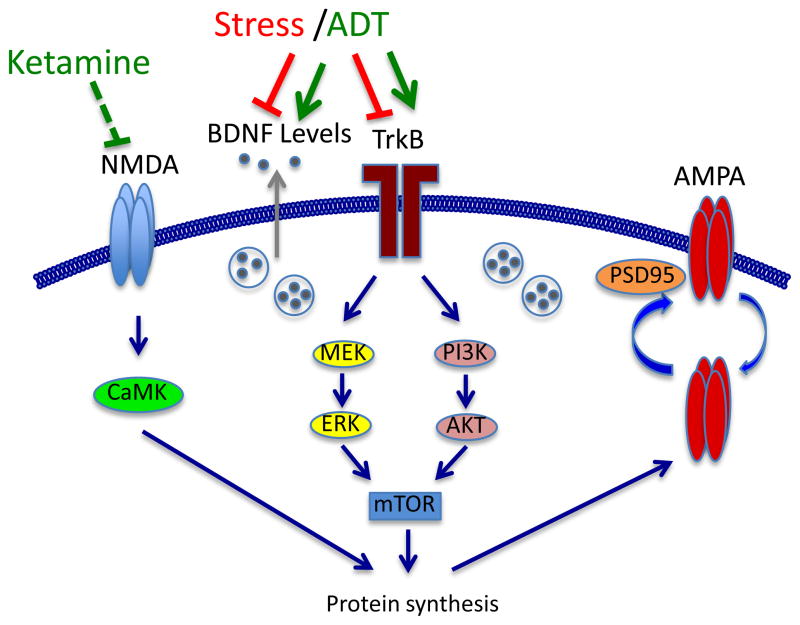
Dendritic spines provide a compartment for assembly and functional organization of synaptic machinery that plays a fundamental role in neuronal communication and neuroplasticity. Studies in humans as well as in animal models have demonstrated abnormal spine architecture in several psychiatric disorders, including depression and other stress-related illnesses. The negative impact of stress on the density and organization of spines is thought to contribute to the behavioral deficits caused by stress exposure. Moreover, there is now evidence that medication-induced recovery involves changes in synaptic plasticity and dendrite morphology, including increased expression of pre- and postsynaptic plasticity-related proteins, as well as the density and function of axo-spinous synapses. Here we review the evidence from brain imaging and postmortem studies demonstrating that depression is accompanied by structural and functional alterations of cortical and limbic brain regions, including the prefrontal cortex, hippocampus and amygdala. In addition, we present more direct evidence from basic research studies that exposure to stress alters spine morphology, function and plasticity and that antidepressants, particularly new rapid acting agents, reverse these effects. Elucidation of the signaling pathways and molecular mechanisms that control spine synapse assembly and plasticity will contribute to a better understanding of the pathophysiology of depression and development of novel, more effective therapeutic agents.
Keywords: ACd; BD; BDNF; CREB; CRS; CUS; ECS; ERK; FST; HPA; IES; IL; LH; LTD; LTP; MAOI; MAPK-phosphatase 1; MDD; MKP-1; MRI; MS; N-methyl-d-aspartate glutamate receptor; NMDA; OFC; PS; PTSD; SSRI; antidepressant; bipolar disorder; brain-derived neurotrophic factor; cAMP response element-binding protein; chronic restraint stress; chronic unpredictable stress; dorsal anterior cingulate; electroconvulsive seizure; extracellular-signal-regulated kinase; fMRI; forced swim test; functional magnetic resonance imaging; glutamate; hippocampus; hypothalamic–pituitary–adrenal; inescapable stress; infralimbic; learned helplessness; long-term depression; long-term potentiation; mPFC; mTOR; magnetic resonance imaging; major depressive disorder; mammalian target of rapamycin; maternal separation; medial prefrontal cortex; monoamine oxidase inhibitor; neurotrophic factor; orbitofrontal cortex; posttraumatic stress disorder; prefrontal cortex; prenatal stress; selective serotonin re-uptake inhibitor; stress.
Copyright © 2012 IBRO. Published by Elsevier Ltd. All rights reserved.
Figures
References
-
- Ackermann M, Matus A. Activity-induced targeting of profilin and stabilization of dendritic spine morphology. Nat Neurosci. 2003;6:1194–1200. - PubMed
-
- Alcantara-Gonzalez F, Juarez I, Solis O, Martinez-Tellez I, Camacho-Abrego I, Masliah E, Mena R, Flores G. Enhanced dendritic spine number of neurons of the prefrontal cortex, hippocampus, and nucleus accumbens in old rats after chronic donepezil administration. Synapse. 2010;64:786–793. - PMC - PubMed
-
- Allain H, Lieury A, Brunet-Bourgin F, Mirabaud C, Trebon P, Le Coz F, Gandon JM. Antidepressants and cognition: comparative effects of moclobemide, viloxazine and maprotiline. Psychopharmacology (Berl) 1992;106(Suppl):S56–61. - PubMed
-
- Almeida OP, Lautenschlager N, Vasikaram S, Leedman P, Flicker L. Association between physiological serum concentration of estrogen and the mental health of community-dwelling postmenopausal women age 70 years and over. Am J Geriatr Psychiatry. 2005;13:142–149. - PubMed
-
- Alvarez VA, Sabatini BL. Anatomical and physiological plasticity of dendritic spines. Annu Rev Neurosci. 2007;30:79–97. - PubMed
Publication types
MeSH terms
Substances
Grants and funding
LinkOut - more resources
Full Text Sources
Other Literature Sources
Medical
Miscellaneous
