. 2013 Jan;1832(1):29-38.
doi: 10.1016/j.bbadis.2012.09.010. Epub 2012 Oct 2.

Lysophosphatidic acid induces increased BACE1 expression and Aβ formation

Affiliations

Lysophosphatidic acid induces increased BACE1 expression and Aβ formation

Jing Shi et al. Biochim Biophys Acta. 2013 Jan.

Abstract

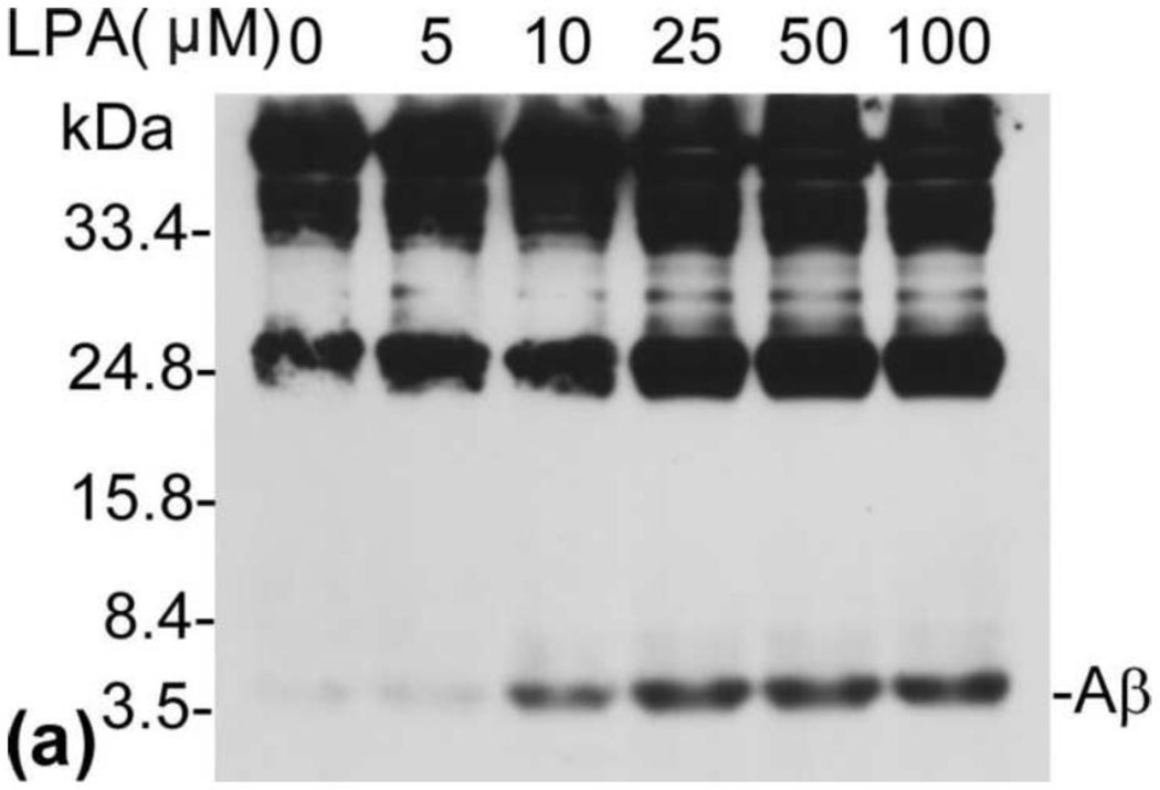
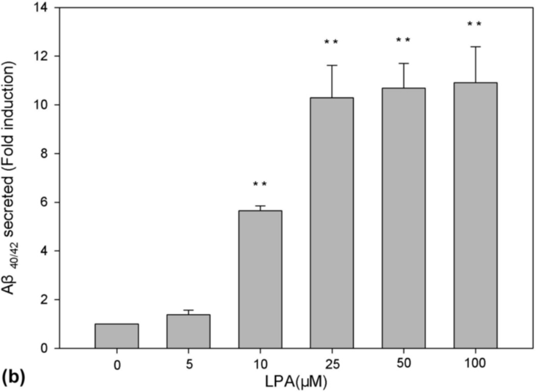
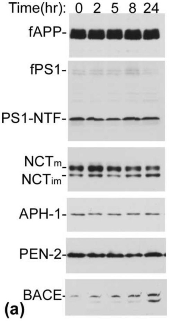
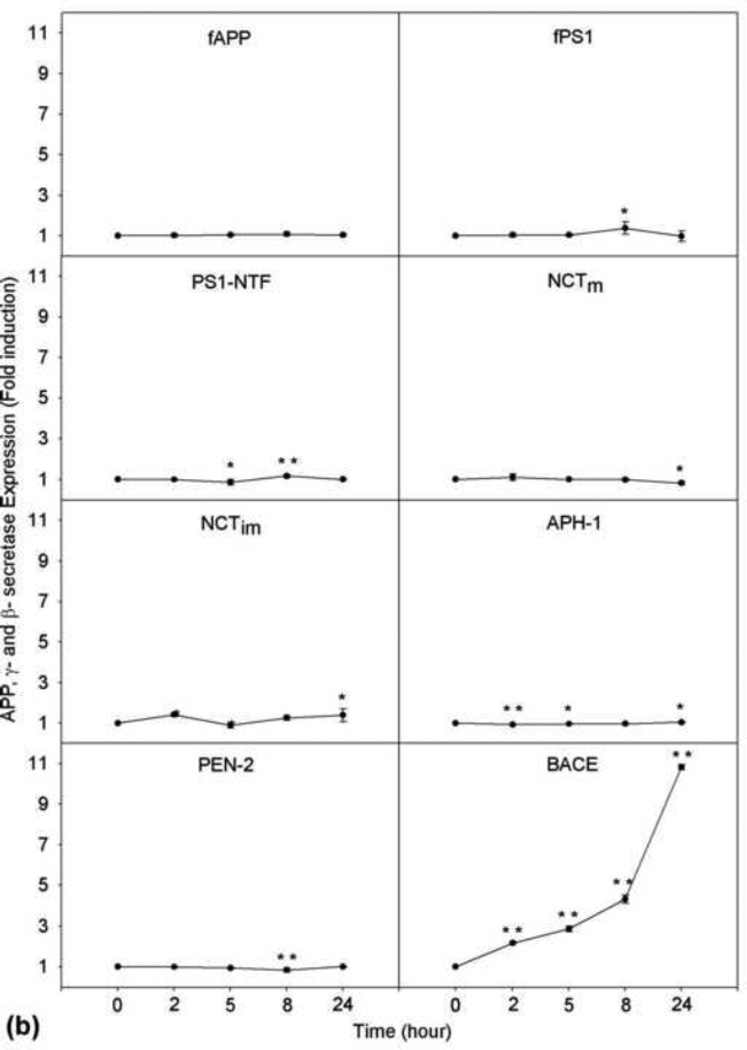
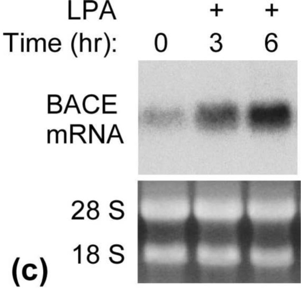
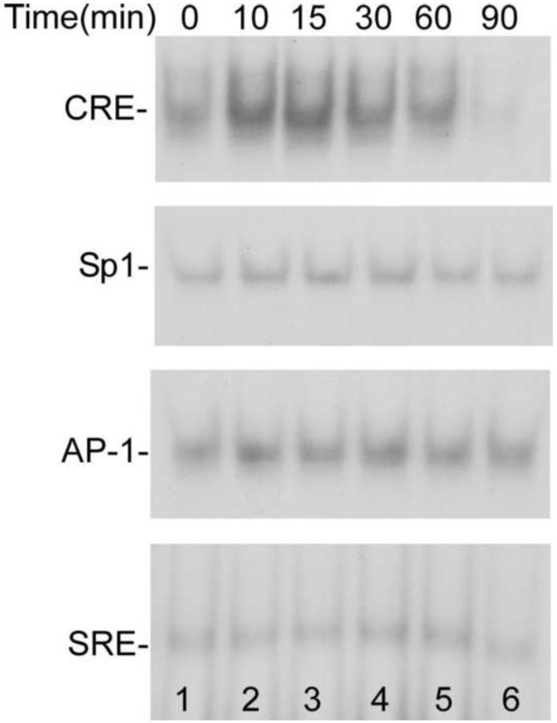
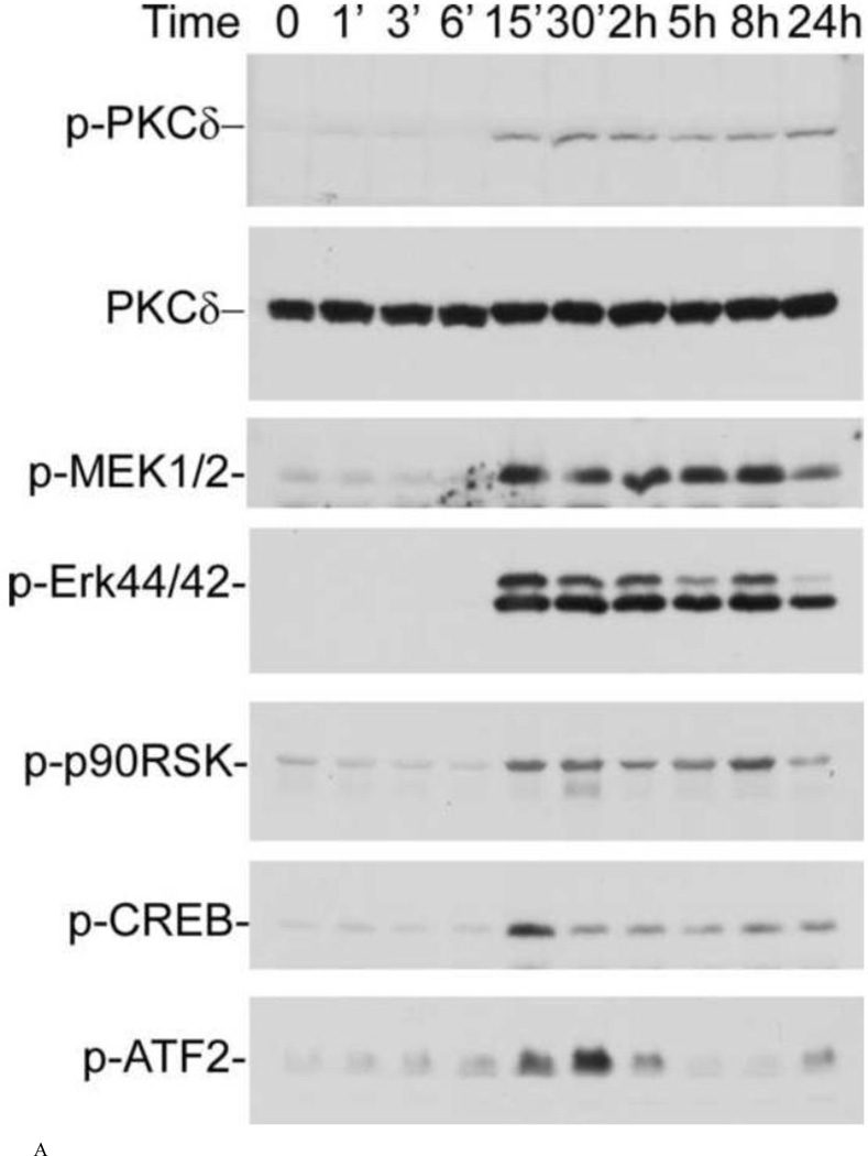
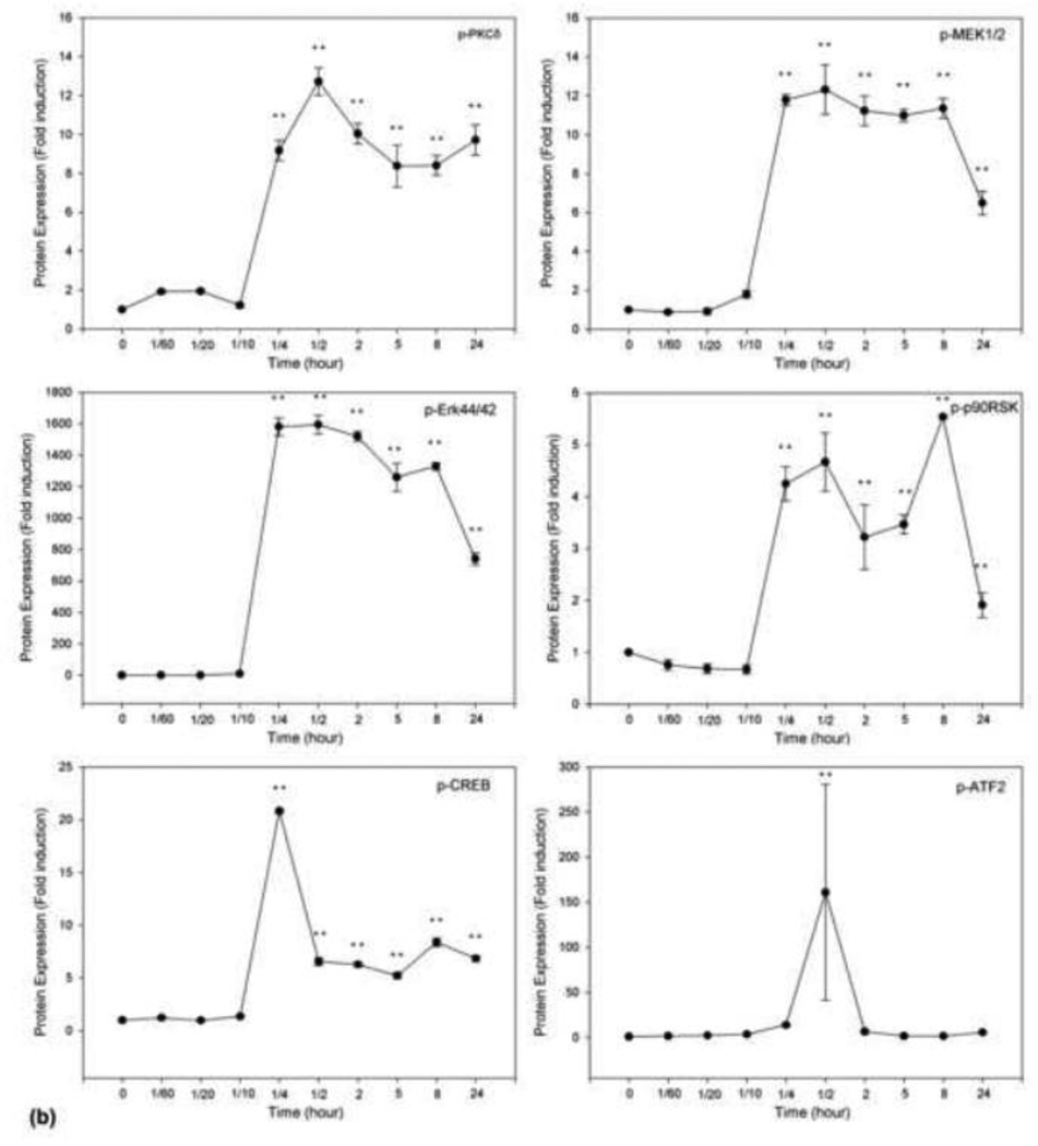
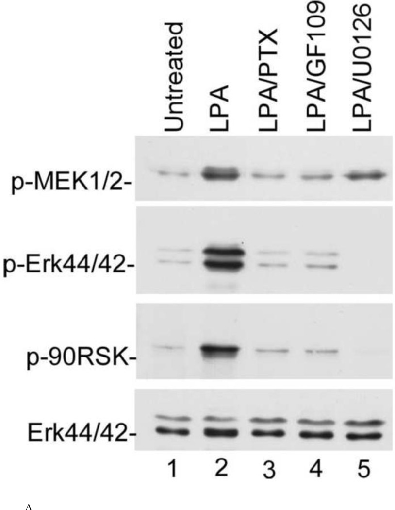
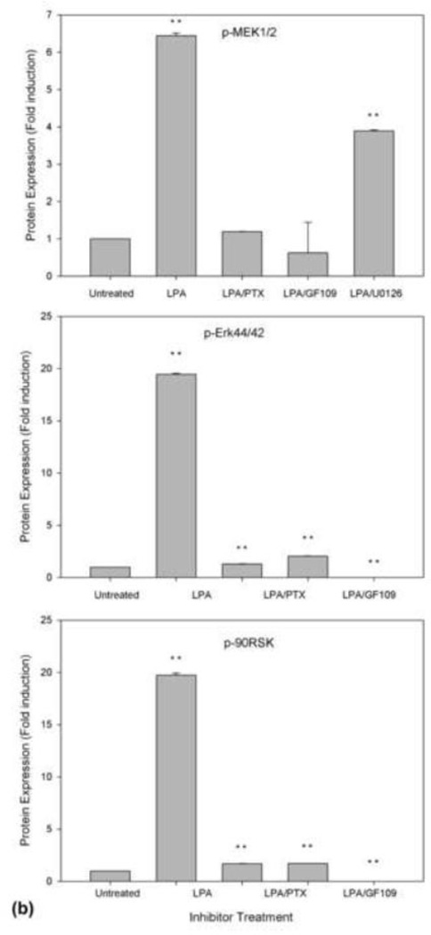
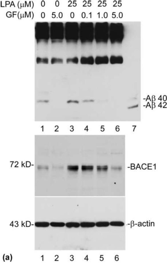
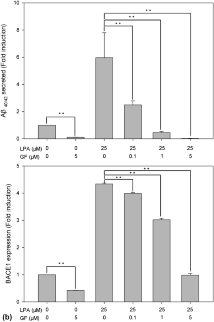
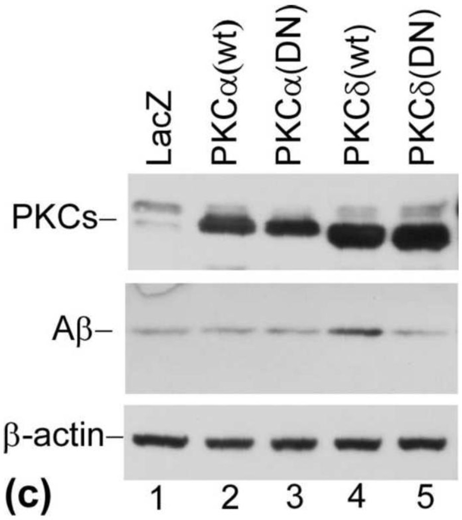
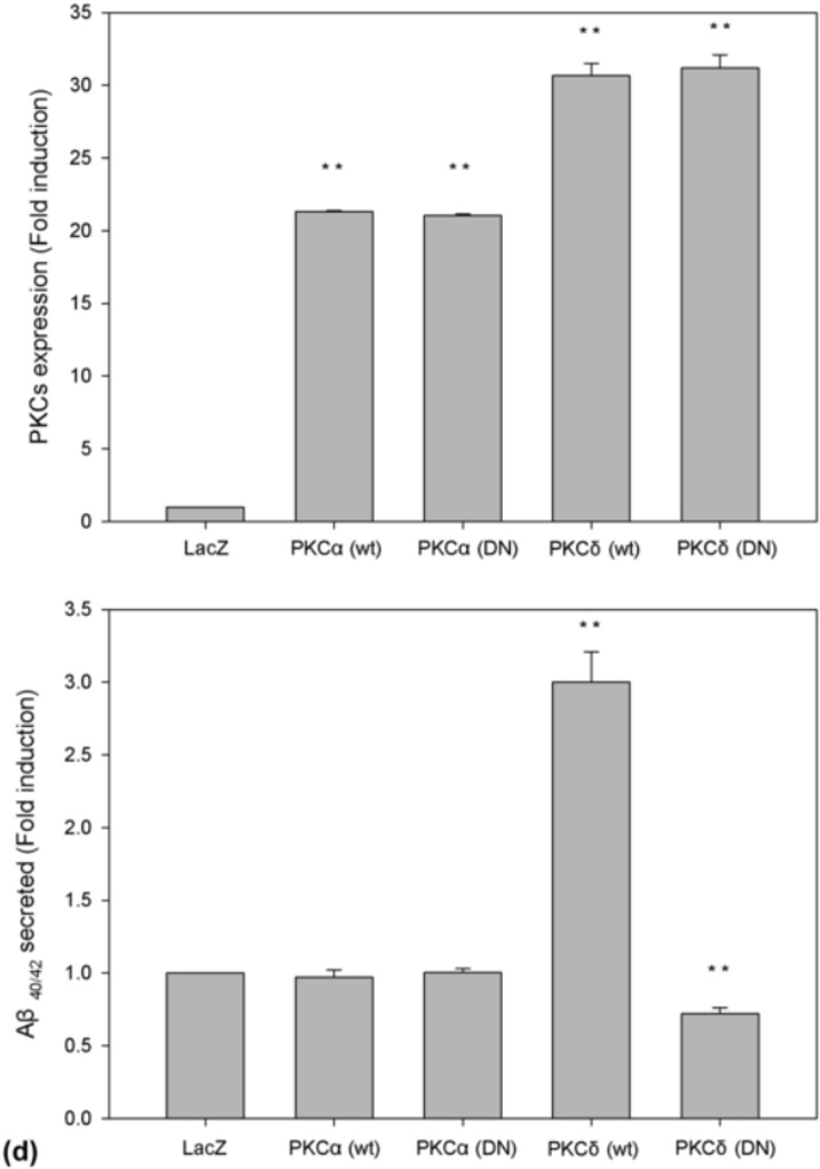
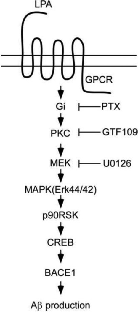
The abnormal production and accumulation of β-amyloid peptide (Aβ), which is produced from amyloid precursor protein (APP) by the sequential actions of β-secretase and γ-secretase, are thought to be the initial causative events in the development of Alzheimer's disease (AD). Accumulating evidence suggests that vascular factors play an important role in the pathogenesis of AD. Specifically, studies have suggested that one vascular factor in particular, oxidized low density lipoprotein (oxLDL), may play an important role in regulating Aβ formation in AD. However, the mechanism by which oxLDL modulates Aβ formation remains elusive. In this study, we report several new findings that provide biochemical evidence suggesting that the cardiovascular risk factor oxLDL may contribute to Alzheimer's disease by increasing Aβ production. First, we found that lysophosphatidic acid (LPA), the most bioactive component of oxLDL induces increased production of Aβ. Second, our data strongly indicate that LPA induces increased Aβ production via upregulating β-secretase expression. Third, our data strongly support the notion that different isoforms of protein kinase C (PKC) may play different roles in regulating APP processing. Specifically, most PKC members, such as PKCα, PKCβ, and PKCε, are implicated in regulating α-secretase-mediated APP processing; however, PKCδ, a member of the novel PKC subfamily, is involved in LPA-induced upregulation of β-secretase expression and Aβ production. These findings may contribute to a better understanding of the mechanisms by which the cardiovascular risk factor oxLDL is involved in Alzheimer's disease.


Figures

Fig. 1
Fig. 1
LPA induces a dose-dependent increase in Aβ formation. (a) N2a cells were starved in serum-free medium containing 0.5% serum for 24 h and then treated with LPA at various concentrations as indicated. Secreted Aβ was immunoprecipitated from CM with 6E10 and analyzed by urea (8M) SDS-PAGE (13%) followed by Western blot using antibody 6E10. All Western blot data presented in this study are representative of the results obtained from at least three repeated experiments. (b) Quantitative analysis of Western blotting signals of secreted Aβ shown in (a). ANOVA identified a significant induction in secreted Aβ in dose curve experiment. **p≤0.01 vs. untreated control.
Fig. 1
Fig. 1
LPA induces a dose-dependent increase in Aβ formation. (a) N2a cells were starved in serum-free medium containing 0.5% serum for 24 h and then treated with LPA at various concentrations as indicated. Secreted Aβ was immunoprecipitated from CM with 6E10 and analyzed by urea (8M) SDS-PAGE (13%) followed by Western blot using antibody 6E10. All Western blot data presented in this study are representative of the results obtained from at least three repeated experiments. (b) Quantitative analysis of Western blotting signals of secreted Aβ shown in (a). ANOVA identified a significant induction in secreted Aβ in dose curve experiment. **p≤0.01 vs. untreated control.
Fig. 2
Fig. 2
LPA induces increased expression of β-secretase protein. (a) Starved cells were treated with 25 µM LPA for different lengths of time as indicated. Cell lysates collected at each time point were analyzed by SDS-PAGE followed by Western blotting assay. Top panel was probed with C15 raised against the C-terminal 15 amino acids to determine the level of full-length APP. Second panel is a blot probed with anti-PS1N to determine the expression level of PS1. Third, fourth, and fifth panels are blots probed with anti-NCT, anti-Aph-1α, and anti-Pen-2 antibodies, respectively, to determine the expression levels of these proteins. The bottom panel is the blot probed with anti-BACE1 antibody to determine the expression level of β-secretase. NCTm and NCTim indicate the mature and immature forms of Nicastrin. (b) Quantitative analysis of Western blotting signals shown in (a) identified a significant induction of BACE1 but not of components of γ-secretase or APP. *p≤0.05; **p≤0.01 vs. untreated control. (c) LPA induction of BACE1 mRNA. Starved cells were treated with 25 μM LPA. At the time points indicated, total RNA was isolated, and Northern blot analysis was performed using mouse BACE1 cDNA as a probe as described in our previous study [19]. Visualized bands of 28S and 18S ribosomal RNA served as loading controls.
Fig. 2
Fig. 2
LPA induces increased expression of β-secretase protein. (a) Starved cells were treated with 25 µM LPA for different lengths of time as indicated. Cell lysates collected at each time point were analyzed by SDS-PAGE followed by Western blotting assay. Top panel was probed with C15 raised against the C-terminal 15 amino acids to determine the level of full-length APP. Second panel is a blot probed with anti-PS1N to determine the expression level of PS1. Third, fourth, and fifth panels are blots probed with anti-NCT, anti-Aph-1α, and anti-Pen-2 antibodies, respectively, to determine the expression levels of these proteins. The bottom panel is the blot probed with anti-BACE1 antibody to determine the expression level of β-secretase. NCTm and NCTim indicate the mature and immature forms of Nicastrin. (b) Quantitative analysis of Western blotting signals shown in (a) identified a significant induction of BACE1 but not of components of γ-secretase or APP. *p≤0.05; **p≤0.01 vs. untreated control. (c) LPA induction of BACE1 mRNA. Starved cells were treated with 25 μM LPA. At the time points indicated, total RNA was isolated, and Northern blot analysis was performed using mouse BACE1 cDNA as a probe as described in our previous study [19]. Visualized bands of 28S and 18S ribosomal RNA served as loading controls.
Fig. 3
Fig. 3
Time course of LPA-induced increase in binding activity of transcriptional factor CREB. LPA-induced binding activities of various transcription factors were analyzed by EMSA. Nuclear proteins from cells untreated (0) or treated with 25 μM LPA for indicated times were incubated with radiolabeled oligonucleotides that contain one of the following possible transcription factor binding sites of BACE1 promoter for 20 min: CRE site (top panel), Sp1 site (second panel), Ap-1 site (third panel), and SRE site (bottom panel). Then, proteins were separated by a 6% TBE acrylamide gel. The protein-nucleic acid complexes were determined by autoradiography of 32P-labeled nucleic acid.
Fig. 4
Fig. 4
Time course of LPA-induced activation of signaling cascades. N2a cells were stimulated with 25 μM LPA, and, at the time points indicated, cell lysates were analyzed by 10% SDS-PAGE and transferred to a PVDF membrane. Phosphorylated PKCδ (top panel), MEK1/2 (third panel), MAPK (Erk44/42, fourth panel), p90RSK (fifth panel), CREB (sixth panel), and ATF2 (seventh panel) were detected with antibodies against phospho-MEK1/2 (PMEK1/2) (Ser217/221), phospho-MAPK (P-Erk44/42, Thr202/Tyr204), and phospho-p90RSK (P-p90RSK, Ser381), phosphor-CREB (p-CREB), and phosphor-ATF2 (p-ATF-2), respectively (Cell Signaling Technology). The membranes in the top panel were stripped and re-probed with anti-PKCδ antibody to ensure equal loading. (a) representative of three repeated Western blot results; (b) quantification and statistical analysis of the western blot results shown in (a).
Fig. 4
Fig. 4
Time course of LPA-induced activation of signaling cascades. N2a cells were stimulated with 25 μM LPA, and, at the time points indicated, cell lysates were analyzed by 10% SDS-PAGE and transferred to a PVDF membrane. Phosphorylated PKCδ (top panel), MEK1/2 (third panel), MAPK (Erk44/42, fourth panel), p90RSK (fifth panel), CREB (sixth panel), and ATF2 (seventh panel) were detected with antibodies against phospho-MEK1/2 (PMEK1/2) (Ser217/221), phospho-MAPK (P-Erk44/42, Thr202/Tyr204), and phospho-p90RSK (P-p90RSK, Ser381), phosphor-CREB (p-CREB), and phosphor-ATF2 (p-ATF-2), respectively (Cell Signaling Technology). The membranes in the top panel were stripped and re-probed with anti-PKCδ antibody to ensure equal loading. (a) representative of three repeated Western blot results; (b) quantification and statistical analysis of the western blot results shown in (a).
Fig. 5
Fig. 5
Effect of PTX, GF109, and U0126 on the activation of MEK (top panel), MAPK (second panel), and p90RSK (third panel). N2a cells were pretreated with 100 ng/ml pertussis toxin for 16 h or pretreated with 5 µM GF109 or 10 μM U0126 for 30 min prior to the addition of 25 μM LPA for 15 min. The cell lysates were analyzed as described in Fig. 4. The membranes of the second panel were stripped and re-probed with anti-Erk antibody to ensure equal loading. (a) representative of three repeated Western blot results; (b) quantification and statistical analysis of the western blot results shown in (a).
Fig. 5
Fig. 5
Effect of PTX, GF109, and U0126 on the activation of MEK (top panel), MAPK (second panel), and p90RSK (third panel). N2a cells were pretreated with 100 ng/ml pertussis toxin for 16 h or pretreated with 5 µM GF109 or 10 μM U0126 for 30 min prior to the addition of 25 μM LPA for 15 min. The cell lysates were analyzed as described in Fig. 4. The membranes of the second panel were stripped and re-probed with anti-Erk antibody to ensure equal loading. (a) representative of three repeated Western blot results; (b) quantification and statistical analysis of the western blot results shown in (a).
Fig. 6
Fig. 6
PKCδ regulates Aβ production. (a) PKC inhibitor GF109203X (GF) blocks LPA-induced Aβ formation. Prior to treatment with LPA, cells were pretreated with PKC inhibitor GF at various concentrations for 30 min and then treated with LPA at indicated concentrations for 24 h. Secreted Aβ was immunoprecipitated from CM (top panel) was analyzed by 13 % urea SDS-PAGE followed by Western blotting using 6E10. BACE1 expression was determined by direct analyzing the cell lysates (second panel). Third panel is the reprobe of the membrane in the second with anti-β-actin to ensure even loading of the samples. Lane 7 is the Aβ40/42 standard mix. (b) Western blotting signals from three repeated experiments shown in (a) were quantified, and the control and LPA-treated samples compared with GF-treated samples. (c) Overexpression of PKCδ increases Aβ production. Twenty-four hours after infection with recombinant adenovirus-expressing PKC variants, cell lysates were separated by 10% SDS-PAGE, followed by Western blot analysis using a mix of anti-PKCα and PKCδ antibodies (top panel, Cell Signaling Technology). (d) Quantification and statistical analysis of the Western blot results shown in (c). Aβ was immunoprecipitated from CM (middle panel). Bottom panel is the reprobe of the membrane in the top panel for β-actin. Statistical significances were calculated by GF-treated and non-GF-treated samples. Statistically significant results are indicated: **p≤0.01.
Fig. 6
Fig. 6
PKCδ regulates Aβ production. (a) PKC inhibitor GF109203X (GF) blocks LPA-induced Aβ formation. Prior to treatment with LPA, cells were pretreated with PKC inhibitor GF at various concentrations for 30 min and then treated with LPA at indicated concentrations for 24 h. Secreted Aβ was immunoprecipitated from CM (top panel) was analyzed by 13 % urea SDS-PAGE followed by Western blotting using 6E10. BACE1 expression was determined by direct analyzing the cell lysates (second panel). Third panel is the reprobe of the membrane in the second with anti-β-actin to ensure even loading of the samples. Lane 7 is the Aβ40/42 standard mix. (b) Western blotting signals from three repeated experiments shown in (a) were quantified, and the control and LPA-treated samples compared with GF-treated samples. (c) Overexpression of PKCδ increases Aβ production. Twenty-four hours after infection with recombinant adenovirus-expressing PKC variants, cell lysates were separated by 10% SDS-PAGE, followed by Western blot analysis using a mix of anti-PKCα and PKCδ antibodies (top panel, Cell Signaling Technology). (d) Quantification and statistical analysis of the Western blot results shown in (c). Aβ was immunoprecipitated from CM (middle panel). Bottom panel is the reprobe of the membrane in the top panel for β-actin. Statistical significances were calculated by GF-treated and non-GF-treated samples. Statistically significant results are indicated: **p≤0.01.
Fig. 7
Fig. 7
A schematic summary of the molecular pathway by which LPA induces Aβ production.

References

    1. Walsh DM, Klyubin I, Fadeeva JV, Rowan MJ, Selkoe DJ. Amyloid-beta oligomers: their production, toxicity and therapeutic inhibition. Biochem. Soc. Trans. 2002;30:552–557. - PubMed
    1. Selkoe DJ. Alzheimer's disease is a synaptic failure. Science. 2002;298:789–791. - PubMed
    1. Hardy J, Selkoe DJ. The amyloid hypothesis of Alzheimer's disease: progress and problems on the road to therapeutics. Science. 2002;297:353–356. - PubMed
    1. Selkoe DJ. Alzheimer's disease: genes, proteins, and therapy. Physiol. Rev. 2001;81:741–766. - PubMed
    1. Yan R, Bienkowski MJ, Shuck ME, Miao H, Tory MC, Pauley AM, Brashier JR, Stratman NC, Mathews WR, Buhl AE, Carter DB, Tomasselli AG, Parodi LA, Heinrikson RL, Gurney ME. Membrane-anchored aspartyl protease with Alzheimer's disease beta-secretase activity. Nature. 1999;402:533–537. - PubMed

Publication types

MeSH terms