. 2012 Nov 23;287(48):40414-24.
doi: 10.1074/jbc.M112.421404. Epub 2012 Oct 4.

Production of selenoprotein P (Sepp1) by hepatocytes is central to selenium homeostasis

Affiliations

Production of selenoprotein P (Sepp1) by hepatocytes is central to selenium homeostasis

Kristina E Hill et al. J Biol Chem. .

Abstract

Background: Sepp1 transports selenium, but its complete role in selenium homeostasis is not known.

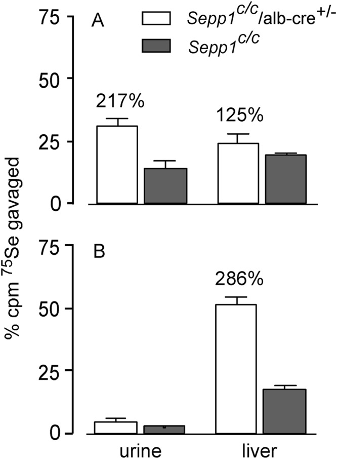
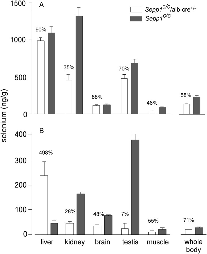
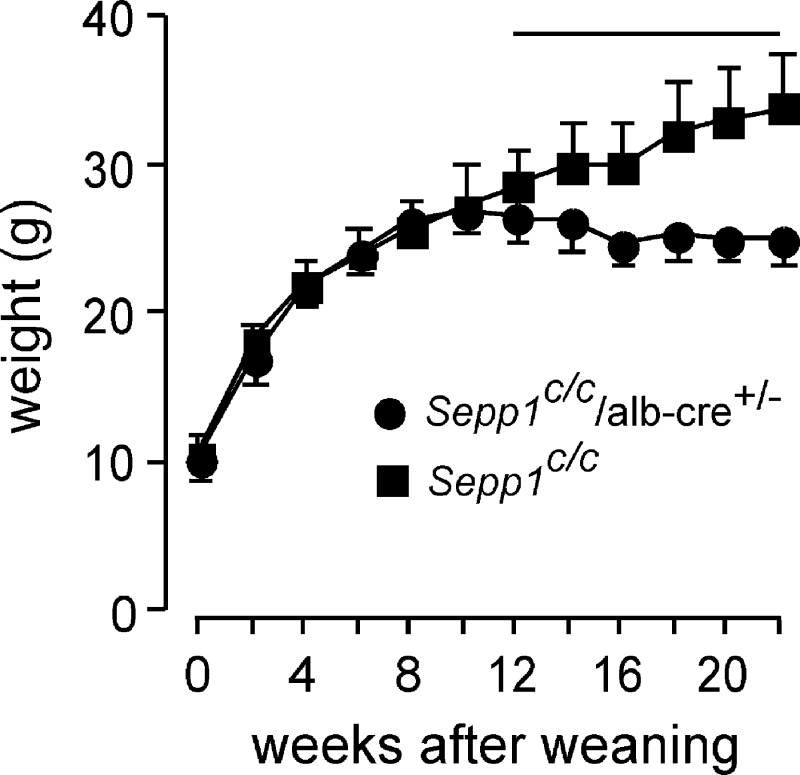
Results: Deletion of Sepp1 in hepatocytes increases liver selenium at the expense of other tissues and decreases whole-body selenium by increasing excretion.

Conclusion: Sepp1 production by hepatocytes retains selenium in the organism and distributes it from the liver to peripheral tissues.

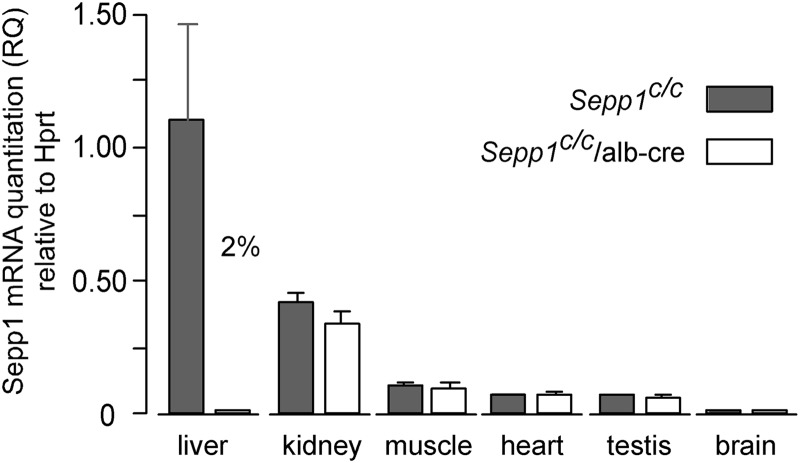
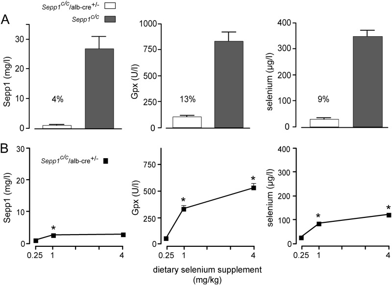
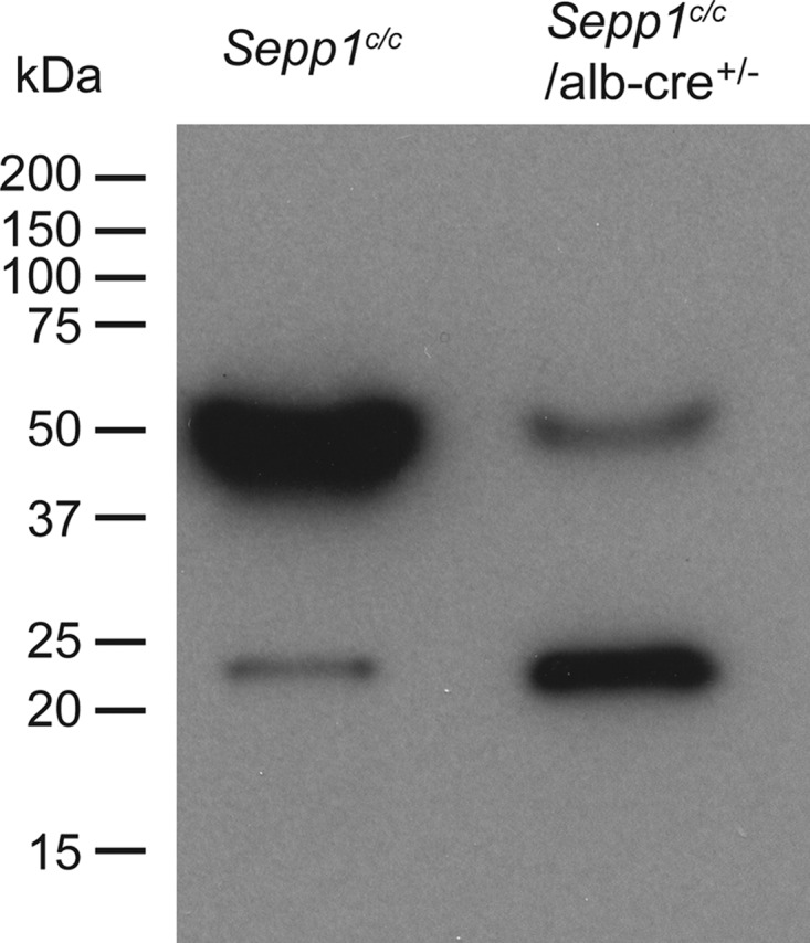
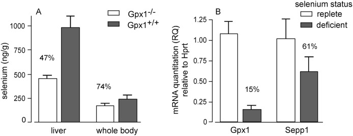
Significance: Sepp1 is central to selenium homeostasis. Sepp1 is a widely expressed extracellular protein that in humans and mice contains 10 selenocysteine residues in its primary structure. Extra-hepatic tissues take up plasma Sepp1 for its selenium via apolipoprotein E receptor-2 (apoER2)-mediated endocytosis. The role of Sepp1 in the transport of selenium from liver, a rich source of the element, to peripheral tissues was studied using mice with selective deletion of Sepp1 in hepatocytes (Sepp1(c/c)/alb-cre(+/-) mice). Deletion of Sepp1 in hepatocytes lowered plasma Sepp1 concentration to 10% of that in Sepp1(c/c) mice (controls) and increased urinary selenium excretion, decreasing whole-body and tissue selenium concentrations. Under selenium-deficient conditions, Sepp1(c/c)/alb-cre(+/-) mice accumulated selenium in the liver at the expense of extra-hepatic tissues, severely worsening clinical manifestations of dietary selenium deficiency. These findings are consistent with there being competition for metabolically available hepatocyte selenium between the synthesis of selenoproteins and the synthesis of selenium excretory metabolites. In addition, selenium deficiency down-regulated the mRNA of the most abundant hepatic selenoprotein, glutathione peroxidase-1 (Gpx1), to 15% of the selenium-replete value, while reducing Sepp1 mRNA, the most abundant hepatic selenoprotein mRNA, only to 61%. This strongly suggests that Sepp1 synthesis is favored in the liver over Gpx1 synthesis when selenium supply is limited, directing hepatocyte selenium to peripheral tissues in selenium deficiency. We conclude that production of Sepp1 by hepatocytes is central to selenium homeostasis in the organism because it promotes retention of selenium in the body and effects selenium distribution from the liver to extra-hepatic tissues, especially under selenium-deficient conditions.


Figures

FIGURE 1.
FIGURE 1.
Generation of a conditional knock-out mouse line for Sepp1. To generate a conditional knock-out allele for Sepp1, a genomic fragment (bold black line) containing sequence from 5′-CTGAAGCAACAGCTAAAAGA-3′ to 5′-AACACTCCATGCAAACTACA-3′ of the Sepp1 gene was used for constructing the targeting vector. A, genomic structure of the wild-type (WT) Sepp1 gene, with its five exons shown in gray. The coding exons are gray and outlined in black. The second exon contains the start codon ATG. Sepp1 has 10 selenocysteines, each encoded by UGA in the mRNA. The first in-frame TGA is in the second exon. The remaining nine in-frame TGA codons are in exon 5. The 3′UTR of exon 5, where the two selenocysteine insertion sequence elements are located, is gray and is not outlined in black. B, loxP sites are represented by the open arrowheads in the targeting vector. The 5′ loxP site was inserted into the BglI site between exon 1 and exon 2. The 3′ loxP site and an FRT-flanked neo selection cassette were inserted into the SpeI site after exon 5. The arrows show the location of primers WS1004 and WS1005 used to assess the presence of the conditional allele. C, mating a heterozygous mouse with an Flp deleter mouse will delete the neo sequence between the two FRT sites (represented by the open hexagon), resulting in a clean Sepp1 conditional allele.
FIGURE 2.
FIGURE 2.
Expression of Sepp1 mRNA in tissues from mice with deletion of hepatocyte Sepp1 (Sepp1c/c/alb-cre+/− mice) and in floxed (Sepp1c/c) controls. Values are means with 1 S.D. indicated by a bracket, n = 3–4. The effect of gene deletion was assessed in each tissue by Student's t test. The only significant difference (p < 0.05) was in liver, in which the deleted value was 2% of the control. RQ, relative quantitation.
FIGURE 3.
FIGURE 3.
In situ hybridization showing Sepp1 mRNA (dark stain) in mouse liver. Portal veins are indicated by filled squares and central veins by asterisks. Mice had been fed a diet supplemented with 0.25 mg of selenium/kg. A depicts liver from a C57BL/6 mouse and B depicts liver from a congenic Sepp1−/− mouse.
FIGURE 4.
FIGURE 4.
Effect of dietary selenium on plasma selenium biomarkers in Sepp1c/c/alb-cre+/− mice. A depicts plasma selenium biomarkers in Sepp1c/c/alb-cre+/− (n = 5) and Sepp1c/c (n = 4) mice that had been fed a diet supplemented with 0.25 mg of selenium/kg for 4 weeks beginning at weaning. B depicts plasma selenium biomarkers in mice fed diets supplemented with 0.25, 1, and 4 mg of selenium/kg. In that experiment, mice that had been fed 0.25 mg of selenium/kg diet for 1–2 months beginning at weaning were used for the study. One group (n = 4) continued to be fed the same diet, and two other groups (n = 5 in each group) were switched to the 1 and 4 mg/kg diets for 4 weeks before plasma was obtained. Values in both panels are means with 1 S.D. indicated by a bracket. Percentages in A indicate that values were significantly different (p < 0.05) by Student's t test. Asterisks in B indicate that values were different from the preceding value (p < 0.05) by Tukey's multiple comparison test.
FIGURE 5.
FIGURE 5.
Effect of hepatocyte Sepp1 deletion on the presence of plasma selenoproteins. Mice fed the diet supplemented with 0.25 mg of selenium/kg were injected intraperitoneally with 10 μCi of 75Se-labeled selenite, and plasma was obtained 4 h later. After SDS-PAGE had been performed, the gel was dried and used for autoradiography. Migrations of protein standards are indicated to the left. Sepp1 migrates at 50 kDa and Gpx3 migrates at 23 kDa (25).
FIGURE 6.
FIGURE 6.
Effect of hepatocyte Sepp1 deletion on urinary excretion and liver incorporation of gavaged 75Se administered as 75Se-labeled selenite. A shows results in mice fed a diet supplemented with 0.15 mg of selenium/kg for 5 weeks beginning at weaning, and B shows results in mice fed a selenium-deficient diet for 20 weeks beginning at weaning. Values are means with 1 S.D. indicated by the bracket, n = 4–6. Percentage differences are given for pairs of values that were significantly different from each other by Student's t test (p < 0.05).
SCHEME 1.
SCHEME 1.
Proposed fate of metabolically available selenium (‘Se’) in the hepatocyte. Hepatocytes have several sources of selenium that are immediately available for further metabolism. Synthesis of sec-tRNA[ser]sec ① competes for selenium with methylation reactions ② that produce excretory metabolites. Synthesis of Sepp1 ③ for export to the plasma competes for sec-tRNA[ser]sec with synthesis of intracellular selenoproteins ④. Liver selenoproteins turn over ⑤ to release selenium. The asterisks on steps ① and ③ indicate the path of selenium favored under conditions of selenium deficiency.
FIGURE 7.
FIGURE 7.
Comparison of Gpx1 and Sepp1 in the liver. A depicts liver and whole-body selenium concentrations of Gpx1−/− and Gpx1+/+ mice fed a diet supplemented with 0.25 mg of selenium/kg for 4 weeks beginning at weaning. B depicts relative Gpx1 and Sepp1 mRNA in livers of mice fed selenium-deficient or selenium-adequate (0.25 mg of selenium/kg) diets for 16 weeks beginning at weaning. Values are means with 1 S.D. indicated by a bracket (n = 5). All pairs were significantly different by Student's t test (p < 0.05).
FIGURE 8.
FIGURE 8.
Effect of hepatocyte Sepp1 deletion on tissue and whole-body selenium concentrations in selenium-adequate (A) and selenium-deficient (B) mice. Selenium-adequate mice were fed a diet supplemented with 0.25 mg of selenium/kg for 4 weeks beginning at weaning. Selenium-deficient mice were fed a selenium-deficient diet for 12 weeks beginning at weaning. Values are means with the bracket indicating 1 S.D. (n = 3–4). Values in every pair were significantly different from each other (p < 0.05) by Student's t test. Percentages indicate the differences. Plasma biomarker values of the selenium-adequate animals (A) are shown in Fig. 4A. Plasma Sepp1 was not detectable (<0.5 mg/liter) in any of the four selenium-deficient Sepp1c/c/alb-cre+/− mice (B) and was 2.2 ± 0.1 mg/liter, n = 3, in the Sepp1c/c mice.
FIGURE 9.
FIGURE 9.
Body weights of Sepp1c/c/alb-cre+/− (n = 8) and Sepp1c/c (n = 12) mice fed a selenium-deficient diet for 22 weeks beginning at week 0 (weaning). Values are means with 1 S.D. indicated by a bracket. The horizontal line is above the portion of the weight curves in which the pairs of values differ by Student's t test (p < 0.05).
FIGURE 10.
FIGURE 10.
Effect of feeding selenium-deficient diet for 24 weeks on the morphology of spermatozoa in the cauda epididymis from C57BL/6 (A) and Sepp1c/c/alb-cre+/− (B) mice. Arrows indicate kinks in spermatozoa at the junction of the midpiece and the principal piece.
FIGURE 11.
FIGURE 11.
Selenium deficiency affects hind limbs and gait of Sepp1c/c/alb-cre+/− mice. Mice were fed a selenium-deficient diet for 61 weeks beginning at weaning. A shows curling of hind limbs when the mouse was picked up by the tail. B shows splaying of hind limbs but not fore limbs.

References

    1. Kryukov G. V., Castellano S., Novoselov S. V., Lobanov A. V., Zehtab O., Guigó R., Gladyshev V. N. (2003) Characterization of mammalian selenoproteomes. Science 300, 1439–1443 - PubMed
    1. Kobayashi Y., Ogra Y., Ishiwata K., Takayama H., Aimi N., Suzuki K. T. (2002) Selenosugars are key and urinary metabolites for selenium excretion within the required to low-toxic range. Proc. Natl. Acad. Sci. U.S.A. 99, 15932–15936 - PMC - PubMed
    1. Burk R. F., Hill K. E. (2009) Selenoprotein P-expression, functions, and roles in mammals. Biochim. Biophys. Acta 1790, 1441–1447 - PMC - PubMed
    1. Ma S., Hill K. E., Caprioli R. M., Burk R. F. (2002) Mass spectrometric characterization of full-length rat selenoprotein P and three isoforms shortened at the C terminus. Evidence that three UGA codons in the mRNA open reading frame have alternative functions of specifying selenocysteine insertion or translation termination. J. Biol. Chem. 277, 12749–12754 - PubMed
    1. Kurokawa S., Hill K. E., McDonald W. H., Burk R. F. (2012) Long isoform mouse selenoprotein P (Sepp1) supplies rat myoblast L8 cells with selenium via endocytosis mediated by heparin binding properties and apolipoprotein E receptor-2 (ApoER2). J. Biol. Chem. 287, 28717–28726 - PMC - PubMed

Publication types

LinkOut - more resources