

Review
doi: 10.1074/jbc.R112.408229.
Epub 2012 Oct 4.
Inhibitory glycine receptors: an update
Affiliations
- PMID: 23038260
- PMCID: PMC3504737
- DOI: 10.1074/jbc.R112.408229
Item in Clipboard
Review
Inhibitory glycine receptors: an update
J Biol Chem.
.
Abstract
Strychnine-sensitive glycine receptors (GlyRs) mediate synaptic inhibition in the spinal cord, brainstem, and other regions of the mammalian central nervous system. In this minireview, we summarize our current view of the structure, ligand-binding sites, and chloride channel of these receptors and discuss recently emerging functions of distinct GlyR isoforms. GlyRs not only regulate the excitability of motor and afferent sensory neurons, including pain fibers, but also are involved in the processing of visual and auditory signals. Hence, GlyRs constitute promising targets for the development of therapeutically useful compounds.
Figures
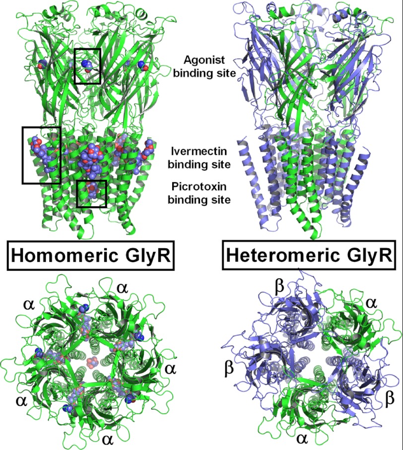
Structures of homomeric α1- and heteromeric α1β-GlyRs modeled using GluCl as a template.
Left, α1-GlyR shown with five glycine and ivermectin molecules each and a single picrotoxin molecule bound (ligands depicted as van der Waals spheres). Right, unliganded heteromeric α1β-GlyR with the different interfaces indicated.
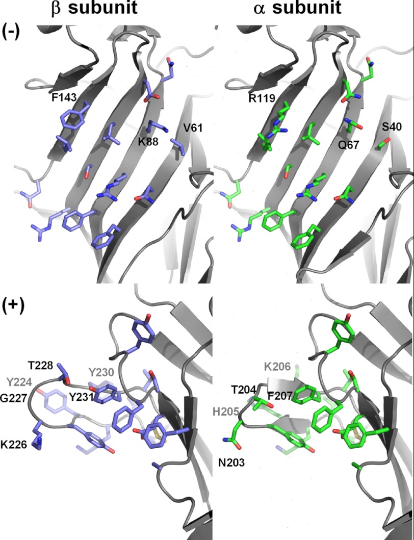
Models of the principal (+) and complementary (−) surfaces of the GlyRα1 and GlyRβ agonist-binding regions. In heteromeric GlyRs, the + and − surfaces of the α1 and β subunit ECDs generate non-equivalent agonist-binding sites at the α-β, β-α, or β-β interface, which are all functional. Note the significant sequence divergence (up to eight substitutions) between α1 and β, in particular within loop C.
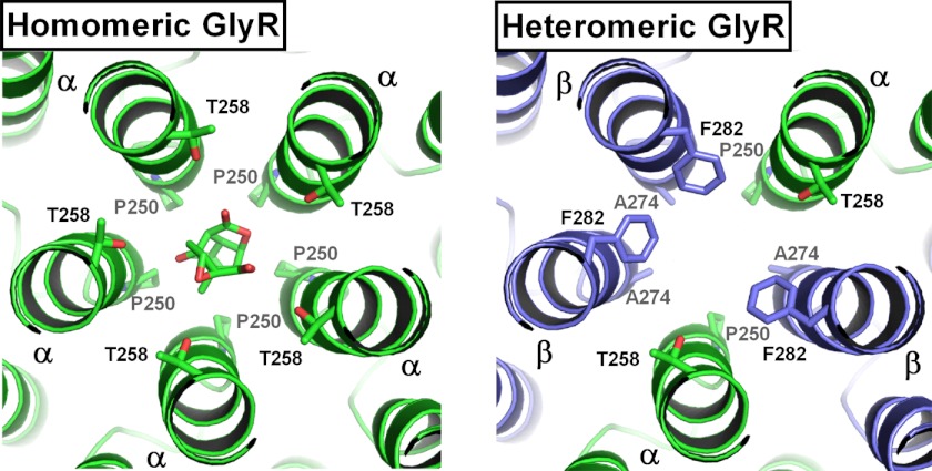
Transmembrane domains of homo- and heteromeric GlyRs.
Left, picrotoxin bound in the chloride channel of the homomeric α1-GlyR. Binding determinants include five Pro-250 and Thr-258 residues each. Right, in the α1β-GlyR, three of the Thr-258 residues are replaced by Phe-282, and three of the Pro-250 residues by Ala-274 of the β subunit. These substitutions explain the ∼50-fold lower affinity of picrotoxin for heteromeric GlyRs.
References
-
- Pfeiffer F., Graham D., Betz H. (1982) Purification by affinity chromatography of the glycine receptor of rat spinal cord. J. Biol. Chem. 257, 9389–9393 - PubMed
-
- Betz H., Laube B. (2006) Glycine receptors: recent insights into their structural organization and functional diversity. J. Neurochem. 97, 1600–1610 - PubMed
-
- Lynch J. W. (2004) Molecular structure and function of the glycine receptor chloride channel. Physiol. Rev. 84, 1051–1095 - PubMed
Publication types
MeSH terms
Substances
LinkOut - more resources
Full Text Sources
Other Literature Sources
Molecular Biology Databases
