

An L-glucose catabolic pathway in Paracoccus species 43P
- PMID: 23038265
- PMCID: PMC3504760
- DOI: 10.1074/jbc.M112.403055
An L-glucose catabolic pathway in Paracoccus species 43P
Abstract
Background: L-Glucose, the enantiomer of D-glucose, was believed not to be utilized by any organisms.
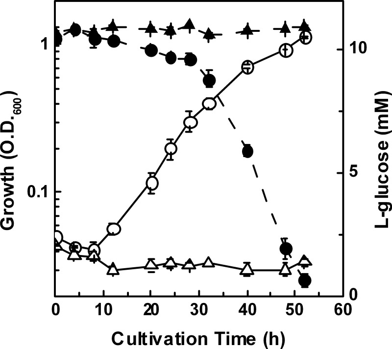
Results: An L-glucose-utilizing bacterium was isolated, and its L-glucose catabolic pathway was identified genetically and enzymatically.
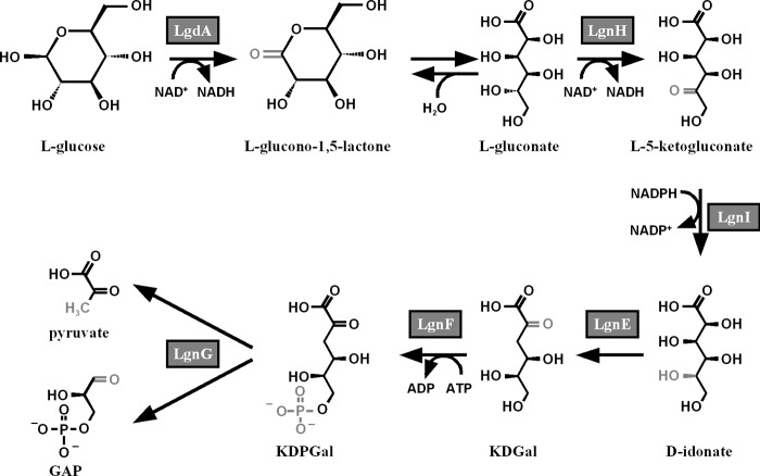
Conclusion: L-Glucose was utilized via a novel pathway to pyruvate and D-glyceraldehyde 3-phosphate.
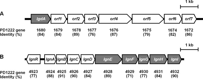
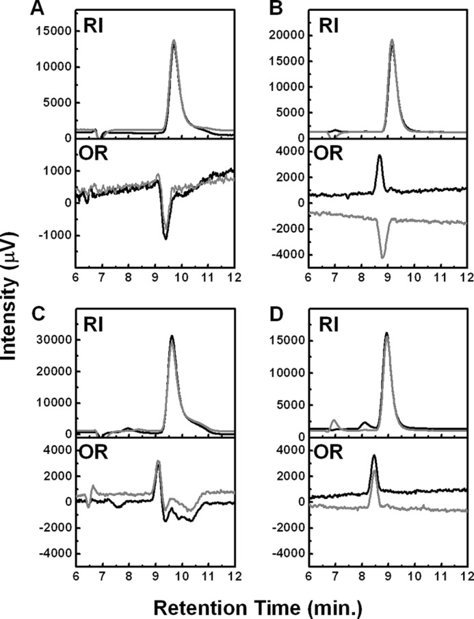
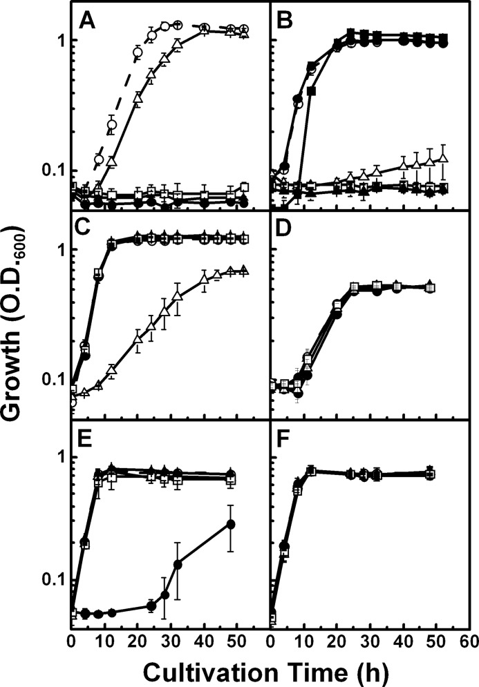
Significance: This might lead to an understanding of homochirality in sugar metabolism. An L-glucose-utilizing bacterium, Paracoccus sp. 43P, was isolated from soil by enrichment cultivation in a minimal medium containing L-glucose as the sole carbon source. In cell-free extracts from this bacterium, NAD(+)-dependent L-glucose dehydrogenase was detected as having sole activity toward L-glucose. This enzyme, LgdA, was purified, and the lgdA gene was found to be located in a cluster of putative inositol catabolic genes. LgdA showed similar dehydrogenase activity toward scyllo- and myo-inositols. L-Gluconate dehydrogenase activity was also detected in cell-free extracts, which represents the reaction product of LgdA activity toward L-glucose. Enzyme purification and gene cloning revealed that the corresponding gene resides in a nine-gene cluster, the lgn cluster, which may participate in aldonate incorporation and assimilation. Kinetic and reaction product analysis of each gene product in the cluster indicated that they sequentially metabolize L-gluconate to glycolytic intermediates, D-glyceraldehyde-3-phosphate, and pyruvate through reactions of C-5 epimerization by dehydrogenase/reductase, dehydration, phosphorylation, and aldolase reaction, using a pathway similar to L-galactonate catabolism in Escherichia coli. Gene disruption studies indicated that the identified genes are responsible for L-glucose catabolism.
Figures
Similar articles
-
Physiological, Biochemical, and Structural Bioinformatic Analysis of the Multiple Inositol Dehydrogenases from Corynebacterium glutamicum.Microbiol Spectr. 2022 Oct 26;10(5):e0195022. doi: 10.1128/spectrum.01950-22. Epub 2022 Sep 12. Microbiol Spectr. 2022. PMID: 36094194 Free PMC article.
-
Characterization of LgnR, an IclR family transcriptional regulator involved in the regulation of L-gluconate catabolic genes in Paracoccus sp. 43P.Microbiology (Reading). 2014 Mar;160(Pt 3):623-634. doi: 10.1099/mic.0.074286-0. Epub 2013 Dec 12. Microbiology (Reading). 2014. PMID: 24336464
-
The Bacillus subtilis yqjI gene encodes the NADP+-dependent 6-P-gluconate dehydrogenase in the pentose phosphate pathway.J Bacteriol. 2004 Jul;186(14):4528-34. doi: 10.1128/JB.186.14.4528-4534.2004. J Bacteriol. 2004. PMID: 15231785 Free PMC article.
-
Single amino acid mutation altered substrate specificity for L-glucose and inositol in scyllo-inositol dehydrogenase isolated from Paracoccus laeviglucosivorans.Biosci Biotechnol Biochem. 2020 Apr;84(4):734-742. doi: 10.1080/09168451.2019.1702870. Epub 2019 Dec 16. Biosci Biotechnol Biochem. 2020. PMID: 31842701
-
Catabolism of 2-keto-3-deoxy-galactonate and the production of its enantiomers.Appl Microbiol Biotechnol. 2024 Jul 2;108(1):403. doi: 10.1007/s00253-024-13235-x. Appl Microbiol Biotechnol. 2024. PMID: 38954014 Free PMC article. Review.
Cited by
-
A Factor Produced by Kaistia sp. 32K Accelerated the Motility of Methylobacterium sp. ME121.Biomolecules. 2020 Apr 16;10(4):618. doi: 10.3390/biom10040618. Biomolecules. 2020. PMID: 32316239 Free PMC article.
-
Structural basis of L-glucose oxidation by scyllo-inositol dehydrogenase: Implications for a novel enzyme subfamily classification.PLoS One. 2018 May 25;13(5):e0198010. doi: 10.1371/journal.pone.0198010. eCollection 2018. PLoS One. 2018. PMID: 29799855 Free PMC article.
-
Construction of a synthetic metabolic pathway for biosynthesis of 2,4-dihydroxybutyric acid from ethylene glycol.Nat Commun. 2023 Apr 6;14(1):1931. doi: 10.1038/s41467-023-37558-x. Nat Commun. 2023. PMID: 37024485 Free PMC article.
-
Complete genome sequence of Shinella sp. strain 1A1, an ʟ-glucose-utilizing bacterium isolated from soil.Microbiol Resour Announc. 2025 Aug 14;14(8):e0021725. doi: 10.1128/mra.00217-25. Epub 2025 Jul 7. Microbiol Resour Announc. 2025. PMID: 40621944 Free PMC article.
-
Physiological, Biochemical, and Structural Bioinformatic Analysis of the Multiple Inositol Dehydrogenases from Corynebacterium glutamicum.Microbiol Spectr. 2022 Oct 26;10(5):e0195022. doi: 10.1128/spectrum.01950-22. Epub 2022 Sep 12. Microbiol Spectr. 2022. PMID: 36094194 Free PMC article.
References
-
- Rudney H. (1940) The utilization of l-glucose by mammalian tissue and bacteria. Science 92, 112–113 - PubMed
-
- Livesey G., Brown J. C. (1995) Whole body metabolism is not restricted to d-sugars because energy metabolism of l-sugars fits a computational model in rats. J. Nutr. 125, 3020–3029 - PubMed
-
- Sun H. J., Saccomanno V., Hedlund B., Mckay C. P. (2009) Stereo-specific glucose consumption may be used to distinguish between chemical and biological reactivity on Mars: a preliminary test on Earth. Astrobiol. 9, 443–446 - PubMed
-
- Sasajima K. I., Sinskey A. J. (1979) Oxidation of lglucose by a Pseudomonad. Biochim. Biophys. Acta 571, 120–126 - PubMed
-
- Kovach M. E., Elzer P. H., Hill D. S., Robertson G. T., Farris M. A., Roop R. M., 2nd, Peterson K. M. (1995) Four new derivatives of the broad-host-range cloning vector pBBR1MCS, carrying different antibiotic resistance cassettes. Gene 166, 175–176 - PubMed
Publication types
MeSH terms
Substances
Associated data
- Actions
- Actions
- Actions
LinkOut - more resources
Full Text Sources
Molecular Biology Databases
Miscellaneous
