Comparative Study
. 2013 Jan;153(1):31-41.
doi: 10.1093/jb/mvs116. Epub 2012 Oct 3.

Post-weaning increases in the milk-fat globule EGF-factor VIII on fat globules in mouse milk and in the uptake of the fat globules by HC11 mammary epithelial cells

Affiliations
Comparative Study

Post-weaning increases in the milk-fat globule EGF-factor VIII on fat globules in mouse milk and in the uptake of the fat globules by HC11 mammary epithelial cells

Hajime Nakatani et al. J Biochem. 2013 Jan.

Abstract

Milk fat globules (MFGs) secreted by lactating mammary gland are unique lipid surrounded by a phospholipid bi-layer. We report here post-weaning changes in MFG EGF factor VIII (MFG-E8) and annexin V-accessible phosphatidyl-l-serine on the surface of MFGs. The MFG content in milk markedly decreased to about one-half within 2 days after forced weaning, despite a slight increase in milk protein content. Immunofluorescence-staining of MFGs using anti-MFG-E8 and annexin V indicated that MFG-E8 was present on some, but not all, MFGs before weaning, whereas most of MFGs were MFG-E8-positive and annexin V-negative after weaning. Free MFG-E8 with binding activity to phosphatidyl-l-serine was present abundantly in the post-weaning milk, and indeed exhibited binding to MFGs in pre-weaning milk. MFGs were taken up by HC11 mouse mammary epithelial cells in vitro, and those from post-weaning milk were remarkable for such cellular uptake. Moreover, the uptake of MFGs by the cells was inhibited by an anti-MFG-E8 antibody. Taken together, these findings suggest that MFG-E8 plays a critical role in regulation of MFG dynamics after weaning or during the suckling interval through the control of MFG-epithelial cell interaction in lactating mammary glands.


Figures
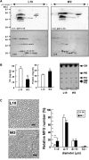
Fig. 1
Fig. 1
Analysis of proteins, lipids and MFGs in mouse milk collected before and after forced weaning. (A) Proteins in L10 (day 10 of lactation) and W2 (2 days after forced weaning) milk samples were analysed by IEF/SDS 2D-PAGE with CBB-staining (bottom), followed by immunoblotting with a mixture of anti-MFG-E8 and anti-ovalbumin (top). The long and short forms (horizontal bars 1 and 2, respectively) of MFG-E8 as well as an internal control, ovalbumin (Control) were identified by independent immunoblotting using each specific antibody. (B) Triglyceride (TG) and protein concentrations in L10 and W2 milk were determined, and the data were expressed as mean ± SD of independent milk samples from four mice (left). Alternatively, polar lipids in L10 and W2 milk were analysed by thin layer chromatography. CH, cholesterol; PS, phosphatidyl-l-serine (Ptd-Ser); PE, phosphatidylethanolamine; SM, sphingomyelin; PI, phosphatidylinositol; SD, standard deviation. (C) MFGs in L10 and W2 milk were observed under a phase contrast microscope and the number and size in diameter of MFGs in four independent visual fields (160 µm × 200 µm) were counted and expressed as mean ± SD. Scale bar: 20 μm. Asterisks in (B) and (C) indicate a significant difference (B, TG concentration P < 0.01; B, Protein concentration and C, P < 0.05).
Fig. 2
Fig. 2
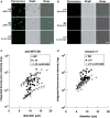
Immuno-fluorescence detection of the MFG-E8 and Ptd-Ser on MFGs. (A) MFG fractions prepared from each milk sample were incubated with anti-MFG-E8 antibody or a control antibody (anti-V8 protease), followed by the incubation with AlexaFluor488-conjugated secondary antibody. Typical images (fluorescence, bright and merged) were presented in each panel. (B) The MFG fractions as above were incubated with annexin V, followed by the immunostaining using anti-annexin V or the control antibody, and AlexaFluor488-conjugated secondary antibody. Typical images (fluorescence, bright and merged ones) were presented in each panel. Scale bar: 20 μm. (C, D) The fluorescence intensity of MFG-E8 and annexin V signals on randomly selected MFGs (40 MFGs) in L10 (closed circles) and W2 (open circles) milk samples was quantified and plotted for the relationship between the diameter of MFGs and the log phase of fluorescence intensity. The intensity of MFGs stained with a control antibody (anti-V8 protease) was also shown with a regression line as a baseline (dotted line).
Fig. 3
Fig. 3
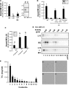
Ptd-Ser binding of MFG-free MFG-E8 in the filtered W2 milk. (A) L10 and W2 milk samples were filtered through cellulose acetate membrane (0.2 µm pore size), and the filtrates regarded as filtered milk were subjected to immunoblotting, Ptd-Ser-binding assay for MFG-E8 and TG content analysis. The data were expressed as mean ± standard deviation (SD) of independent samples from four mice. Values with different superscript letters (a, b and c) indicate significant difference (P < 0.01). (B) L10 and W2 milk samples and their membrane filtrates (filtered milk) were subjected to SDG ultracentrifugation, and MFG-E8 in each density fraction was analysed by SDS-PAGE followed by immunoblotting using anti-MFG-E8 antibody. Horizontal lines in each panel indicate the fractions corresponding to two major peaks of MFG-E8 in L10 milk sample before filtration. (C) MFG-E8 in the filtered W2 milk was fractionated by gel filtration chromatography and each fraction was subjected to Ptd-Ser-binding assay (top) and immunoblotting analysis (bottom) for MFG-E8. A void volume fraction (Vo) and the fractions corresponding to the molecular-mass standard proteins are shown as arrowheads above the MFG-E8 chromatogram as determined by Ptd-Ser-binding.
Fig. 4
Fig. 4
Binding of MFG-E8 in W2 milk to MFGs in L10 milk. (A) Ptd-Ser (PS) coated wells were incubated with W2 milk, L10 milk and the W2 milk mixed and incubated with L10 milk, respectively, and MFG-E8 bound to Ptd-Ser was measured by ELISA. Values with different superscript letters (a, b and c) indicate significant difference (P < 0.01). Alternatively, Ptd-Ser (PS)-, PC-coated or methanol-treated wells were incubated with L10 milk, W2 milk and the W2 milk incubated with L10 milk. After washing of the wells, SDS sample buffer was added to each well and recovered proteins were analysed by immunoblotting with anti-MFG-E8 antibody. (B) L10 milk was separated into milk serum (whey) and MFG fractions by centrifugation as described in Materials and Methods. W2 milk was mixed and incubated with the L10 milk serum, the L10 MFG fraction, the filtered L10 milk (see the legend of Fig. 3), or whole L10 milk. These milk samples were then incubated with Ptd-Ser (PS) or PC-coated wells and MFG-E8 bound to Ptd-Ser was measured by ELISA. Data were shown as mean ± standard deviation (SD) of independent L10 milk from four mice. (C) The W2-milk serum fraction prepared by ultracentrifugation was incubated with Pts-Ser (PS)- and PC-coated wells in the presence and absence of liposome, Ptd-Ser-liposome or PC-liposome. MFG-E8 bound to the coated phospholipids was measured by ELISA. Data are shown as mean ± SD of three determinations. (D) L10 milk, W2 milk and a mixture (1 : 1 by vol.) of both milk samples were fractionated by SDG ultracentrifugation and 12 fractions (900 µl per fraction) were collected from the top (corresponding to lanes 1–12). Distribution of MFG-E8 in these fractions was analysed by immunoblotting. (E) L10 milk was fractionated by SDG ultracentrifugation, the triglyceride concentration of each fraction was determined and expressed as mean ± standard deviation (SD) of four independent ultracentrifugation experiments. The fractions 1, 2, 6 and 7 were subjected to MFG observation under a phase contrast microscope (the right panels).
Fig. 5
Fig. 5
MFG-E8-dependent uptake of MFGs by mammary HC11 cells and integrin expression in the cells. (A) L10 milk was incubated with HC11 cells and the bound MFGs were visualized with Oil red O staining as described in Materials and Methods. In panel d, the L10 milk was filtered before the incubation to remove MFGs. In panels b and c, the milk samples were pre-incubated with anti-MFG-E8 antibody and a control antibody (anti-V8 protease), respectively. (B) L10 and W2 milk samples were incubated with HC11 cells, in which triglyceride concentration in those samples was adjusted to equal. Bound MFGs on HC11 cells were visualized by Nile red staining, and observed under a fluorescence microscope. The number of MFGs on HC11 cells was counted using the Image-J software (the right panel). Asterisk indicates a significant difference (P < 0.01). (C) mRNA of several integrins in HC11 cells cultured on plate or in Engelbreth-Holm-Swarm sarcoma gel (3D) as well as mammary gland (MG) of L10 and W2 mice was analysed by RT-PCR. β-Actin mRNAs as internal control were also analysed for comparison. (D) L10-milk samples were incubated with HC11 cells in the presence of RGE-peptide (Control) and RGD-peptide. Bound MFGs on HC11 cells were visualized by Nile red staining, and observed under a fluorescence microscope. The number of MFGs on HC11 cells was counted using the Image-J software (the right panel).

Similar articles

Cited by

  • Characterization and comparison of human and mouse milk cells.
    Doerfler R, Yerneni S, Newby A, Chaudhary N, Shu A, Fein K, Hofstatter Azambuja J, Whitehead KA. Doerfler R, et al. PLoS One. 2024 Jan 31;19(1):e0297821. doi: 10.1371/journal.pone.0297821. eCollection 2024. PLoS One. 2024. PMID: 38295101 Free PMC article.

References

    1. Franke WW, Heid HW, Grund C, Winter S, Freudenstein C, Schmid E, Jarasch ED, Keenan TW. Antibodies to the major insoluble milk fat globule membrane-associated protein specific location in apical regions of lactating epithelial cells. J. Cell Biol. 1981;89:485–494. - PMC - PubMed
    1. Blasiole DA, Davis RA, Attie AD. The physiological and molecular regulation of lipoprotein assembly and secretion. Mol. Biosyst. 2007;3:608–619. - PubMed
    1. Monks J, Geske FJ, Lehman L, Fadok VA. Do inflammatory cells participate in mammary gland involution? J. Mammary Gland Biol. Neoplasia. 2002;7:163–176. - PubMed
    1. Nakatani H, Aoki N, Nakagawa Y, Jin-No S, Aoyama K, Oshima K, Ohira S, Sato C, Nadano D, Matsuda T. Weaning-induced expression of a milk-fat globule protein, MFG-E8, in mouse mammary glands, as demonstrated by the analyses of its mRNA, protein and phosphatidylserine-binding activity. Biochem. J. 2006;395:21–30. - PMC - PubMed
    1. Oshima K, Aoki N, Negi M, Kishi M, Kitajima K, Matsuda T. Lactation-dependent expression of an mRNA splice variant with an exon for a multiply O-glycosylated domain of mouse milk fat globule glycoprotein MFG-E8. Biochem. Biophys. Res. Commun. 1999;254:522–528. - PubMed

Publication types

MeSH terms