. 2012 Dec;153(12):6041-53.
doi: 10.1210/en.2012-1670. Epub 2012 Oct 4.

The Scribble/Lgl/Dlg polarity protein complex is a regulator of blood-testis barrier dynamics and spermatid polarity during spermatogenesis

Affiliations

The Scribble/Lgl/Dlg polarity protein complex is a regulator of blood-testis barrier dynamics and spermatid polarity during spermatogenesis

Wenhui Su et al. Endocrinology. 2012 Dec.

Abstract

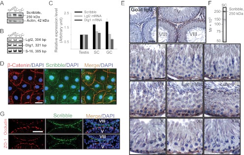
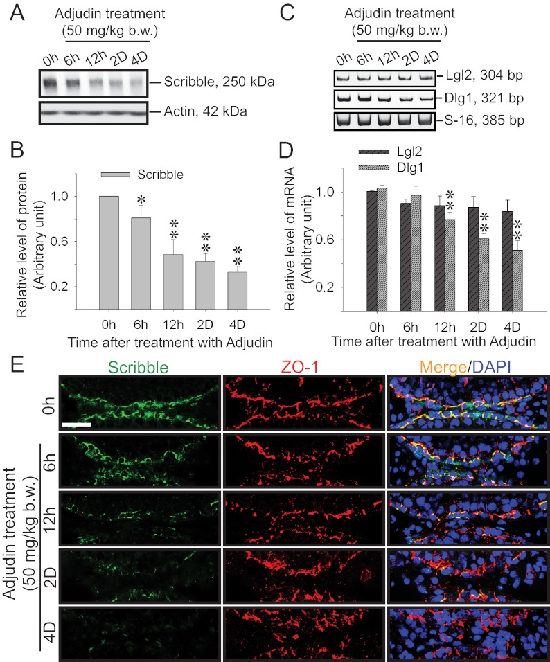
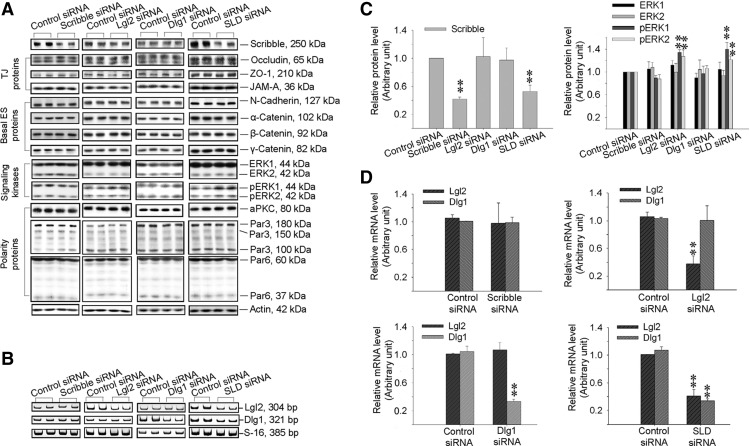
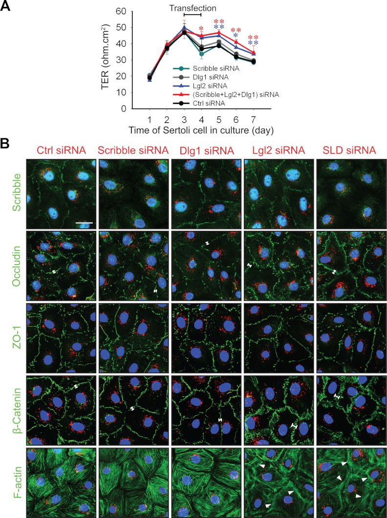
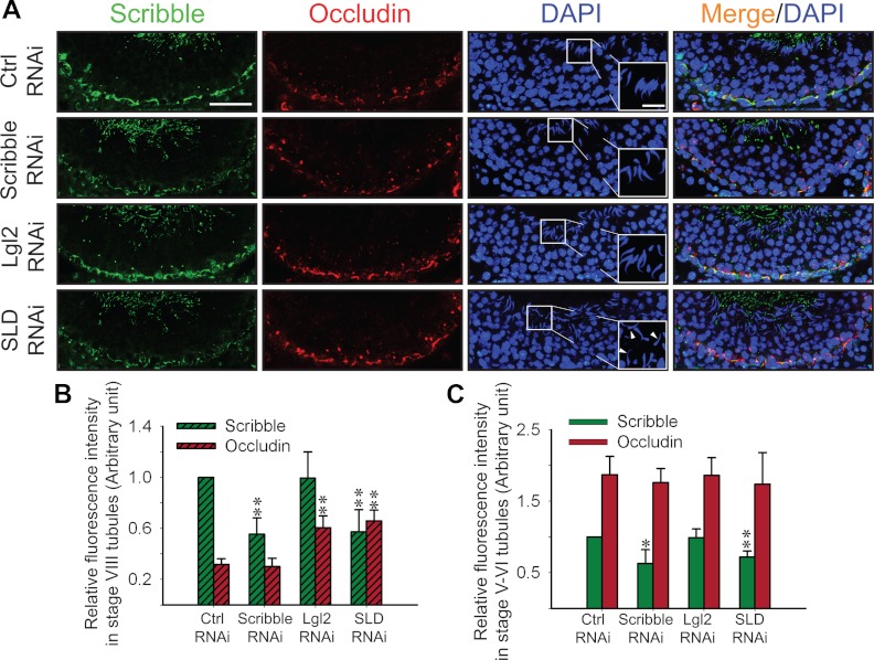
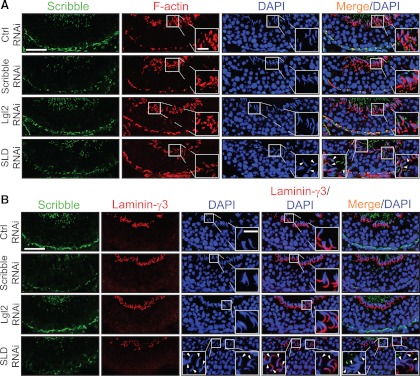
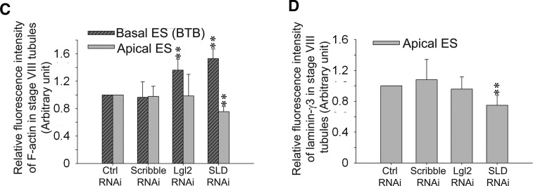
During spermatogenesis, spermiogenesis that releases sperm into the tubule lumen and restructuring of the blood-testis barrier (BTB) that accommodates the transit of preleptotene spermatocytes take place simultaneously, but at the opposite ends of the seminiferous epithelium. These events are tightly regulated and coordinated; however, neither the underlying mechanism(s) nor the involving molecules are known. Herein, the Scribble/Lgl (Lethal giant larvae)/Dlg (Discs large) polarity complex was shown to regulate spermatid polarity during spermiogenesis and tight junction (TJ)-permeability barrier via changes in protein distribution at the apical ectoplasmic specialization and the BTB during the epithelial cycle, respectively. Scribble, Lgl2, and Dlg1 were found to be expressed by Sertoli and germ cells. Scribble also displayed stage-specific expression at the BTB, being highest at stages VII-VIII, colocalizing with TJ proteins occludin and ZO-1. Unlike components of other polarity complex modules, such as partitioning-defective 6, the knockdown of which by RNA interference was found to impede Sertoli cell TJ barrier, a knockdown of the Scribble complex (i.e. simultaneous knockdown of Scribble, Lgl and Dlg or Lgl alone; but not Scribble or Dlg alone) both in vitro and in vivo promoted the TJ integrity. This was mediated by reorganizing actin filament network at the Sertoli cell-cell interface, which, in turn, affected changes in the localization and/or distribution of occludin and/or β-catenin at the BTB. These knockdowns also perturbed F-actin organization at the Sertoli cell-spermatid interface, thereby modulating spermatid adhesion and polarity at the apical ectoplasmic specialization. In summary, the Scribble/Lgl/Dlg complex participates in the regulation of BTB dynamics and spermatid adhesion/polarity in the testis.


Figures

Fig. 1.
Fig. 1.
Expression of components of the Scribble polarity complex in cultured Sertoli and germ cells and the testis, and stage-specific localization of Scribble at the BTB during the epithelial cycle of spermatogenesis. A, Immunoblotting of Scribble in lysates of adult rat testis, Sertoli cells (SC), and germ cells (GC) with actin served as a loading control (50 μg protein/lane). B, RT-PCR results depicting the relative steady-state mRNA levels of Lgl2 and Dlg1 in lysates of testis, SC, and GC, with S-16 serving as a control. C, Histogram summarizes results of immunoblotting and RT-PCR shown in panels A and B after normalizing each data point against actin (for immunoblot) or S-16 (for RT-PCR). Protein (Scribble) or mRNA (Lgl2 or Dlg1) level in the testis was arbitrarily set as 1. Each bar is the mean ± sd of n = 3. D, Colocalization of Scribble (green) and basal ES protein β-catenin (red) to the Sertoli cell-cell interface by dual-labeled immunofluorescence analysis in cells cultured for 4 d. Nuclei were stained with DAPI (blue). Scale bar, 5 μm, which applies to other micrographs. E, Stage-specific expression and localization of Scribble at the BTB in the epithelium during the epithelial cycle. Scribble was expressed predominantly at the basal region near the basement membrane, consistent with its localization at the BTB, with the highest level in stages VII–VIII, at the time of BTB restructuring to facilitate the transit of preleptotene spermatocytes at the site. Negative control was shown in which the anti-Scribble antibody was substituted by normal goat IgG; scale bar, 40 μm, which applies to the micrograph on the right. Scale bar, 20 μm, in the staged I–II tubule, which applies to all other micrographs. F, Immunoblot analysis illustrating the specificity of the goat anti-Scribble antibody using Sertoli cell (SC) lysates. G, Colocalization of Scribble (green) with tight junction proteins occludin (red) or ZO-1 (red), predominantly at the BTB. Nuclei were stained with DAPI (blue). Scale bar, 30 μm, which applies to other micrographs.
Fig. 2.
Fig. 2.
Changes in the expression and localization of Scribble and/or its partners during adjudin-induced junction restructuring in adult rat testes. Adjudin was administered to adult rats at 0 h (hr), and testes were obtained at specified time points for immunoblot analysis (A and B), RT-PCR (C), qPCR (D), dual-labeled immunofluorescence analysis (E), and IHC (Supplemental Fig. 1). A, Changes in the protein level of Scribble after adjudin treatment vs. actin, which served as a protein loading control. B, Histogram summarizes the result shown in panel A after normalizing each data point against actin. Protein level at 0 h was arbitrarily set at 1. Each bar, mean ± sd of n = 3 rats. *, P < 0.05; **, P < 0.01. C, RT-PCR results illustrating changes in the steady-state mRNA levels of Lgl2 and Dlg1 after adjudin treatment, with S-16 serving as a control. D, Results of qPCR on the steady-state mRNA levels of Lgl2 and Dlg1, validating the findings in panel C. Each bar, mean ± sd of n = 3 rats. **, P < 0.01. E, Changes in expression and colocalization of Scribble (green) with TJ-protein ZO-1 (red) after adjudin treatment. Nuclei were stained with DAPI (blue). Scale bar, 50 μm, which applies to all micrographs.
Fig. 3.
Fig. 3.
Changes in the steady-state protein or mRNA levels of Scribble complex components and BTB-associated proteins after single knockdown of either Scribble, Lgl2, or Dlg1 vs. triple knockdown of Scribble, Lgl2, and Dlg1 by RNAi. Sertoli cells cultured in vitro for 4 d with an established TJ barrier were transfected with specific siRNA duplexes targeting 1) Scribble, 2) Lgl2, 3) Dlg1, or 4) Scribble, Lgl2 and Dlg1 (SLD, triple knockdown) vs. nontargeting control for 24 h. On d 7 (i.e. 2 d after transfection), cultures were terminated for immunoblotting (A and C), RT-PCR (B), and qPCR (D) to assess the efficacy of the knockdown and any off-target effects. A, Immunoblotting showed a knockdown of Scribble by approximately 60% in Scribble single-silencing group and by about 55% in triple Scribble/Lgl2/Dlg1-silencing group (SLD) vs. nontargeting control. In Lgl2 single-silencing and SLD triple-silencing groups, levels of pERK1 and pERK2 were induced vs. control. No off-target effect was detected. Also, the single silencing of either Lgl2 or Dlg1 did not affect the steady-state protein level of Scribble. B, RT-PCR result illustrating the steady-state mRNA levels of Lgl2 and Dlg1 in the three single-silencing groups, and the triple-silencing group vs. the nontargeting control group. This experiment was done to assess the specificity of each knockdown because satisfactory antibodies against Lgl2 and Dlg1 were not available commercially. C, Histograms summarizing part of the data shown in panel A after normalizing each data point against actin (see Supplemental Fig. 4 for remaining data), illustrating specificity of the knockdown. Protein level in the nontargeting control siRNA group was arbitrarily set at 1. Each bar, mean ± sd of n = 3 experiments. **, P < 0.01. D, qPCR results that validated the RT-PCR findings shown in panel B. In Scribble single-silencing group, Lgl2 and Dlg1 mRNA levels were unaffected. In Lgl2 single-silencing group, the level of Lgl2 mRNA was knocked down by about 60% with no off-target effect on Dlg1. In Dlg1 single-silencing group, the level of Dlg1 mRNA was knocked down by approximately 70% with no off-target effect on Lgl2. In SLD triple-silencing group, the levels of Lgl2 and Dlg1 were knocked down by approximately 60% and 70%, respectively. Each bar, mean ± sd of n = 3 experiments. **, P < 0.01.
Fig. 4.
Fig. 4.
Changes in the Sertoli cell TJ-permeability barrier after knockdown of different components of the Scribble protein complex by RNAi. A, The TJ-barrier function was quantified by measuring TER across the Sertoli cell epithelium. Sertoli cells were plated on Matrigel-coated bicameral units at time 0, and on d 3, cells were transfected for 24 h with the corresponding siRNA duplexes. In SLD triple-silencing group, the transfection mix was composed of equal amount of the three siRNA duplexes targeting at Scribble, Lgl2, and Dlg1 at about 67 nm each to a total of 200 nm siRNA duplexes vs. 200 nm for the single knockdown of either Scribble, Lgl2, or Dlg1 and also nontargeting control cells. B, Changes in protein distribution at the cell-cell interface in Sertoli cells cultured on Matrigel-coated coverslips at 0.05 × 10⁶ cells/cm2 after knockdown of different components of the Scribble complex vs. nontargeting control (Ctrl) siRNA duplexes. Transfection for 24 h using siRNA duplexes (at a final concentration of 80 nm for single silencing of Scribble, Dlg1, or Lgl2; or 90 nm for triple silencing of Scribble, Lgl2, and Dlg1) (SLD) with siGLO red transfection indicator (1 nm, to assess successful transfection) was performed on d 3 before immunofluorescence microscopy or F-actin staining (green, using FITC-conjugated phalloidin) on d 6. Single knockdown of Scribble or Dlg1 had no apparent effect on the distribution of BTB proteins (e.g. occludin, ZO-1, and β-catenin, all in green) vs. control at the Sertoli cell-cell interface, as well as F-actin filament organization. In contrast, single knockdown of Scribble or SDL triple knockdown, but not Dlg1- or Lgl2-single knockdown, indeed reduced the signal of Scribble (green) considerably at the Sertoli cell-cell interface, illustrating the efficacy and specificity of the knockdown. However, single knockdown of Lgl2 and triple SLD knockdown caused a more focused localization of occludin and β-catenin (annotated by white brackets), but not ZO-1 (green), at the cell-cell interface, concomitant with a redistribution of F-actin, which was more intensely localized at the cell-cell interface (see white arrowheads). Cell nuclei were stained with DAPI (blue). Scale bar, 5 μm, which applies to other micrographs.
Fig. 5.
Fig. 5.
Changes in the distribution of TJ-protein occludin and spermatid polarity in the seminiferous epithelium after knockdown of Scribble complex components by RNAi in adult rat testes in vivo. Rats were treated with corresponding siRNA duplexes to silence different components of the Scribble complex vs. nontargeting controls. Control, Scribble-silenced, Lgl2-silenced, and Scribble/Lgl2/Dlg1 triple-silenced testes were immunostained for Scribble (panel A, green) and occludin (panel A, red) using frozen sections from rats 3 d after the last siRNA transfection mix injection. A, In stage VIII tubules, the signals of Scribble in the epithelium were considerably weakened in Scribble single- and SLD triple-silenced (but not in Lgl2 single-silenced group) groups vs. controls (Ctrl). The signal of occludin in Scribble single-silenced tubules was similar to controls; however, it became more intense in Lgl2 single-silenced and SLD triple-silenced tubules vs. control. Nuclei were stained with DAPI (blue). The boxed area in micrographs in the DAPI panel was enlarged and shown in insets. White arrowheads denote misoriented spermatids with incorrect polarity, only found in SLD triple-silenced group. Scale bar, 80 μm, which applies to all other micrographs; scale bar in inset, 20 μm, which applies to all other insets. B and C, Semiquantitative analysis of data shown in panel A and in Supplemental Fig. 5. Fluorescence intensity of Scribble and occludin in the epithelium was quantified by ImageJ 1.44I in each treatment group vs. control in stage VIII (B) or stage V–VI (C) tubules. The signal intensity of Scribble in the control group was arbitrarily set at 1. At least 50 staged tubules were randomly selected from three rats and quantified. Each bar, mean ± sd of n = 3 rats. *, P < 0.05; **, P < 0.01.
Fig. 6.
Fig. 6.
Changes in F-actin organization, apical ES protein distribution, and spermatid polarity in the seminiferous epithelium in stage VIII tubules after knockdown of components of Scribble complex in adult rat testes in vivo. A, F-actin (red) in the epithelium was visualized by using rhodamine-conjugated phalloidin in siRNA-transfected rat testes in nontargeting control (Ctrl) group vs. treatment groups. In both Lgl2 single- and SLD triple-silenced groups, F-actin staining at the BTB increased considerably in stage VIII tubules, but not in Scribble single-silenced group, when compared with the control group. Scribble (green) staining in the epithelium was found to diminish considerably in the Scribble single-silenced and SLD triple-silenced groups, but not the Lgl2 single-silenced group vs. the control group, because of the Scribble knockdown. Also, the organization of F-actin at the apical ES appeared to be disrupted as illustrated by the diffused and weakened F-actin signals at the site in the SLD triple-silenced group (but not in the single Scribble- or even the Lgl2-silenced group) vs. the control group, illustrating the actin filament bundles at the ES were reorganized, with these F-actin bundles being shifted from the apical to the basal ES at the BTB. Thus, cell polarity in the step 19 spermatids in stage VIII tubules from testes of the SLD triple-silenced group was found to be disrupted, possibly due to the diminished actin filament bundles at the apical ES, with these elongated spermatids no longer pointing toward the basement membrane and became misoriented, unlike the nontargeting or single-silenced Scribble and Lgl2 groups. Indeed, F-actin staining was considerably diminished in misoriented spermatids with a loss of cell polarity (see insets in SLD RNAi panel which magnified spermatids in boxed areas vs. control and other treatment groups). Cell nuclei were stained with DAPI (blue). White arrowheads denote mis-oriented spermatids that had lost their polarity in SLD triple-silenced group with diminished F-actin. B, Due to the loss of spermatid polarity as noted in panel A, apical ES protein laminin-γ3 (red) vs. Scribble (green) was also examined in the epithelium of these rat testes and shown herein. Distribution of laminin-γ3 at the apical ES surrounding the spermatid head in Scribble or Lgl2 single-silenced group remained unaltered, similar to the nontargeting control group, but laminin-γ3 was considerably weakened at the apical ES in SLD triple-silenced group. It was also noted that in step 19 spermatids that had lost their polarity and became misoriented, laminin-γ3 staining was either considerably weakened or virtually absent at the apical ES in elongated spermatids (see insets in the SLD RNAi panel wherein step 19 spermatids in boxed areas were magnified). White arrowheads denote misoriented spermatids that had lost the polarity in SLD triple-silenced group with either diminished or a virtual loss of laminin-γ3 staining. Scale bar, 80 μm in panels A and B, which applies to all micrographs in panels A and B; scale bar, 20 μm in inset, which applies to all insets in panels A and B. Imaging analysis of fluorescence signals of F-actin (C) and laminin-γ3 (D) from micrographs obtained from dual-labeled immunofluorescence analysis, which was used to investigate changes in the expression and localization of Scribble and F-actin in the seminiferous epithelium after knockdown of either Scribble, Lgl2, SLD vs. nontargeting control, such as those shown in Fig. 6 in stage VIII tubules. About 90 stage VIII tubules were randomly selected from cross-sections of three rat testes in different silencing groups vs. control group. A significant increase in F-actin fluorescence at the BTB was noted in the Lgl2 single-knockdown (but not the Scribble single-knockdown) and SDL triple-knockdown groups vs. nontargeting control group; however, a significant decrease in F-actin and laminin-γ3 was also noted at the apical ES in the SLD triple-knockdown group, but not in either single-knockdown group.**, P < 0.01, compared with nontargeting control (Ctrl) group.
Fig. 6.
Fig. 6.
Changes in F-actin organization, apical ES protein distribution, and spermatid polarity in the seminiferous epithelium in stage VIII tubules after knockdown of components of Scribble complex in adult rat testes in vivo. A, F-actin (red) in the epithelium was visualized by using rhodamine-conjugated phalloidin in siRNA-transfected rat testes in nontargeting control (Ctrl) group vs. treatment groups. In both Lgl2 single- and SLD triple-silenced groups, F-actin staining at the BTB increased considerably in stage VIII tubules, but not in Scribble single-silenced group, when compared with the control group. Scribble (green) staining in the epithelium was found to diminish considerably in the Scribble single-silenced and SLD triple-silenced groups, but not the Lgl2 single-silenced group vs. the control group, because of the Scribble knockdown. Also, the organization of F-actin at the apical ES appeared to be disrupted as illustrated by the diffused and weakened F-actin signals at the site in the SLD triple-silenced group (but not in the single Scribble- or even the Lgl2-silenced group) vs. the control group, illustrating the actin filament bundles at the ES were reorganized, with these F-actin bundles being shifted from the apical to the basal ES at the BTB. Thus, cell polarity in the step 19 spermatids in stage VIII tubules from testes of the SLD triple-silenced group was found to be disrupted, possibly due to the diminished actin filament bundles at the apical ES, with these elongated spermatids no longer pointing toward the basement membrane and became misoriented, unlike the nontargeting or single-silenced Scribble and Lgl2 groups. Indeed, F-actin staining was considerably diminished in misoriented spermatids with a loss of cell polarity (see insets in SLD RNAi panel which magnified spermatids in boxed areas vs. control and other treatment groups). Cell nuclei were stained with DAPI (blue). White arrowheads denote mis-oriented spermatids that had lost their polarity in SLD triple-silenced group with diminished F-actin. B, Due to the loss of spermatid polarity as noted in panel A, apical ES protein laminin-γ3 (red) vs. Scribble (green) was also examined in the epithelium of these rat testes and shown herein. Distribution of laminin-γ3 at the apical ES surrounding the spermatid head in Scribble or Lgl2 single-silenced group remained unaltered, similar to the nontargeting control group, but laminin-γ3 was considerably weakened at the apical ES in SLD triple-silenced group. It was also noted that in step 19 spermatids that had lost their polarity and became misoriented, laminin-γ3 staining was either considerably weakened or virtually absent at the apical ES in elongated spermatids (see insets in the SLD RNAi panel wherein step 19 spermatids in boxed areas were magnified). White arrowheads denote misoriented spermatids that had lost the polarity in SLD triple-silenced group with either diminished or a virtual loss of laminin-γ3 staining. Scale bar, 80 μm in panels A and B, which applies to all micrographs in panels A and B; scale bar, 20 μm in inset, which applies to all insets in panels A and B. Imaging analysis of fluorescence signals of F-actin (C) and laminin-γ3 (D) from micrographs obtained from dual-labeled immunofluorescence analysis, which was used to investigate changes in the expression and localization of Scribble and F-actin in the seminiferous epithelium after knockdown of either Scribble, Lgl2, SLD vs. nontargeting control, such as those shown in Fig. 6 in stage VIII tubules. About 90 stage VIII tubules were randomly selected from cross-sections of three rat testes in different silencing groups vs. control group. A significant increase in F-actin fluorescence at the BTB was noted in the Lgl2 single-knockdown (but not the Scribble single-knockdown) and SDL triple-knockdown groups vs. nontargeting control group; however, a significant decrease in F-actin and laminin-γ3 was also noted at the apical ES in the SLD triple-knockdown group, but not in either single-knockdown group.**, P < 0.01, compared with nontargeting control (Ctrl) group.

References

    1. Cheng CY, Mruk DD. 2012. The blood-testis barrier and its implication in male contraception. Pharmacol Rev 64:16–64 - PMC - PubMed
    1. Cheng CY, Mruk DD. 2010. A local autocrine axis in the testes that regulates spermatogenesis. Nat Rev Endocrinol 6:380–395 - PMC - PubMed
    1. Hess RA, de Franca L. 2008. Spermatogenesis and cycle of the seminiferous epithelium. Adv Exp Med Biol 636:1–15 - PubMed
    1. Mruk DD, Silvestrini B, Cheng CY. 2008. Anchoring junctions as drug targets: role in contraceptive development. Pharmacol Rev 60:146–180 - PMC - PubMed
    1. O'Donnell L, Nicholls PK, O'Bryan MK, McLachlan RI, Stanton PG. 2011. Spermiation: the process of sperm release. Spermatogenesis 1:14–35 - PMC - PubMed

Publication types