. 2012 Dec 15;125(Pt 24):5984-97.
doi: 10.1242/jcs.109066. Epub 2012 Oct 4.

Drosophila embryos close epithelial wounds using a combination of cellular protrusions and an actomyosin purse string

Affiliations

Drosophila embryos close epithelial wounds using a combination of cellular protrusions and an actomyosin purse string

Maria Teresa Abreu-Blanco et al. J Cell Sci. .

Abstract

The repair of injured tissue must occur rapidly to prevent microbial invasion and maintain tissue integrity. Epithelial tissues in particular, which serve as a barrier against the external environment, must repair efficiently in order to restore their primary function. Here we analyze the effect of different parameters on the epithelial wound repair process in the late stage Drosophila embryo using in vivo wound assays, expression of cytoskeleton and membrane markers, and mutant analysis. We define four distinct phases in the repair process, expansion, coalescence, contraction and closure, and describe the molecular dynamics of each phase. Specifically, we find that myosin, E-cadherin, Echinoid, the plasma membrane, microtubules and the Cdc42 small GTPase respond dynamically during wound repair. We demonstrate that perturbations of each of these components result in specific impairments to the wound healing process. Our results show that embryonic epithelial wound repair is mediated by two simultaneously acting mechanisms: crawling driven by cellular protrusions and actomyosin ring contraction along the leading edge of the wound.


Figures

Fig. 1.
Fig. 1.
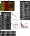
Phases of epithelial wound repair in Drosophila. (A) Schematic diagram of multicellular wound repair. Damaged cells at the leading edge of the wound will be either repaired (gray) or removed (white). Wound closure can then proceed by lamellipodial crawling or contraction of an actin purse string. (B–E) Drosophila embryo ectoderm is a single layer epithelium. Confocal images of the lateral side (B), ventral side (C) and cross-section (E) of stage 15 embryos expressing actin and nuclear markers (sGMCA and His2Av-mRFP). In B and C anterior is left; in B dorsal is up. (D) Cross-sections of a stage 15 embryo expressing GFP-α-tubulin in the epithelium only. (F) Schematic diagram of a stage 15 embryo showing the organization of the germ layers (adapted from Hartenstein, 1993). (G,G′) Time-lapse series of surface projections (G) and cross-sections (G′) of a stage 15 embryo expressing actin and nuclei markers (sGMCA and His2Av-mRFP). The phases of wound repair are depicted. White arrows and arrowheads indicate leading edge cells retracting and accumulating actin. Collapsing cells are marked with asterisks. Filopodial extensions (blue arrows) and tethering (blue arrowhead) are indicated. (G′) Yellow arrowheads mark the extent of wound expansion. Hemocytes (yellow arrows) and the actin cable (green arrows) are indicated. (H) Analysis of the wound healing response (n = 10; results are given as means ± s.e.m.), showing the wound repair phases. (I) Time-lapse series of an embryo expressing the caspase biosensor Apoliner. GFP is not translocated to the nuclei of rounded cells (arrows). (J–L) Time-lapse series of surface projections (J,K,L) and cross-sections (I′,J′) of a stage 15 embryo expressing actin (sGMCA). High magnification images (100×) show filopodia probing the wound environment during the coalescence (L) and contraction phases (K,K′; arrows). Scale bars: 20 µm (C,G,I,J); 10 µm (B,D,E,G′,J′,K,K′); and 5 µm (L).
Fig. 2.
Fig. 2.
Parameters of epithelial wound repair. (A–I) Parameters of the epithelial wound response (all results are given as means ± s.e.m.). (A) Effects of wound size on wound repair dynamics (small, n = 5; medium, n = 10; large, n = 5). Analysis of each phase of repair: expansion and coalescence by area (B,D,F) and duration (C,E,G); contraction phase by area (H) and duration (I). (J–L) Effects of embryo mounting techniques on wound repair. Single slice micrograph at the plane of the wound in membrane (J) and coverslip (K) mounted embryos expressing actin (sGMCA). (L) Analysis of wound repair dynamics using membrane (blue) and coverslip (red) mounting. Scale bar: 20 µm.
Fig. 3.
Fig. 3.
Myosin II is required for assembly of an actin purse string. (A–B′) Time-lapse series of surface projections (A,A′) and cross-sections (A″) of a stage 15 embryo expressing myosin and actin markers (sqh-GFP and sChMCA). Myosin accumulates at the leading edge of epithelium wounds overlapping with the actin cable. (B,B′) High magnification views (white squares in A) showing myosin and actin overlapping at the leading edge. Myosin II is absent in the protrusions (arrows). (C,C′) Time-lapse series of stage 15 zip1 mutant embryo expressing an actin marker (sGMCA), and showing incomplete actin cable assembly. Surface projections (C) and cross-sections (C′) are shown. (D,E) Quantification of wound area over time (wild type, n = 10; zip1, n = 4; results are given as means ± s.e.m.) for all phases of wound repair (D) and an expansion of the first 20 minutes (E). (F) High magnification (100×) views of protrusions in wild-type and zip1 wounds at time-points representing 100, 75 and 50% of the maximum wound area. (G) Quantification of the area of protrusions (wild type, n = 4; zip1, n = 4), P-values are indicated. Scale bars: 20 µm (A,A′,C) and 10 µm (A″,C′,F).
Fig. 4.
Fig. 4.
Intact adherens junctions are required to anchor the actomyosin cable. (A–B′) Time-lapse series of surface projections (A–A″,B,B′) and cross-sections (A′″) in wild-type embryos expressing DE-cadherin-GFP and actin (sChMCA). Cells retaining E-cadherin but lacking actin are indicated (white arrowheads). (B,B′) High magnification views (white squares in A) showing actin and E-cadherin overlaps at the leading edge junctions. (C–D) Wound repair in a shgk03401 mutant embryo expressing an actin marker (sGMCA). Time-lapse series of surface projections (C,D) and cross-sections (C′). High magnification views of shgk03401 embryos showing repair by localized protrusion zippering between neighbor leading edge cells (D, white brackets). (E,F) Quantification of wound area over time (wild type, n = 10; shgk03401, n = 6; results are given as means ± s.e.m.) for all phases of wound repair (E) and an expansion of the first 20 minutes (F). (G–H) Time-lapse series of surface projections (G,H) and cross-sections (G′) of an edM/Z mutant embryo expressing an actin marker (sGMCA). edM/Z protrusions contact one another, resulting in local contractions of the wound edge (H, arrows). (I,J) Quantification of wound area over time (wild type, n = 10; edM/Z, n = 5) for all phases of wound repair (I) and an expansion of the first 20 minutes (J). (K–L′) edM/Z mutants assemble partial actomyosin cables at the wound leading edge. Myosin II (α-non muscle myosin, MHC) accumulates at the wound edge of wild-type (K,K′) and edM/Z mutants (L,L′) overlapping with actin (phalloidin). Scale bars: 20 µm (A–A″,C,G); 10 µm (A″′,C′,D,H,G′); and 5 µm (K–L′).
Fig. 5.
Fig. 5.
Dynamic protrusions are assembled at the leading edge of epithelial wounds. (A–C″) Time-lapse series of surface projections (A,A″,B,C,C″) and cross-sections (A′,C′) of embryos expressing the membrane reporters PH-PLC-GFP (A–B) or Venus-GAP43 (C–C″). High magnification view of protrusions in embryos expressing PH-PLC-GFP (A″,B) and Venus-GAP43 (C″). Membrane in protrusions (arrows) and accumulation at cell-cell junctions (arrowheads) are indicated. (D–F) Time-lapse series of embryos expressing GFP-α-tubulin in the epithelium, showing microtubules in cell protrusions (arrows). High magnification views in E,F. (G,G′) Time-lapse series of surface projections (G) and cross-sections (G′) of a stage 15 eb12 embryo expressing an actin marker (sGMCA) showing delayed actin cable assembly. (H,I) Quantification of the wound area over time (wild type, n = 10; eb12, n = 6; results are given as means ± s.e.m.) for all phases of wound repair (H) and an expansion of the first 20 minutes (I). Scale bars: 20 µm (A,C,D,G) and 10 µm (A′,A″,B,C′,C″,E,F,G′).
Fig. 6.
Fig. 6.
Cdc42 is required for normal protrusion activity and, together with Myosin II, for proper epithelial repair. (A–A″) Confocal time series following wound repair in stage 15 embryos coexpressing ChFP-Cdc42 (A′) and actin (sGMCA). (B,B′) Time-lapse series of wound repair in Cdc424/Cdc426 mutant embryos expressing an actin marker (sGMCA). Notice the lack of protrusions at the leading edge cells. (C,D) Quantification of the wound area over time (wild type, n = 10; Cdc424/Cdc426, n = 5; results are given as means ± s.e.m.) for all phases of wound repair (C) and an expansion of the first 20 minutes (D). (E) High magnification views (100×) showing the protrusions at the leading edge cells in wild-type and Cdc424/Cdc426 embryos at 100, 75 and 50% maximum wound area. (F) Quantification of the area of protrusions, P-values are indicated (wild type, n = 4; Cdc424/Cdc426, n = 4). (G,G′) Time-lapse series of wound repair in zip1 Cdc42RNAi double mutant embryos followed with an actin marker (sGMCA). Actin cable assembly, as well as cellular protrusions, are severely disrupted. (H) Quantification of the wound area over time (wild type, n = 10; zip1, n = 4; Cdc424/Cdc426, n = 5, zip1 Cdc42RNAi, n = 4; results are given as means ± s.e.m.). Scale bars: 20 µm (A,A′,B,G) and 10 µm (A″,B′,E,G′).
Fig. 7.
Fig. 7.
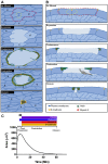
Working model for epithelial wound repair in the Drosophila embryo. (A,B) Schematic of surface projection (A) and cross-section (B) views depicting the dynamic behavior of embryo epithelium cells in response to wounds, as well as the molecular components recruited during the wound repair process. Shape changes and behavior for different cells (indicated by letters a–e) at the wound edge are indicated. (C) Phases and components of the epithelial wound response.

References

    1. Abreu–Blanco M. T., Verboon J. M., Parkhurst S. M. (2011). Cell wound repair in Drosophila occurs through three distinct phases of membrane and cytoskeletal remodeling. J. Cell Biol. 193, 455–464 10.1083/jcb.201011018 - DOI - PMC - PubMed
    1. Bardet P. L., Kolahgar G., Mynett A., Miguel–Aliaga I., Briscoe J., Meier P., Vincent J. P. (2008). A fluorescent reporter of caspase activity for live imaging. Proc. Natl. Acad. Sci. USA 105, 13901–13905 10.1073/pnas.0806983105 - DOI - PMC - PubMed
    1. Bement W. M., Forscher P., Mooseker M. S. (1993). A novel cytoskeletal structure involved in purse string wound closure and cell polarity maintenance. J. Cell Biol. 121, 565–578 10.1083/jcb.121.3.565 - DOI - PMC - PubMed
    1. Brock J., Midwinter K., Lewis J., Martin P. (1996). Healing of incisional wounds in the embryonic chick wing bud: characterization of the actin purse-string and demonstration of a requirement for Rho activation. J. Cell Biol. 135, 1097–1107 10.1083/jcb.135.4.1097 - DOI - PMC - PubMed
    1. Buck R. C. (1979). Cell migration in repair of mouse corneal epithelium. Invest. Ophthalmol. Vis. Sci. 18, 767–784 - PubMed

Publication types