. 2013 Mar;26(4):327-38.
doi: 10.3109/14767058.2012.735724. Epub 2012 Nov 23.

Interleukin-33 in the human placenta

Affiliations

Interleukin-33 in the human placenta

Vanessa Topping et al. J Matern Fetal Neonatal Med. 2013 Mar.

Abstract

Objective: Interleukin-33 (IL-33) is the newest member of the IL-1 cytokine family, a group of key regulators of inflammation. The purpose of this study was to determine whether IL-33 is expressed in the human placenta and to investigate its expression in the context of acute and chronic chorioamnionitis.

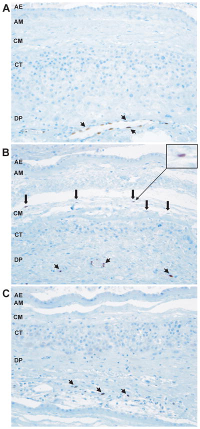
Methods: Placental tissues were obtained from five groups of patients: 1) normal pregnancy at term without labor (n = 10); 2) normal pregnancy at term in labor (n = 10); 3) preterm labor without inflammation (n = 10); 4) preterm labor with acute chorioamnionitis and funisitis (n = 10); and 5) preterm labor with chronic chorioamnionitis (n = 10). Immunostaining was performed to determine IL-33 protein expression patterns in the placental disk, chorioamniotic membranes, and umbilical cord. mRNA expression of IL-33 and its receptor IL1RL1 (ST2) was measured in primary amnion epithelial and mesenchymal cells (AECs and AMCs, n = 4) and human umbilical vein endothelial cells (HUVECs, n = 4) treated with IL-1β (1 and 10 ng/ml) and CXCL10 (0.5 and 1 or 5 ng/ml).

Results: 1) Nuclear IL-33 expression was found in endothelial and smooth muscle cells in the placenta, chorioamniotic membranes, and umbilical cord; 2) IL-33 was detected in the nucleus of CD14+ macrophages in the chorioamniotic membranes, chorionic plate, and umbilical cord, and in the cytoplasm of myofibroblasts in the Wharton's jelly; 3) acute (but not chronic) chorioamnionitis was associated with the presence of IL-33+ macrophages in the chorioamniotic membranes and umbilical cord; 4) expression of IL-33 or IL1RL1 (ST2) mRNA in AECs was undetectable; 5) IL-33 mRNA expression increased in AMCs and HUVECs after IL-1β treatment but did not change with CXCL10 treatment; and 6) IL1RL1 (ST2) expression decreased in AMCs and increased in HUVECs after IL-1β but not CXCL10 treatment.

Conclusions: IL-33 is expressed in the nucleus of placental endothelial cells, CD14+ macrophages, and myofibroblasts in the Wharton's jelly. IL-1β can induce the expression of IL-33 and its receptor. Protein expression of IL-33 is detectable in macrophages of the chorioamniotic membranes in acute (but not chronic) chorioamnionitis.


Conflict of interest statement

Declaration of Interest

The authors report no declarations of interest. This work was supported by the Perinatology Research Branch, Division of Intramural Research, Eunice Kennedy Shriver National Institute of Child Health and Human Development, National Institutes of Health, U.S. Department of Health and Human Services.

Figures

Figure 1
Figure 1. IL-33 protein expression in the placenta and chorioamniotic membranes of normal term pregnancies
(A) IL-33 is expressed in the nuclei of endothelial (arrowhead) and smooth muscle cells (arrow) of blood vessels in the chorionic plate and chorionic villi. CP = chorionic plate; CV = chorionic villi. x200 magnification. (B) Immunofluorescence staining of CD31 (red), IL-33 (green) and DAPI (blue) shows nuclear IL-33 staining of vascular endothelial cells in chorionic villi. x900 magnification. (C) IL-33 is expressed in the nuclei of decidual endothelial cells (arrowhead) in blood vessels in chorioamniotic membranes. AE = amnion epithelium; AM = amnion mesoderm; CM = chorionic mesoderm; CT = chorionic trophoblast layer; DP = decidua parietalis. x200 magnification. (D) Immunofluorescence staining of CD31 (red), IL-33 (green) and DAPI (blue) show nuclear IL-33 staining in endothelial cells of decidual blood vessels. x400 magnification. (E) Immunofluorescence staining of CD14 (red), IL-33 (green) and DAPI (blue) shows nuclear IL-33 staining in macrophages in the mesodermal layer. x1500 magnification.
Figure 2
Figure 2. IL-33 protein expression in the umbilical cord of normal term pregnancies
(A) IL-33 is expressed in the umbilical artery (B) and Wharton’s jelly (C). x100 magnification. (B) IL-33 is expressed in the nuclei of endothelial (arrowhead) and smooth muscle cells (arrow) in the umbilical vessels. x200 magnification. (C) IL-33 is expressed in the nuclei of stromal cells in the Wharton’s jelly (inset). x100 magnification. (D) Immunofluorescence staining of procollagen (red), IL-33 (green) and DAPI (blue) shows cytoplasmic IL-33 staining in myofibroblasts in the Wharton’s jelly. x630 magnification. (E) Immunofluorescence staining of CD14 (red), IL-33 (green) and DAPI (blue) shows cytoplasmic IL-33 staining in macrophages in the Wharton’s jelly. x630 magnification. UA = umbilical artery; WJ = Wharton’s jelly.
Figure 3
Figure 3. IL-33 expression in pathologic pregnancies
(A) IL-33 is expressed in the nuclei of endothelial cells of decidual blood vessels in patients with preterm labor. IL-33 is not expressed in the mesodermal layer of the amnion. x200 magnification. (B) IL-33 is expressed in the nuclei of macrophages in the chorioamniotic membranes of patients with preterm labor and acute chorioamnionitis. x200 magnification. (C) IL-33 is not expressed in macrophages in the chorioamnionic membranes of patients with preterm labor and chronic chorioamnionitis. x200 magnification. Arrowheads are endothelial cells and thick arrows are macrophages for all. AE = amnion epithelium; AM = amnion mesoderm; CM = chorionic mesoderm; CT = chorionic trophoblast layer; DP = decidua parietalis.
Figure 4
Figure 4. IL-33 and IL1RL1 (ST2) mRNA expression in amnion mesenchymal cells
(A) Amnion mesenchymal cells (AMCs) treated with IL-1β expressed significantly increased IL-33 mRNA (p<0.001 for both 1 ng/ml and 10 ng/ml) and significantly decreased IL1RL1 (ST2) mRNA (p=0.007 for 1 ng/ml and p=0.003 for 10 ng/ml). (B) IL-33 and IL1RL1 (ST2) mRNA expression did not change in AMCs treated with CXCL10 (p>0.05 for both concentrations).
Figure 5
Figure 5. IL-33 and IL1RL1 (ST2) mRNA expression in HUVECs
(A) HUVECs treated with IL-1β expressed significantly increased IL-33 mRNA at a concentration of 1 ng/ml (p=0.02) and a slight increase at 10 ng/ml although this difference was not significant (p=0.11). IL1RL1 (ST2) mRNA expression significantly increased with IL-1β treatment (p=0.001 for 1 ng/ml and p=0.003 for 10 ng/ml. (B) IL-33 and IL1RL1 (ST2) mRNA expression did not change in HUVECs treated with CXCL10.

References

    1. Romero R. Prenatal medicine: the child is the father of the man. 1996. J Matern Fetal Neonatal Med. 2009;22:636–639. - PubMed
    1. Di Renzo GC. The great obstetrical syndromes. J Matern Fetal Neonatal Med. 2009;22:633–635. - PubMed
    1. Gervasi MT, Chaiworapongsa T, Pacora P, Naccasha N, Yoon BH, Maymon E, Romero R. Phenotypic and metabolic characteristics of monocytes and granulocytes in preeclampsia. Am J Obstet Gynecol. 2001;185:792–797. - PubMed
    1. Sacks GP, Studena K, Sargent K, Redman CW. Normal pregnancy and preeclampsia both produce inflammatory changes in peripheral blood leukocytes akin to those of sepsis. Am J Obstet Gynecol. 1998;179:80–86. - PubMed
    1. Romero R, Espinoza J, Kusanovic JP, Gotsch F, Hassan S, Erez O, Chaiworapongsa T, Mazor M. The preterm parturition syndrome. BJOG. 2006;113(Suppl 3):17–42. - PMC - PubMed

Publication types

MeSH terms