

In vivo reflectance confocal microscopy of Basal cell carcinoma: feasibility of preoperative mapping of cancer margins
- PMID: 23039159
- PMCID: PMC3546396
- DOI: 10.1111/j.1524-4725.2012.02587.x
In vivo reflectance confocal microscopy of Basal cell carcinoma: feasibility of preoperative mapping of cancer margins
Abstract
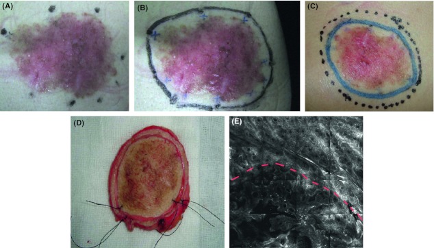
Background: Reflectance confocal microscopy (RCM) images skin at cellular resolution and has shown utility for the diagnosis of nonmelanoma skin cancer in vivo. It has the potential to define lesion margins before surgical therapy.
Objectives: To investigate the feasibility of RCM in defining the margins of basal cell carcinoma before surgery.
Methods: The margins of 10 lesions were evaluated using RCM. Biopsies of the margins were used to confirm the results. A protocol was constructed to define margins. RCM was used to delineate preoperative surgical margins in 13 patients. Intraoperative frozen biopsy was used to confirm the margins.
Results: In seven of 10 (70.0%) cases, the margins of the cancer were identified suing RCM. The tumor island was the critical feature in identifying the margins. In 12 of 13 (92.3%) cases, frozen biopsy corroborated that the surgical margins delineated by RCM were clear.
Conclusion: RCM imaging of the margins is feasible and demonstrates the possibility of preoperative mapping of cancer margins.
© 2012 by the American Society for Dermatologic Surgery, Inc. Published by Wiley Periodicals, Inc.
Figures

References
-
- English DR, Armstrong BK, Kricker A, Fleming C. Sunlight, cancer. Cancer Causes Control. 1997;8:271–83. - PubMed
-
- Jih MH, Friedman PM, Goldberg LH, Kimyai-Asadi A. Curettage prior to Mohs' micrographic surgery for previously biopsied nonmelanoma skin cancers: what are we curetting? Retrospective, prospective, comparative study. Dermatol Surg. 2005;31:10–5. - PubMed
-
- Calzavara-Pinton P, Longo C, Venturini M, Sala R, et al. Reflectance confocal microscopy for in vivo skin imaging. Photochem Photobiol. 2008;84:1421–30. - PubMed
-
- Rishpon A, Kim N, Scope A, Oliviero MC, et al. Reflectance confocal microscopy criteria for squamous cell carcinomas, actinic keratoses. Arch Dermatol. 2009;145:766–72. - PubMed
Publication types
MeSH terms
LinkOut - more resources
Full Text Sources
Medical
