. 2012 Nov;170(2):222-30.
doi: 10.1111/j.1365-2249.2012.04652.x.

Bead-size directed distribution of Pseudomonas aeruginosa results in distinct inflammatory response in a mouse model of chronic lung infection

Affiliations

Bead-size directed distribution of Pseudomonas aeruginosa results in distinct inflammatory response in a mouse model of chronic lung infection

L J Christophersen et al. Clin Exp Immunol. 2012 Nov.

Abstract

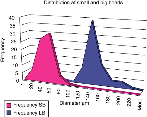
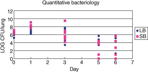
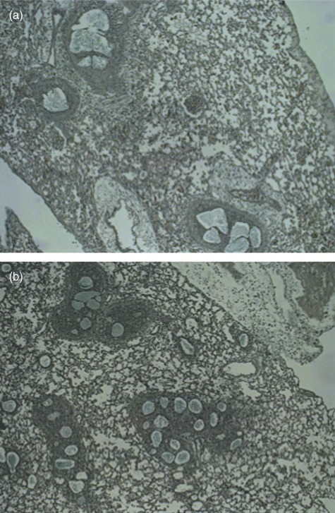
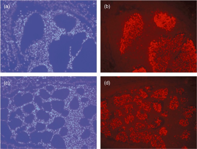
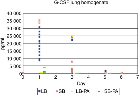
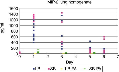
Chronic Pseudomonas aeruginosa lung infection in cystic fibrosis (CF) patients is characterized by biofilms, tolerant to antibiotics and host responses. Instead, immune responses contribute to the tissue damage. However, this may depend on localization of infection in the upper conductive or in the peripheral respiratory zone. To study this we produced two distinct sizes of small alginate beads (SB) and large beads (LB) containing P. aeruginosa. In total, 175 BALB/c mice were infected with either SB or LB. At day 1 the quantitative bacteriology was higher in the SB group compared to the LB group (P < 0·003). For all time-points smaller biofilms were identified by Alcian blue staining in the SB group (P < 0·003). Similarly, the area of the airways in which biofilms were identified were smaller (P < 0·0001). A shift from exclusively endobronchial to both parenchymal and endobronchial localization of inflammation from day 1 to days 2/3 (P < 0·05), as well as a faster resolution of inflammation at days 5/6, was observed in the SB group (P < 0·03). Finally, both the polymorphonuclear neutrophil leucocyte (PMN) mobilizer granulocyte colony-stimulating factor (G-CSF) and chemoattractant macrophage inflammatory protein-2 (MIP-2) were increased at day 1 in the SB group (P < 0·0001). In conclusion, we have established a model enabling studies of host responses in different pulmonary zones. An effective recognition of and a more pronounced host response to infection in the peripheral zones, indicating that increased lung damage was demonstrated. Therefore, treatment of the chronic P. aeruginosa lung infection should be directed primarily at the peripheral lung zone by combined intravenous and inhalation antibiotic treatment.


Figures

Fig. 1
Fig. 1
Size distribution of seaweed alginate beads. Different sizes of Pseudomonas aeruginosa containing seaweed alginate beads were produced using different nozzle size, airway pressure and volume speed of alginate. Mean value of two diameters measured at right angles for each bead (n = 72). Two distinct populations of beads were produced.
Fig. 2
Fig. 2
Number of Pseudomonas aeruginosa in the lungs of BALB/c mice infected with two different sizes of P. aeruginosa containing seaweed alginate beads. All mice received the same number of bacteria and the same amount of alginate in the left lung. The number of colony-forming units (CFU)/ml was significantly higher at day 1 after infection in the group of mice infected with the small beads (SB). At other time-points, no significant difference in quantitative bacteriology could be observed.
Fig. 3
Fig. 3
The distribution and sizes of Pseudomonas aeruginosa-containing seaweed alginate beads was estimated after Alcian blue staining. At all time-points smaller beads were identified in the small beads (SB) challenge group (P < 0·0001), although statistical analysis could not be performed for days 5/6 due to too few identifiable beads and representative bronchioles in the SB group. At days 2/3 the beads were identified in significantly smaller airways of the BALB/c mice lungs infected with the SB (P < 0·002). The same trend was observed at day 1, but did not reach statistical significance.
Fig. 4
Fig. 4
Alcian blue staining of lung slides prepared from mouse lungs obtained at day 1 after infection with either large beads (LB) in (a) and small beads (SB) in (b). Alcian blue staining was performed to identify the biofilm-like alginate-containing structures in the lungs. Magnification ×400.
Fig. 5
Fig. 5
The presence of Pseudomonas aeruginosa in the biofilm-like structures in the airways of BALB/c mice was confirmed by specific peptide nucleic acid–fluorescence in-situ hybridization (PNA–FISH) technique staining P. aeruginosa red. PMNs are stained blue by 4′, 6-diamidino-2-phenylindole (DAPI). (a) (LB) and (c) (SB) show DAPI-stained lungs and (b) (LB) and (d) (SB) the corresponding PNA–FISH-stained lungs obtained at day 1 after infection with P. aeruginosa-containing alginate beads. All magnification ×400.
Fig. 6
Fig. 6
Serum were obtained from all mice by cardiac puncture. Concentrations of the polymorphonuclear leucocytes (PMN) mobilizer granulocyte colony-stimulating factor (G-CSF) were determined by enzyme-linked immunosorbent assay (ELISA). G-CSF concentrations were increased significantly in the small beads (SB) group compared to the large beads (LB) group at day 1 (P < 0·0001). No statistically significant differences were observed at other time-points.
Fig. 7
Fig. 7
Concentrations of the polymorphonuclear leucocytes (PMN) mobilizer granulocyte colony-stimulating factor (G-CSF) in the supernatants of the lung homogenates were determined by enzyme-linked immunosorbent assay (ELISA). G-CSF concentrations were increased significantly in the small beads (SB) group at day 1 compared to the large beads (LB) group (P < 0·0001). No statistically significant differences were observed at other time-points.
Fig. 8
Fig. 8
Concentrations of the polymorphonuclear leucocytes (PMN) chemoattractant macrophage inflammatory protein-2 (MIP-2) in the supernatants of the lung homogenates were determined by enzyme-linked immunosorbent assay (ELISA). MIP-2 concentrations were increased significantly in the small beads (SB) group at day 1 compared to the large beads (LB) group (P < 0·0001). No statistically significant differences were observed at other time-points.

References

    1. Jensen PO, Givskov M, Bjarnsholt T, Moser C. The immune system vs. Pseudomonas aeruginosa biofilms. FEMS Immunol Med Microbiol. 2010;59:292–305. - PubMed
    1. Knowles MR, Boucher RC. Mucus clearance as a primary innate defense mechanism for mammalian airways. J Clin Invest. 2002;109:571–7. - PMC - PubMed
    1. Craig A, Mai J, Cai S, Jeyaseelan S. Neutrophil recruitment to the lungs during bacterial pneumonia. Infect Immun. 2009;77:568–75. - PMC - PubMed
    1. Schmiedl A, Kerber-Momot T, Munder A, Pabst R, Tschernig T. Bacterial distribution in lung parenchyma early after pulmonary infection with Pseudomonas aeruginosa. Cell Tissue Res. 2010;342:67–73. - PubMed
    1. McCullagh A, Rosenthal M, Wanner A, Hurtado A, Padley S, Bush A. The bronchial circulation – worth a closer look: a review of the relationship between the bronchial vasculature and airway inflammation. Pediatr Pulmonol. 2010;45:1–13. - PubMed

MeSH terms