. 2013 Mar;63(3):551-60.
doi: 10.1016/j.eururo.2012.09.034. Epub 2012 Sep 24.

Intratunical injection of human adipose tissue-derived stem cells prevents fibrosis and is associated with improved erectile function in a rat model of Peyronie's disease

Affiliations

Intratunical injection of human adipose tissue-derived stem cells prevents fibrosis and is associated with improved erectile function in a rat model of Peyronie's disease

Fabio Castiglione et al. Eur Urol. 2013 Mar.

Erratum in

  • Eur Urol. 2013 Jul;64(1):e21

Abstract

Background: Peyronie's disease (PD) is a connective tissue disorder of the tunica albuginea (TA). Currently, no gold standard has been developed for the treatment of the disease in its active phase.

Objective: To test the effects of a local injection of adipose tissue-derived stem cells (ADSCs) in the active phase of a rat model of PD on the subsequent development of fibrosis and elastosis of the TA and underlying erectile tissue.

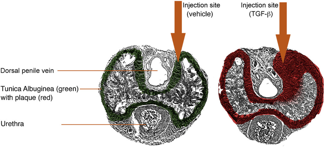
Design, setting, and participants: A total of 27 male 12-wk-old Sprague-Dawley rats were divided in three equal groups and underwent injection of vehicle (sham), 0.5-μg [corrected] transforming growth factor (TGF)-β1 in a 50-μl vehicle in either a PD or a PD plus ADSC group in the dorsal aspect of the TA.

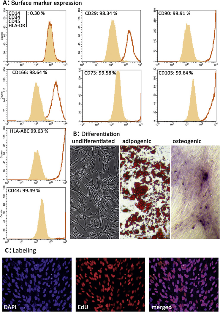
Intervention: The sham and PD groups were treated 1 d after TGF-β1 injection with intralesional treatment of vehicle, and the PD plus ADSC group received 1 million human-labeled ADSCs in the 50-μl vehicle. Five weeks after treatment, six rats per group underwent erectile function measurement. Following euthanasia, penises were harvested for histology and Western blot.

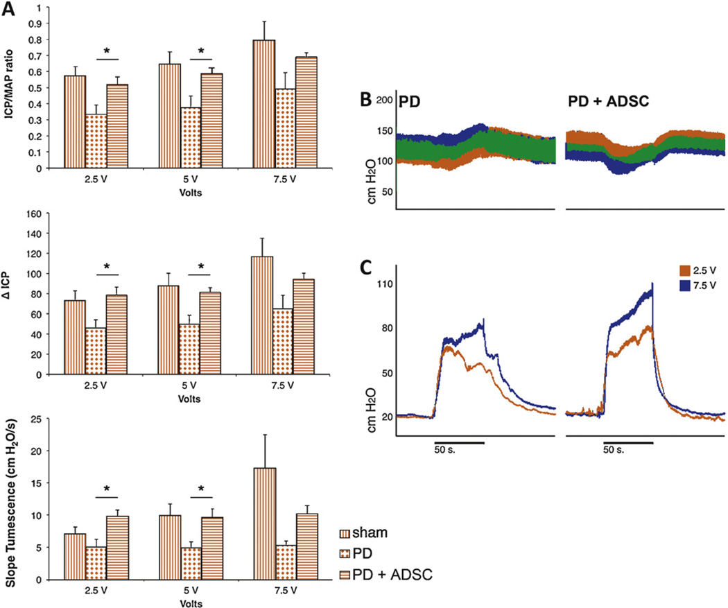
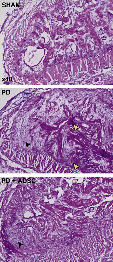
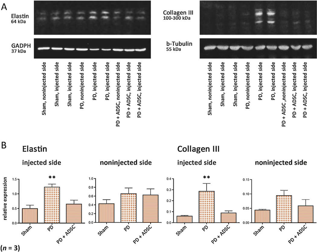
Outcome measurements and statistical analysis: The ratio of intracavernous pressure to mean arterial pressure (ICP/MAP) upon cavernous nerve stimulation, elastin, and collagen III protein expression and histomorphometric analysis of the penis. Statistical analysis was performed by analysis of variance followed by the Tukey-Kramer test for post hoc comparisons or the Mann-Whitney test when applicable.

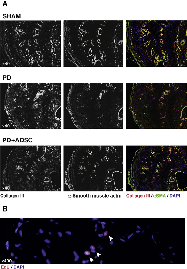
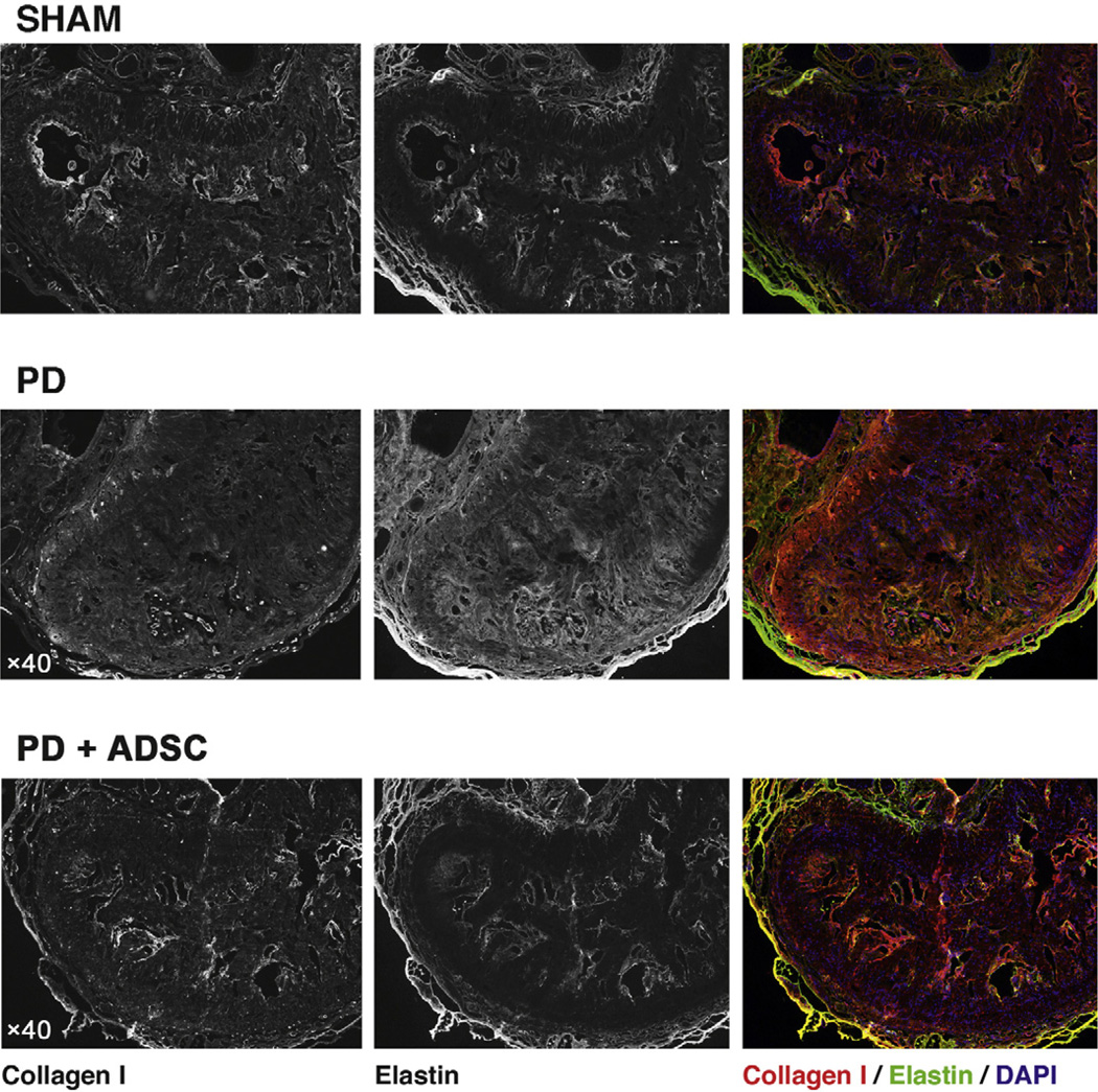
Results and limitations: Erectile function significantly improved after ADSC treatment (ICP/MAP 0.37 in PD vs 0.59 in PD plus ADSC at 5-V stimulation; p=0.03). PD animals developed areas of fibrosis and elastosis with a significant upregulation of collagen III and elastin protein expression. These fibrotic changes were prevented by ADSC treatment.

Conclusions: This study is the first to test stem cell therapy in an animal model of PD. Injection of ADSCs into the TA during the active phase of PD prevents the formation of fibrosis and elastosis in the TA and corpus cavernosum.


Conflict of interest statement

Financial disclosures: Petter Hedlund certifies that all conflicts of interest, including specific financial interests and relationships and affiliations relevant to the subject matter or materials discussed in the manuscript (eg, employment/affiliation, grants or funding, consultancies, honoraria, stock ownership or options, expert testimony, royalties, or patents filed, received, or pending), are the following: None.

Figures

Fig. 1
Fig. 1
Establishment of the model. Schematic figure depicts the anatomy of a transversally transected penile midshaft specimen and injection site of 50-µl vehicle or 50-µg transforming growth factor (TGF)-β1 in 50-µl vehicle in the dorsal-dorsomedial tunica albuginea resulting in thickening of the tunica albuginea and plaque formation (red) after TGF-β1 injection.
Fig. 2
Fig. 2
Characterization, differentiation, and labeling efficacy of adipose tissue-derived stem cells (ADSCs). (A) Flow cytometry analysis on ADSCs isolated from a female human donor shows that the vast majority of cells possess a CD14, CD34, CD45, HLA-DR, and CD29+, CD90+, CD166+, CD73+, CD105+, CD44+, HLA-ABC+ phenotype, consistent with the typical mesenchymal multipotent stromal cell surface marker profile (light gray area: isotype control; black line: sample). (B) Cultured human ADSCs display a typical fibroblast-like morphology and are able to differentiate between adipogenic and osteogenic lineages in vitro, as demonstrated by positive oil red O staining for lipid droplet formation and alizarin red staining for calcium deposit formation. (C) ADSCs were incubated with the thymidine analog 5-ethynyl-2-deoxyuridine (EdU) for 48 h, resulting in complete labeling of attached ADSCs.
Fig. 3
Fig. 3
Erectile function measurement. (A) Summarized data comparing erectile function measurements in sham (black bars) PD rats (blocked bars) and rats treated with adipose tissue-derived stem cells (ADSCs) (open bars) at various voltages during cavernous nerve electrostimulation. Top: Intracavernous pressure (ICP) change from baseline to peak ICP (ΔICP); middle: ICP normalized over mean arterial pressure (MAP); bottom: steepness of slope of tumescence phase of erection. * p < 0.05 in analysis of variance with post hoc Student-Newman-Keuls analysis. (B) Representative example traces of MAP; (C) ICP recordings during 2.5- and 7.5-V cavernous nerve electrostimulation in PD and PD plus ADSC rats.
Fig. 4
Fig. 4
Hematoxylin and eosin staining in midshaft section of rat penis. Original magnification ×40. Photographs depict the dorsal part of the corpus cavernosum and tunica albuginea in sham, Peyronie’s disease (PD), and PD plus adipose tissue-derived stem cells (ADSC) rats. Note the open cavernous sinusoids in the sham rats and the surrounding normal bilayered structure of the tunica albuginea. In PD rats, there is deposition of amorphic extracellular matrix material with scattered high numbers of cells, which are expected to be fibroblasts based on their spindle-shaped morphology and relationship with the extracellular matrix (black arrowhead). Also note the haphazard organization of trabecular bundles arising from the dorsomedial tunica albuginea (white arrowheads). In the PD plus ADSC group, there is an increase in fibroblasts and extracellular matrix deposition; however, collagen fibers seem better organized and sinusoid structure is largely preserved.
Fig. 5
Fig. 5
Western blot analysis for collagen III and elastin. (A) Representative chemiluminescence images of blotted membranes containing protein extracts of all three groups, both injected/treated sides and noninjected sides serving as internal control for elastin and collagen III expression. Double bands are due to binding of antibodies to glycosylated and nonglycosylated forms of these molecules. (B) Summarized protein expression levels for elastin and collagen III. **p < 0.05 versus both sham and Peyronie’s disease (PD) plus adipose tissue-derived stem cells (ADSCs) in analysis of variance with post hoc Student-Newman-Keuls analysis.
Fig. 6
Fig. 6
Smooth muscle, collagen III, and adipose tissue-derived stem cells (ADSCs) in midshaft section of rat penis. Original magnification (a) ×40; (b) ×400. (A) Top: Note how in sham animals α-smooth muscle actin (αSMA; green) and collagen III (red) colocalize in the smooth muscle layers surrounding the cavernous sinusoids and the dorsal penile arteries. In normal tunica albuginea (TA), expression of these proteins is very low. Middle: In Peyronie’s disease (PD) animals, 5 wk following injection of transforming growth factor-β1, sinusoidal structure has largely disappeared near the injection site. αSMA is largely absent in the dorsal part of the corpus cavernosum (by means of internal antibody control, expression is preserved in the dorsal penile vessels). Note how collagen III is now deposited in an amorphic fashion in regions of the corpus cavernosum adjacent to the injection site in the dorsomedial TA. Bottom: The changes observed in PD rats are present in a mild or moderate degree after ADSC treatment. However, overall structure and sinusoids are largely preserved, and αSMA-positive sinusoids have undergone less dramatic reorganization than those in PD animals and collagen III. (B) High-magnification image of nuclear double staining of 4′,6-diamidino-2-phenylindole (DAPI) and 5-ethynyl-2-deoxyuridine (EdU) in a few cells in the corpus cavernosum of PD plus ADSC rats. These double-labeled cells were scarcely found.
Fig. 7
Fig. 7
Collagen I and elastin in midshaft section of rat penis. Original magnification, ×40. (A) Top: In sham animals, collagen I (red) is localized around the sinusoids and mainly in the internal layer of the tunica albuginea. Elastin (green) displays a similar expression pattern and is highly present in the Buck fascia (outer layer of tissue), and by means of internal control it is visible in the internal and external elastic laminae of the dorsal penile vessels. Middle: A destruction of the normal anatomy of the dorsal tunica is observed in Peyronie’s disease (PD) animals, with collagen I present in all layers of the tunica and collagen fibers extending in a disorganized fashion into the erectile tissue. Also, elastin staining is very bright and fibers are haphazardly organized. A clear overgrowth of elastin fibers in the tunica and into the corpus cavernosum is observed. Bottom: When adipose tissue-derived stem cells (ADSCs) were administered to these animals in the acute phase of PD, the observed changes were prevented and the sinusoidal structure of the dorsal parts of the corpus cavernosum was observed.

Comment in

Similar articles

Cited by

References

    1. Lin CS, Lin G, Wang Z, Maddah SA, Lue TF. Upregulation of monocyte chemoattractant protein 1 and effects of transforming growth factor-beta 1 in Peyronie’s disease. Biochem Biophys Res Commun. 2002;295:1014–1019. - PubMed
    1. Brock G, Hsu GL, Nunes L, von Heyden B, Lue TF. The anatomy of the tunica albuginea in the normal penis and Peyronie’s disease. J Urol. 1997;157:276–281. - PubMed
    1. Hatzimouratidis K, Eardley I, Giuliano F, et al. Guidelines on penile curvature. Eur Urol. 2012;62:543–552. - PubMed
    1. Smith JF, Walsh TJ, Conti SL, Turek P, Lue T. Risk factors for emotional and relationship problems in Peyronie’s disease. J Sex Med. 2008;5:2179–2184. - PMC - PubMed
    1. Ferretti L, Giuliani M, Bessède T, et al. Tissue engineering for penile surgery: comparative study of noncellular and cell-seeded synthetic grafts for tunica albuginea replacement. J Sex Med. 2012;9:625–631. - PubMed

Publication types

MeSH terms