. 2012 Oct 5;11(4):554-66.
doi: 10.1016/j.stem.2012.07.002.

Signaling from the sympathetic nervous system regulates hematopoietic stem cell emergence during embryogenesis

Affiliations

Signaling from the sympathetic nervous system regulates hematopoietic stem cell emergence during embryogenesis

Simon R Fitch et al. Cell Stem Cell. .

Abstract

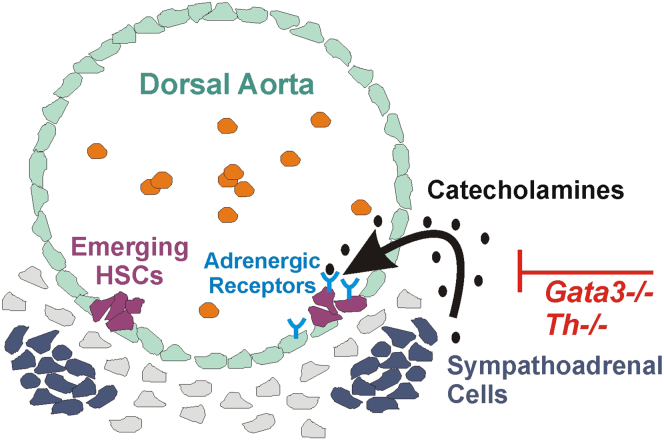
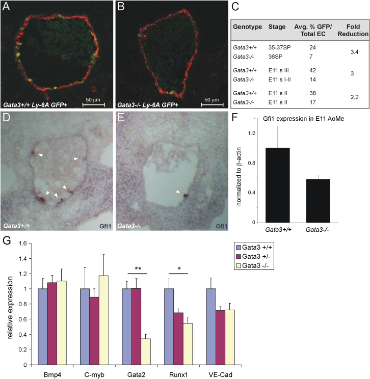
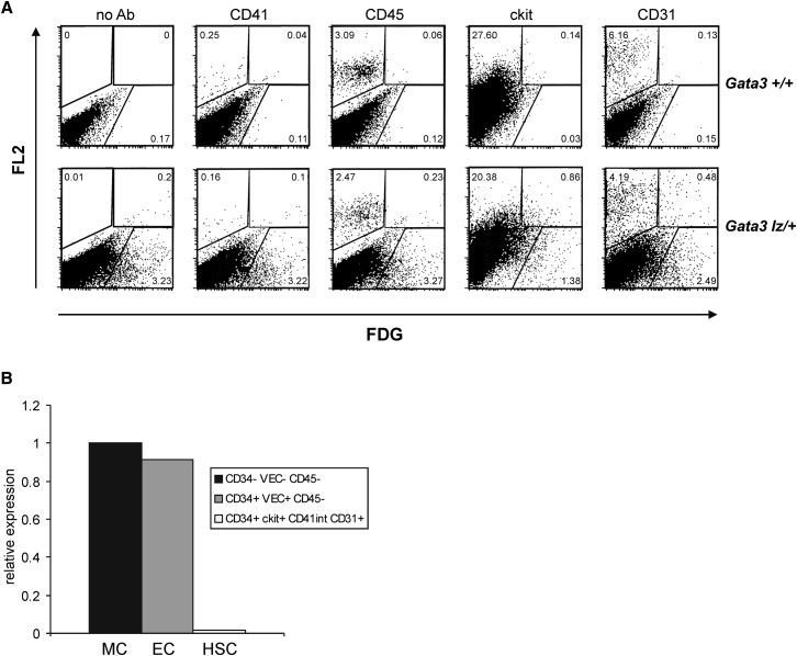
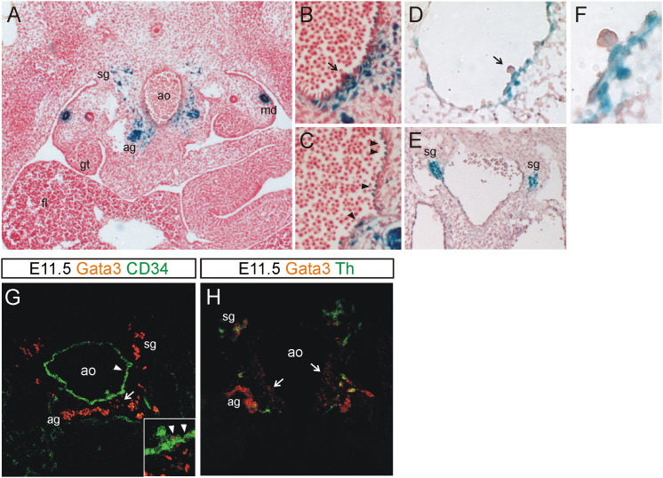
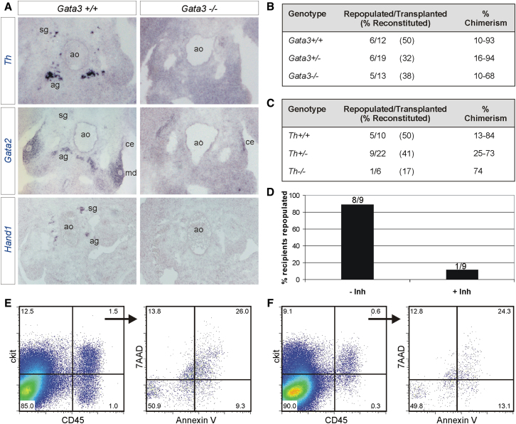
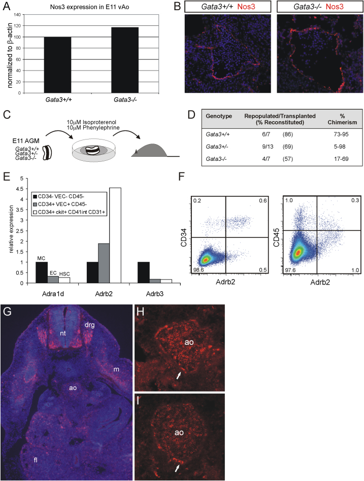
The first adult-repopulating hematopoietic stem cells (HSCs) emerge in the aorta-gonads-mesonephros (AGM) region of the embryo. We have recently identified the transcription factor Gata3 as being upregulated in this tissue specifically at the time of HSC emergence. We now demonstrate that the production of functional and phenotypic HSCs in the AGM is impaired in the absence of Gata3. Furthermore, we show that this effect on HSC generation is secondary to the role of Gata3 in the production of catecholamines, the mediators of the sympathetic nervous system (SNS), thus making these molecules key components of the AGM HSC niche. These findings demonstrate that the recently described functional interplay between the hematopoietic system and the SNS extends to the earliest stages of their codevelopment and highlight the fact that HSC development needs to be viewed in the context of the development of other organs.


Figures

None
Graphical abstract
Figure 1
Figure 1
Phenotypic HSCs Are Reduced in Gata3-Deficient Embryos Sections were prepared from E10.5–E11.5 Gata3+/+Ly-6A GFP+ and Gata3−/−Ly-6A GFP+ embryos that had been staged according to their somite pairs (E10.5) or according to the pigmentation around their eyes (E11). Sections were costained for CD34 and the number of GFP+ cells within the total endothelial cells were counted on confocal images of every tenth section. Representative sections of Gata3+/+Ly-6A GFP+ and Gata3−/−Ly-6A GFP+ embryos are shown in (A) and (B), respectively. GFP, green; CD34, red/Cy5; ventral, down. A summary of the results from three independent experiments is shown in (C). SP, somite pairs; s, stage. Sections of E11 Gata3+/+ (D) and Gata3−/− (E) embryos were stained with a riboprobe for Gfi1. Ventral, down, 10×/0.25 objective. Levels of Gfi1 were determined in subdissected E11 Gata3+/+ and Gata3−/− aorta-mesenchyme by quantitative RT-PCR (F). Error bars represent SD. (G) Quantitative RT-PCR was performed on cDNA prepared from ventral halves of aorta-mesenchymes from E11 Gata3+/+ (n = 3), Gata3+/− (n = 7), and Gata−/− (n = 3) embryos for the named genes and was normalized to Actb and Tbp. p < 0.05, ∗∗p < 0.01; error bars represent the SEM of the normalized relative expressions.
Figure 2
Figure 2
Gata3 Is Not Expressed in Hematopoietic Cells in the E11.5 AGM Region (A) Gata3+/+ (top panels) and Gata3lz/+ (bottom panels) E11.5 AGM cells were loaded with the fluorescent β-galactosidase substrate FDG and analyzed for coexpression of CD41, CD45, ckit, and CD31 (FL2). (B) Mesenchymal (MC), endothelial (EC), and hematopoietic stem cell (HSC) populations were sorted from E11.5 wild-type aorta-mesenchymes using the indicated marker combinations and the levels of Gata3 transcripts (normalized to Actb and Tbp) determined by quantitative RT-PCR.
Figure 3
Figure 3
Gata3 Is Expressed in a Number of Different Cell Types in the AGM E11.5 (A–C) and E10 (D–F) Gata3lz/+ embryos were stained with X-gal (blue) and cryosections were counterstained with Neutral Red. Ventral, down. Objectives were 10×/0.25 (A), 20×/0.45 (B, C, and E), 40×/0.65 (D), or 100×/1.4 (F). Arrows in (B) and (D) point to intra-aortic clusters; arrowheads in  (C) highlight Gata3-expressing endothelial cells. (F) A close-up of the cluster in (D). (G and H) E11.5 wild-type sections were costained for Gata3 (red/Cy5) and CD34 (green/FITC) (G) or Th (green/Alexa 488) (H) and confocal images were obtained. Arrows in (G) and (H) point to Gata3 staining in subaortic mesenchymal cells, and arrowheads in (G) point to Gata3+ endothelial cells. ag, adrenal anlage; ao, dorsal aorta; fl, fetal liver; gt, gonadal tissue; md, mesonephric duct; sg, sympathetic ganglia; Th, tyrosine hydroxylase.
Figure 4
Figure 4
Gata3 Regulation of AGM HSCs Is Secondary to Its Role in the Sympathetic Nervous System (A) In situ hybridization with riboprobes for Th, Gata2, and Hand1 on Gata3+/+ and Gata3−/− E11.5 embryo sections. (B) Summary of repopulation analysis of recipients injected with uncultured E11/11.5 AGM cells (1 ee) from Gata3+/+, Gata3+/−, and Gata3−/− embryos that had received catecholamine derivatives in vivo through the drinking water from E8.5. See also Figure S2. (C) Summary of repopulation analysis of recipients injected with uncultured E11/11.5 AGM cells (1 ee) from Th+/+, Th+/−, and Th−/− embryos. See also Figure S2. (D) Percentage of recipients repopulated with wild-type E11.5 AGMs that had been cultured in the presence or absence of a Th inhibitor. The number of repopulated/total recipients is indicated above each bar. (E and F) Flow cytometry analysis of ckit and CD45 expression on cells from E11.5 wild-type AGMs cultured in the absence (E) or presence (F) of a Th inhibitor. The percentage of apoptotic cells within the ckit+CD45+ population is shown.
Figure 5
Figure 5
Catecholamines Can Rescue HSC Activity In Vitro in the Absence of Circulation (A) Ventral halves of dorsal aortae from E11 Gata3+/+ and Gata3−/− embryos were analyzed for Nos3 expression by quantitative RT-PCR. Data is representative of two independent experiments. (B) E11.5 Gata3+/+ and Gata3−/− embryo sections were stained for Nos3 (red/Alexa 555) and counterstained with DAPI (blue). Ventral, down; 20×/0.45 objective. (C) Schematic outline of catecholamine treatment of AGMs in explant culture. (D) Summary of repopulation analysis of recipients injected with cells (1 ee) from Gata3+/+, Gata3+/−, and Gata3−/− E11/11.5 AGMs that had been exposed to catecholamine derivatives in explant cultures. See also Figure S3. (E) Quantitative RT-PCR expression analysis of the α1d- (Adra1d), β2- (Adrb2), and β3- (Adrb3) adrenergic receptors in mesenchymal (MC), endothelial (EC), and hematopoietic stem cell (HSC) populations sorted from E11.5 aorta-mesenchymes (normalized to Actb and Tbp). (F) Flow cytometry analysis of the expression of the β2-adrenergic receptor on CD34+ or CD45+ E11.5 AGM cells. (G–I) Immunohistochemistry on E11.5 wild-type embryo sections with an antibody to the β2-adrenergic receptor (red, Cy3). Nuclear DAPI staining is shown (blue) in (G). Arrows in (H) and (I) highlight expression on endothelial cells. Ventral, down; objectives were 10×/0.25 (G) or 20×/0.45 (H and I). ao, dorsal aorta; drg, dorsal root ganglia; fl, fetal liver; m, myotome; nt, neural tube.

References

    1. Adamo L., Naveiras O., Wenzel P.L., McKinney-Freeman S., Mack P.J., Gracia-Sancho J., Suchy-Dicey A., Yoshimoto M., Lensch M.W., Yoder M.C. Biomechanical forces promote embryonic haematopoiesis. Nature. 2009;459:1131–1135. - PMC - PubMed
    1. Asselin-Labat M.L., Sutherland K.D., Barker H., Thomas R., Shackleton M., Forrest N.C., Hartley L., Robb L., Grosveld F.G., van der Wees J. Gata-3 is an essential regulator of mammary-gland morphogenesis and luminal-cell differentiation. Nat. Cell Biol. 2007;9:201–209. - PubMed
    1. Benveniste P., Frelin C., Janmohamed S., Barbara M., Herrington R., Hyam D., Iscove N.N. Intermediate-term hematopoietic stem cells with extended but time-limited reconstitution potential. Cell Stem Cell. 2010;6:48–58. - PubMed
    1. Bertrand J.Y., Giroux S., Golub R., Klaine M., Jalil A., Boucontet L., Godin I., Cumano A. Characterization of purified intraembryonic hematopoietic stem cells as a tool to define their site of origin. Proc. Natl. Acad. Sci. USA. 2005;102:134–139. - PMC - PubMed
    1. Buza-Vidas N., Duarte S., Luc S., Bouriez-Jones T., Woll P.S., Jacobsen S.E. GATA3 is redundant for maintenance and self-renewal of hematopoietic stem cells. Blood. 2011;118:1291–1293. - PubMed

Publication types