. 2012 Oct 5;91(4):703-12.
doi: 10.1016/j.ajhg.2012.08.015.

Mutations in calmodulin cause ventricular tachycardia and sudden cardiac death

Affiliations

Mutations in calmodulin cause ventricular tachycardia and sudden cardiac death

Mette Nyegaard et al. Am J Hum Genet. .

Abstract

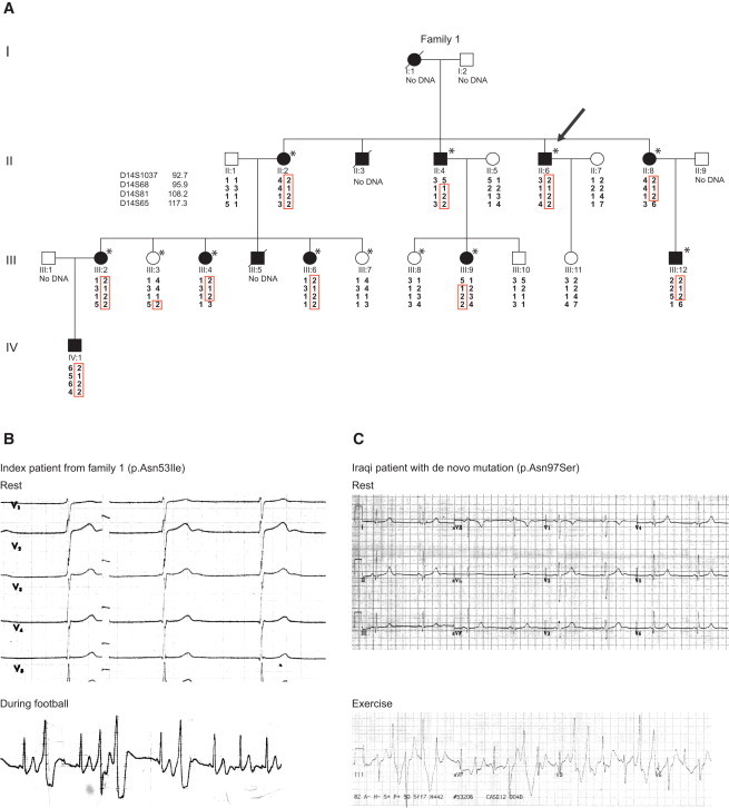
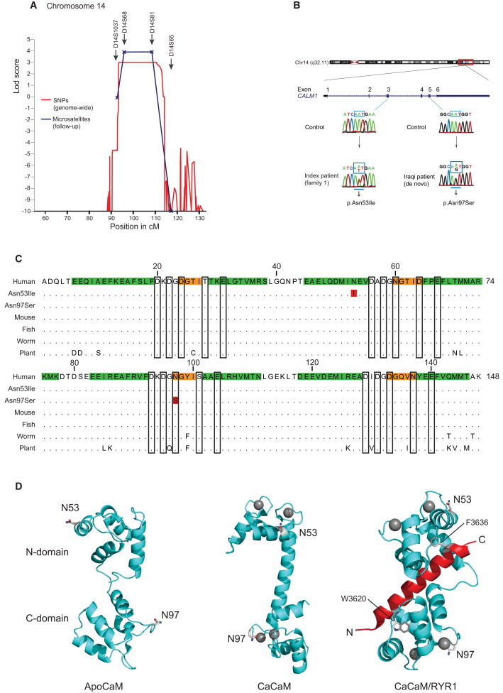
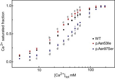
Catecholaminergic polymorphic ventricular tachycardia (CPVT) is a devastating inherited disorder characterized by episodic syncope and/or sudden cardiac arrest during exercise or acute emotion in individuals without structural cardiac abnormalities. Although rare, CPVT is suspected to cause a substantial part of sudden cardiac deaths in young individuals. Mutations in RYR2, encoding the cardiac sarcoplasmic calcium channel, have been identified as causative in approximately half of all dominantly inherited CPVT cases. Applying a genome-wide linkage analysis in a large Swedish family with a severe dominantly inherited form of CPVT-like arrhythmias, we mapped the disease locus to chromosome 14q31-32. Sequencing CALM1 encoding calmodulin revealed a heterozygous missense mutation (c.161A>T [p.Asn53Ile]) segregating with the disease. A second, de novo, missense mutation (c.293A>G [p.Asn97Ser]) was subsequently identified in an individual of Iraqi origin; this individual was diagnosed with CPVT from a screening of 61 arrhythmia samples with no identified RYR2 mutations. Both CALM1 substitutions demonstrated compromised calcium binding, and p.Asn97Ser displayed an aberrant interaction with the RYR2 calmodulin-binding-domain peptide at low calcium concentrations. We conclude that calmodulin mutations can cause severe cardiac arrhythmia and that the calmodulin genes are candidates for genetic screening of individual cases and families with idiopathic ventricular tachycardia and unexplained sudden cardiac death.


Figures

Figure 1
Figure 1
Pedigree and Electrocardiography (A) Pedigree of Swedish family with the chromosome 14 haplotypes presented graphically with HaploPainter. The index case is indicated with an arrow. The individuals included in the initial SNP genome-wide linkage analysis are marked by an asterisk. (B) The ECG during rest and an arrhythmia event (while the individual was playing football) occurring during a 24 hr ECG registration of the index case II:6 of family 1. (C) Resting and exercise ECGs from a second, unrelated individual of Iraqi origin. Both resting ECGs demonstrate prominent U waves in the anterior leads, and bidirectional ventricular ectopy is seen during physical activity or exercise.
Figure 2
Figure 2
Genome-wide Linkage Analysis and Position of the Calmodulin Mutations (A) A locus for a severe dominantly inherited form of CPVT-like arrhythmia is shown at position 14q31-32 (LOD score 3.9) and harboring approximately 70 known genes. (B) CALM1 structure and position of the c.161A>T and c.293A>G mutations identified in two different individuals with ventricular tachycardia and the sequencing electropherograms of exon 3 and 5 in the case and a control subject. (C) Alignment of calmodulin amino acid sequences from different species; the p.Asn53Ile and p.Asn97Ser substitutions are indicated (red residues). Secondary-structure elements are shown in green and orange (α-helix and β strand, respectively), and the Ca2+-binding residues are framed. Dots indicate conserved residues, showing that calmodulin is 100% conserved in all vertebrates and only displays a few changes in round worms and plants. (D) The position of the substituted residues in the three-dimensional structural model of native calmodulin prepared with Pymol (Schrödinger, LLC). Left panel: apo-calmodulin (Protein Data Bank [PDB] ID: 1DMO21), Middle panel: Ca2+-saturated calmodulin (pdb ID: 1CLL22), Right panel: Ca2+ bound calmodulin-RYR1 peptide complex (PDB ID: 2BCX23). Calcium ions are shown as gray spheres; the RYR1 peptide is in red. The two aromatic hydrophobic binding anchors in RYR1 are shown in stick representation. The residue Asn53 is positioned on the solvent-exposed side of α-helix C, not in contact with Ca2+, peptide ligand, or other calmodulin domains. Asn97 is one of the Ca2+-coordinating residues of Ca2+-binding site III. The calmodulin N domain is positioned on top, and the C domain is at the bottom. The Ca2+- and RYR1-complexed calmodulin structures have been rotated roughly 90 and 180 degrees counterclockwise, respectively, around a vertical axis after structural alignment of the N domains with the apo-calmodulin structure.
Figure 3
Figure 3
Calcium Binding The relative Ca2+ binding of the C domain of calmodulin as a function of total Ca2+ concentration, determined as the change in the tyrosine fluorescence intensity. The data presents the average of three independent experiments (+/− SD); in most cases the error bars are smaller than the symbols. In comparison to native calmodulin, p values are as follows: a, p < 0.05; b, p < 0.01; c, p < 0.001; and d, p < 0.0001 (two-way repeated-measures ANOVA with the Bonferroni multiple comparisons post-hoc test).
Figure 4
Figure 4
Interaction with RYR2 Peptide Calmodulin binding to the RYR2-CBD peptide (Arg3581–Leu3611). The RYR2 Trp3586 fluorescence-emission spectrum, normalized to the maximal fluorescence intensity of the peptide alone, is shown without added calmodulin (RYR) and with a saturating amount of the indicated calmodulin variants added at (A) low intracellular (100 nM free Ca2+), (B) moderate (1 μM free Ca2+), and (C) saturating (200 μM Ca2+) concentrations.

References

    1. Badhwar N., Scheinman M.M. Idiopathic ventricular tachycardia: Diagnosis and management. Curr. Probl. Cardiol. 2007;32:7–43. - PubMed
    1. Coumel P., Fidelle J., Lucet V., Attuel P., Bouvrain Y. Catecholaminergic-induced severe ventricular arrhythmias with Adams-Stokes syndrome in children: Report of four cases. Br. Heart J. 1978;40(suppl):28–37.
    1. Napolitano C., Priori S.G. Diagnosis and treatment of catecholaminergic polymorphic ventricular tachycardia. Heart Rhythm. 2007;4:675–678. - PubMed
    1. Aizawa Y., Komura S., Okada S., Chinushi M., Aizawa Y., Morita H., Ohe T. Distinct U wave changes in patients with catecholaminergic polymorphic ventricular tachycardia (CPVT) Int. Heart J. 2006;47:381–389. - PubMed
    1. Viitasalo M., Oikarinen L., Väänänen H., Kontula K., Toivonen L., Swan H. U-waves and T-wave peak to T-wave end intervals in patients with catecholaminergic polymorphic ventricular tachycardia, effects of beta-blockers. Heart Rhythm. 2008;5:1382–1388. - PubMed

Publication types

MeSH terms

Substances