Review
. 2012 Oct 4;76(1):98-115.
doi: 10.1016/j.neuron.2012.09.014.

Neuropeptide transmission in brain circuits

Affiliations
Review

Neuropeptide transmission in brain circuits

Anthony N van den Pol. Neuron. .

Abstract

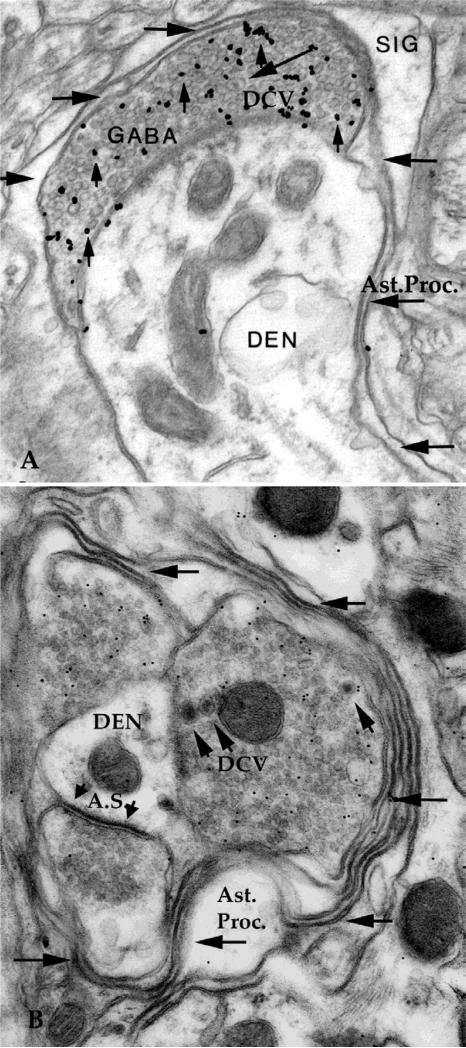
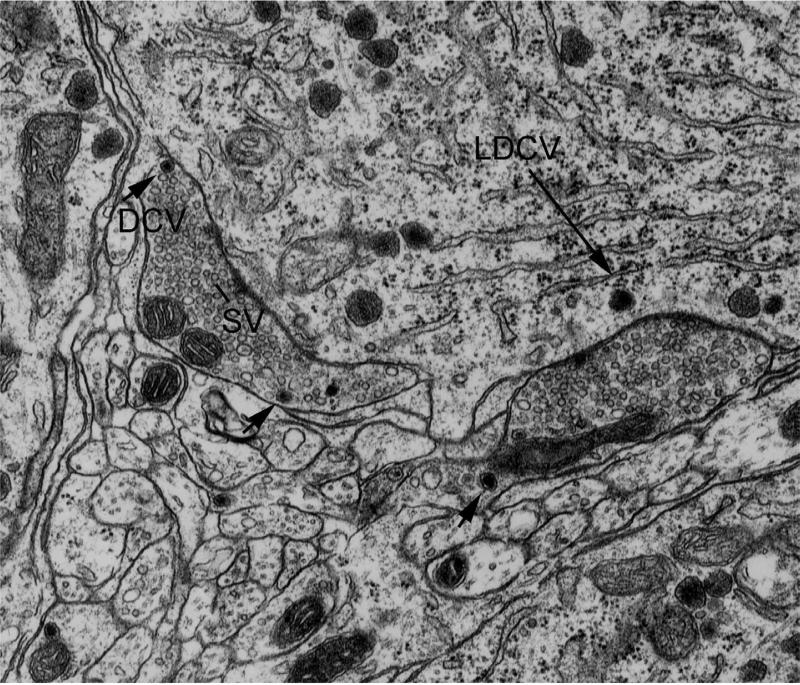
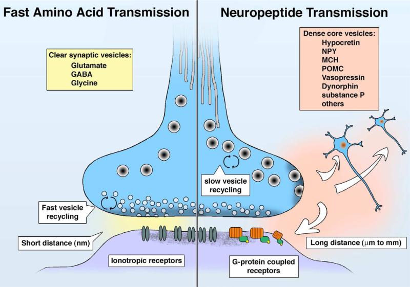
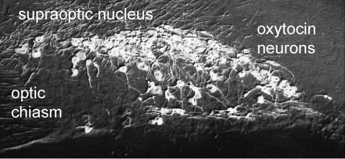
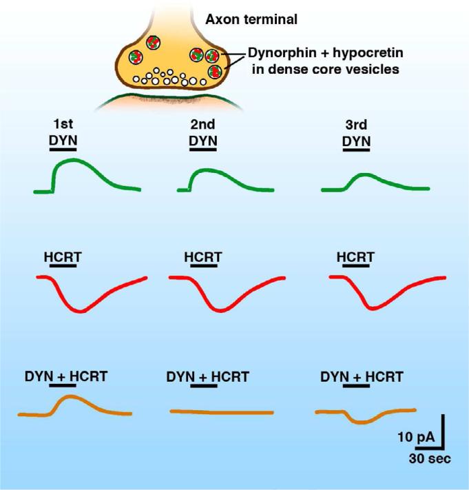
Neuropeptides are found in many mammalian CNS neurons where they play key roles in modulating neuronal activity. In contrast to amino acid transmitter release at the synapse, neuropeptide release is not restricted to the synaptic specialization, and after release, a neuropeptide may diffuse some distance to exert its action through a G protein-coupled receptor. Some neuropeptides such as hypocretin/orexin are synthesized only in single regions of the brain, and the neurons releasing these peptides probably have similar functional roles. Other peptides such as neuropeptide Y (NPY) are synthesized throughout the brain, and neurons that synthesize the peptide in one region have no anatomical or functional connection with NPY neurons in other brain regions. Here, I review converging data revealing a complex interaction between slow-acting neuromodulator peptides and fast-acting amino acid transmitters in the control of energy homeostasis, drug addiction, mood and motivation, sleep-wake states, and neuroendocrine regulation.


Figures

Fig. 1
Fig. 1
A. Astrocytic processes (Ast.Proc.) surround presynaptic axons in contact with central dendrite (DEN). SIG, silver intensified immunogold, DCV, dense core vesicle. Horizontal arrows show astrocytic process surrounding synaptic complex. B. Three boutons contact a central dendrite (DEN). Two GABA boutons are labeled with immunogold, a third bouton makes an asymmetrical synapse (A.S.) typical of glutamate synapses. The synaptic complex is surrounded by several layers of astrocytic processes (Ast.Proc.). Width of micrograph, A 2 um, B 1.7 um.
Fig. 2
Fig. 2
Two axons make symmetrical-type synaptic contact with a magnocellular neuron. The presynaptic axons contain a few dense core vesicles (DCV, arrowheads) and many small clear synaptic vesicles (SV). In the postsynaptic neuron, a large dense core vesicle (LDCV) is shown by long arrow. Width of micrograph, 4 um.
Fig. 3
Fig. 3
Comparison of fast amino acid synaptic transmission (left) and slower neuropeptide transmission (right).
Fig. 4
Fig. 4
This micrograph shows the oxytocin neurons of the hypothalamic supraoptic nucleus. Width of micrograph, 350 um.
Fig. 5
Fig. 5
Whole cell recording of GABA-mediated synaptic currents. TRH substantially increased the frequency of the IPSCs recorded in GABAergic MCH neurons, which recovered after peptide washout. The GABAA receptor antagonist bicuculline (BIC) blocked the synaptic currents. TRH had no effect on miniature PSCs. Recordings were done in the presence of AP5 and CNQX to block responses to synaptically-released glutamate. The recording pipette contained a high concentration of Cl, resulting in GABA-mediated inward currents. (From Zhang and van den Pol,2012).
Fig. 6
Fig. 6
A. A schematic representation of two axons both in synaptic contact with a common dendrite. B. Release of neuropeptide from the bouton on the left diffuses laterally to the other axon and enhances release of the fast amino acid transmitter.
Fig. 7
Fig. 7
NPY inhibits glutamatergic hypocretin cells by multiple mechanisms. NPY acts on Y1 postsynaptic receptors to inhibit the hypocretin cell, and acts on Y2 presynaptic receptors to attenuate release of GABA and glutamate. Different actions of NPY are shown by the red arrows, with a downward arrow indicating a decrease, and upward arrow an increase. These NPY axons also contain GABA. Based on Fu et al, 2004; Horvath et al,1999. NPY probably has similar effect on many of its target neurons, including the POMC cell (Cowley et al, 2001), although the subset of NPY receptors expressed in cell bodies or terminals may differ from cell to cell.
Fig. 8
Fig. 8
Dynorphin attenuates voltage-gated calcium current. A command depolarization of a glutamatergic hypocretin neuron caused a calcium influx (a). Dynorphin reduced the calcium current (b), which recovered after peptide washout. Cd2+ completely blocked the current, consistent with it being calcium-mediated. From Li and van den Pol,2006.
Fig. 9
Fig. 9
Inhibitory dynorphin and excitatory hypocretin are synthesized and released together. Serial application of dynorphin evokes an outward current (green) with an amplitude that desensitizes and deceases substantially with repeated exposure. Serial application of hypocretin evokes an inward current (red) with only modest desensitization. Combined application of dynorphin + hypocretin (orange) gives an initial outward current, then little current, and then an inward current. Based on Li and van den Pol,2006.

References

    1. Acuna-Goycolea C, van den Pol A. Glucagon-like peptide 1 excites hypocretin/orexin neurons by direct and indirect mechanisms: implications for viscera-mediated arousal. J Neurosci. 2004;24:8141–8152. - PMC - PubMed
    1. Acsády L, Arabadzisz D, Freund TF. Correlated morphological and neurochemical features identify different subsets of vasoactive intestinal polypeptide-immunoreactive interneurons in rat hippocampus. Neuroscience. 1996;73:299–315. - PubMed
    1. Acsády L, Katona I, Martínez-Guijarro FJ, Buzsáki G, Freund TF. Unusual target selectivity of perisomatic inhibitory cells in the hilar region of the rat hippocampus. J Neurosci. 2000;20:6907–6919. - PMC - PubMed
    1. Antonucci F, Alpár A, Kacza J, Caleo M, Verderio C, Giani A, Martens H, Chaudhry FA, Allegra M, Grosche J, Michalski D, Erck C, Hoffmann A, Harkany T, Matteoli M, Härtig W. Cracking down on inhibition: selective removal of GABAergic interneurons from hippocampal networks. J Neurosci. 2012;32:1989–2001. - PMC - PubMed
    1. Ariel P, Ryan TA. Optical mapping of release properties in synapses. Front Neural Circuits 4: art. 2010;18:1–10. - PMC - PubMed

Publication types