Review
. 2012 Oct 4;76(1):116-29.
doi: 10.1016/j.neuron.2012.08.036.

Acetylcholine as a neuromodulator: cholinergic signaling shapes nervous system function and behavior

Affiliations
Review

Acetylcholine as a neuromodulator: cholinergic signaling shapes nervous system function and behavior

Marina R Picciotto et al. Neuron. .

Abstract

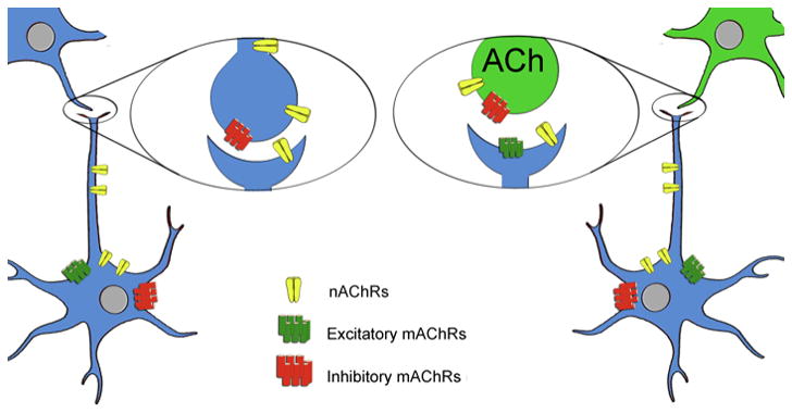
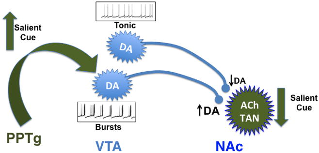
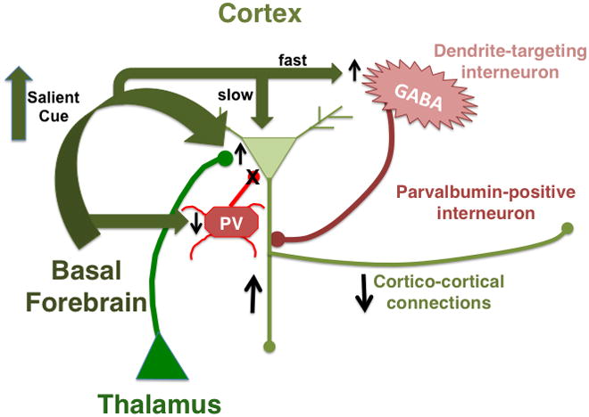
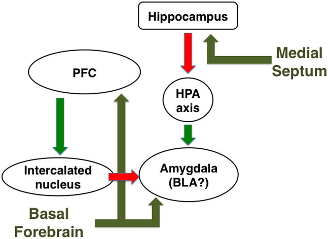
Acetylcholine in the brain alters neuronal excitability, influences synaptic transmission, induces synaptic plasticity, and coordinates firing of groups of neurons. As a result, it changes the state of neuronal networks throughout the brain and modifies their response to internal and external inputs: the classical role of a neuromodulator. Here, we identify actions of cholinergic signaling on cellular and synaptic properties of neurons in several brain areas and discuss consequences of this signaling on behaviors related to drug abuse, attention, food intake, and affect. The diverse effects of acetylcholine depend on site of release, receptor subtypes, and target neuronal population; however, a common theme is that acetylcholine potentiates behaviors that are adaptive to environmental stimuli and decreases responses to ongoing stimuli that do not require immediate action. The ability of acetylcholine to coordinate the response of neuronal networks in many brain areas makes cholinergic modulation an essential mechanism underlying complex behaviors.


Figures

Figure 1
Figure 1. Sites of action for nicotinic and muscarinic acetylcholine receptors
Nicotinic (nAChR) and muscarinic (mAChR) acetylcholine receptors are localized both pre- and post-synaptically. Presynaptic mAChRs (M2, M4) are largely inhibitory, and act as inhibitory autoreceptors on cholinergic terminals, with M2 the predominant autoreceptor in hippocampus and cerebral cortex, and M4 predominant in striatum (Wess, 2003b; Wess et al., 2003). Post-synaptic mAChRs can be either inhibitory (M2, M4) or excitatory (M1, M3, M5) (Wess, 2003b; Wess et al., 2003). Presynaptic nAChRs induce release of a number of neurotransmitters including GABA, glutamate, dopamine, serotonin, norepinephrine and acetylcholine (McGehee et al., 1995; Wonnacott, 1997). Postsynaptic nAChRs depolarize neurons, increase their firing rate and can contribute to long-term potentiation (Bucher and Goaillard, 2011; Ge and Dani, 2005; Ji et al., 2001; Kawai et al., 2007; Mansvelder and McGehee, 2000; Picciotto et al., 1995; Picciotto et al., 1998; Radcliffe and Dani, 1998; Wooltorton et al., 2003).
Figure 2
Figure 2. Effects of acetylcholine on activity of dopamine neurons in the mesolimbic circuit
Salient cues associated with primary rewards increase activity of pedunculopontine tegmental area (PPTg) neurons, inducing acetylcholine release in the ventral tegmental area (VTA) (Futami et al., 1995; Omelchenko and Sesack, 2006). Acetylcholine increases firing of dopamine (DA) neurons in the VTA and is likely to be important for burst firing of these neurons (Maskos, 2008). Salient cues associated with rewards also induce a pause in firing of tonically active cholinergic neurons (ACh TAN) in the nucleus accumbens (NAc) (Goldberg and Reynolds, 2011). Decreased release of ACh onto terminals in NAc attenuates DA release due to tonic firing of DA neurons, while preserving DA release in response to phasic firing (Exley and Cragg, 2008).
Figure 3
Figure 3. Effects of acetylcholine on activity of cortical neurons
Salient cues induce acetylcholine release onto interneurons targeting the apical dendrites of cortical pyramidal neurons, resulting in rapid inhibition of pyramidal cells (Arroyo et al., 2012; Couey et al., 2007; Fanselow et al., 2008; Ferezou et al., 2002; Gulledge et al., 2007; Kawaguchi and Kubota, 1997). Acetylcholine subsequently depolarizes pyramidal neurons through M1 mAChRs (Delmas and Brown, 2005; McCormick and Prince, 1985, 1986). Acetylcholine also activates stimulatory α4β2 nAChRs on glutamatergic thalamocortical terminals (Gil et al., 1997; Lambe et al., 2003; Oldford and Castro-Alamancos, 2003) and inhibitory M2 mAChRs on GABAergic terminals of parvalbumin-expressing (PV) interneurons (Kruglikov and Rudy, 2008). Activation of PV interneurons enhances stimulation of pyramidal neuron firing by thalamocortical inputs (Gabernet et al., 2005; Higley and Contreras, 2006; Kruglikov and Rudy, 2008). Acetylcholine also suppresses cortico-cortical transmission through inhibitory M2 mAChRs on pyramidal cell axon terminals (Gil et al., 1997; Hsieh et al., 2000; Kimura and Baughman, 1997; Oldford and Castro-Alamancos, 2003), reducing intra-cortical communication while preserving responses to thalamic inputs (Kimura et al., 1999).
Figure 4
Figure 4. Effects of acetylcholine on hippocampal-amygdala stress response
Stress increases acetylcholine release in the hippocampus and frontal cortex (Mark et al., 1996) and impairs signaling in the prefrontal cortex (PFC) (Arnsten, 2009). The hippocampus provides inhibitory feedback to the amygdala through inhibition of the hypothalamic-pituitary-adrenal (HPA) axis (Tasker and Herman, 2011) whereas the PFC can normally decrease basolateral amygdala activity through projections to the intercalated nucleus (Manko et al., 2011; Pinard et al., 2012). The effects of stress-induced acetylcholine release on output of hippocampus and cortex is unknown, but cholinergic modulation of cortico-amygdala glutamatergic connections strengthens associations between environmental stimuli and stressful events (Mansvelder et al., 2009).

References

    1. Alkondon M, Pereira EF, Albuquerque EX. alpha-bungarotoxin- and methyllycaconitine-sensitive nicotinic receptors mediate fast synaptic transmission in interneurons of rat hippocampal slices. Brain Res. 1998;810:257–263. - PubMed
    1. Alkondon M, Pereira EF, Barbosa CT, Albuquerque EX. Neuronal nicotinic acetylcholine receptor activation modulates gamma-aminobutyric acid release from CA1 neurons of rat hippocampal slices. J Pharmacol Exp Ther. 1997;283:1396–1411. - PubMed
    1. Angelucci F, Brene S, Mathe AA. BDNF in schizophrenia, depression and corresponding animal models. Molecular psychiatry. 2005;10:345–352. - PubMed
    1. Aramakis V, Metherate R. Nicotine selectively enhances NMDA receptor-mediated synaptic transmission during postnatal development in sensory neocortex. J Neurosci. 1998;18:8485–8495. - PMC - PubMed
    1. Aramakis VB, Hsieh CY, Leslie FM, Metherate R. A critical period for nicotine-induced disruption of synaptic development in rat auditory cortex. J Neurosci. 2000;20:6106–6116. - PMC - PubMed

Publication types