Review
. 2012 Oct 4;76(1):209-22.
doi: 10.1016/j.neuron.2012.09.012.

Neuromodulation of brain states

Affiliations
Review

Neuromodulation of brain states

Seung-Hee Lee et al. Neuron. .

Abstract

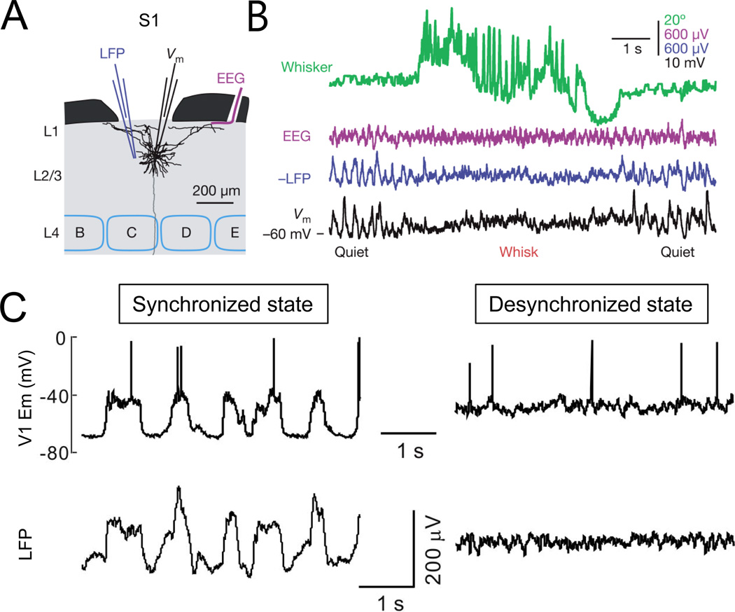
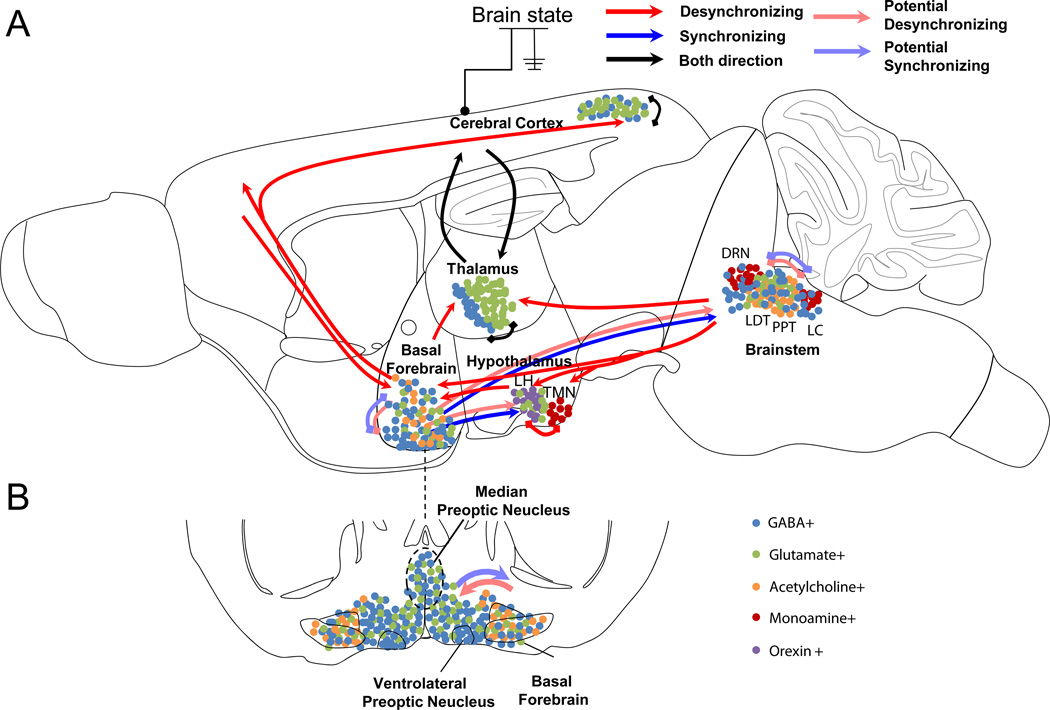
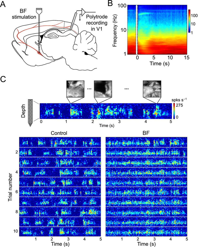
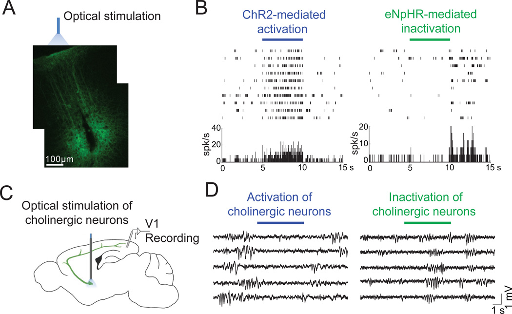
Switches between different behavioral states of the animal are associated with prominent changes in global brain activity, between sleep and wakefulness or from inattentive to vigilant states. What mechanisms control brain states, and what are the functions of the different states? Here we summarize current understanding of the key neural circuits involved in regulating brain states, with a particular emphasis on the subcortical neuromodulatory systems. At the functional level, arousal and attention can greatly enhance sensory processing, whereas sleep and quiet wakefulness may facilitate learning and memory. Several new techniques developed over the past decade promise great advances in our understanding of the neural control and function of different brain states.


Figures

Figure 1
Figure 1. Different methods for monitoring brain states
A, Schematic showing the recording configuration for simultaneous measurement of EEG, LFP, and single-cell membrane potential in the S1 barrel cortex. A pyramidal neuron in layer 2/3 was reconstructed. B, EEG, LFP, and whole-cell recordings show large-amplitude, low-frequency activity during quiet wakefulness and synchronous state change during whisking (Figures adapted and reproduced with permission from Poulet and Petersen, 2008). C, Synchronized (left) and desynchronized (right) brain states observed with simultaneous whole-cell patch clamp recording from a visual cortical neuron and LFP recording 2 mm from the patch electrode. Figures reproduced from Li et al., 2009).
Figure 2
Figure 2. Schematic diagram showing the key circuits involved in regulating brain states
A, Sagital view. Arrows indicate major pathways connecting the brain areas. Red arrow, pathway inducing cortical desynchronization; blue arrow, pathway inducing cortical synchronization, black arrow; pathway that mediate both synchronization and desynchronization; light red, possible pathway for desynchronization; light blue, possible pathway for synchronization. Each cell types were schematically illustrated by colored dots in each brain area. B, Coronal view of the basal forebrain/preoptic area, at the position indicated by dashed line in A. MnPO is in fact more anterior but outlined here for convenience.
Figure 3
Figure 3. Effect of basal forebrain stimulation on multiunit activity in the visual cortex
A, Schematic illustration of experimental setup. B, Time-frequency analysis of LFP before and after basal forebrain stimulation from an example experiment, averaged over 30 trials. Amplitude is color coded. Vertical lines indicate the period of basal forebrain stimulation. C, Multiunit spike rate (color coded) in response to the natural movie stimulation recorded by a multichannel silicon probe plotted against cortical depth. Bottom panel shows the responses to 10 trials of visual stimuli before (control) and 0–5 s after basal forebrain (BF) stimulation. Basal forebrain stimulation decreased correlation between cortical neurons and increased response reliability during visual stimulation (adapted and reproduced from Goard and Dan, 2009).
Figure 4
Figure 4. Optogenetic manipulation of neuronal activity in vivo
A, Channelrhodopsin-2 (ChR2) was expressed in cortical neurons. Optical stimulation was applied to the area of ChR2 expression and recording site (marked by lesion caused by electrode) (Figures reproduced from Lee et al., 2012 and unpublished data from Lee S.H.). B, Bi-directional modulation of neuronal activity by optical stimulation of ChR2 or halorhodopsin (eNpHR) in the same neuron in primary visual cortex expressing both ChR2 and eNpHR. C, Schematic showing simultaneous LFP recording in the visual cortex and optogenetic manipulation of cholinergic neurons in the basal forebrain. D, Activation of cholinergic neurons (left) causes cortical desynchronization while inactivation (right) caused more synchronized activity (unpublished data from Pinto L.).
Figure 5
Figure 5. Effects of thalamic and cortical stimulation on brain states
A, UP or DOWN states were measured in brain slices with intact thalamocortical circuits (TC slice). B, Population data showing probability of triggering a cortical UP state (left) and overall membrane potential depolarization measured by area (right) in response to stimulation of the thalamus, cortex, or both as a function of stimulation intensity. Figures adapted and reproduced with permission from Rigas and Castro-Alamancos, 2007. C, Schematic illustration of simultaneous whole-cell and LFP recordings measuring brain state change in response to single-cell stimulation. D, Brain state switch measured by change in LFP power spectrum from synchronized to desyncrhronized state or vice versa induced by single-cell stimulation (adapted from Li et al., 2009). E, Local excitatory influence of single-cell stimulation in the visual cortex. Blue cross, stimulated cell. Each circle represents a cell and the diameter represents the magnitude of excitation (ΔdF/Fpost-pre) measured by two-photon calcium imaging in layer 2/3 cortical neurons (red, SOM+; green, PV+; black, unidentified neurons; adapted from Kwan and Dan, 2012).
Figure 6
Figure 6. Schematic diagram showing potential pathways for attentional modulation of sensory processing
Arrows indicate major pathways connecting brain areas. Red arrows, top-down connections from prefrontal cortex to sensory areas; green arrows, projections from prefrontal cortex to brainstem and basal forebrain neuromodulatory centers; green arrows; projections from neuromodulatory centers to the cortex.

References

    1. Adamantidis AR, Zhang F, Aravanis AM, Deisseroth K, de Lecea L. Neural substrates of awakening probed with optogenetic control of hypocretin neurons. Nature. 2007;450:420–424. - PMC - PubMed
    1. Amzica F, Steriade M. Disconnection of intracortical synaptic linkages disrupts synchronization of a slow oscillation. The Journal of Neuroscience. 1995;15:4658–4677. - PMC - PubMed
    1. Ascoli GA, Alonso-Nanclares L, Anderson SA, Barrionuevo G, Benavides-Piccione R, Burkhalter A, Buzsaki G, Cauli B, DeFelipe J, Fairen A, et al. Petilla terminology: nomenclature of features of GABAergic interneurons of the cerebral cortex. Nature Reviews Neuroscience. 2008;9:557–568. - PMC - PubMed
    1. Aston-Jones G, Bloom FE. Activity of norepinephrine-containing locus coeruleus neurons in behaving rats anticipates fluctuations in the sleep-waking cycle. The Journal of Neuroscience. 1981;1:876–886. - PMC - PubMed
    1. Atiani S, Elhilali M, David SV, Fritz JB, Shamma SA. Task difficulty and performance induce diverse adaptive patterns in gain and shape of primary auditory cortical receptive fields. Neuron. 2009;61:467–480. - PMC - PubMed

Substances