. 2012 Dec 15;372(2):263-73.
doi: 10.1016/j.ydbio.2012.09.021. Epub 2012 Oct 3.

BMP2 induces segment-specific skeletal regeneration from digit and limb amputations by establishing a new endochondral ossification center

Affiliations

BMP2 induces segment-specific skeletal regeneration from digit and limb amputations by establishing a new endochondral ossification center

Ling Yu et al. Dev Biol. .

Abstract

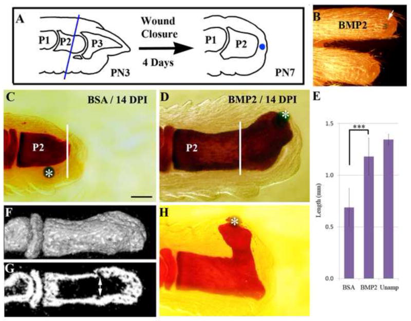
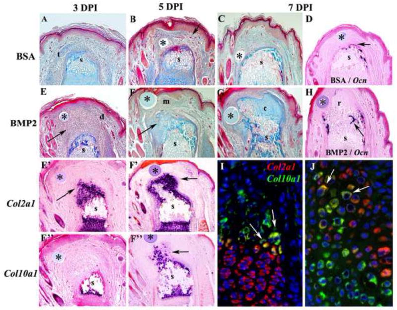
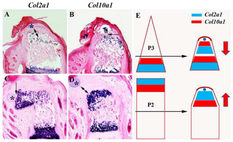
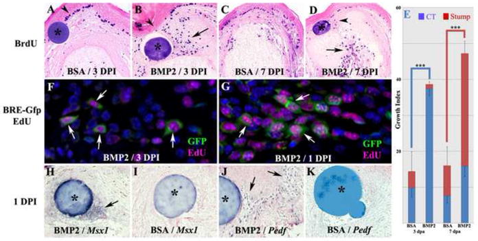
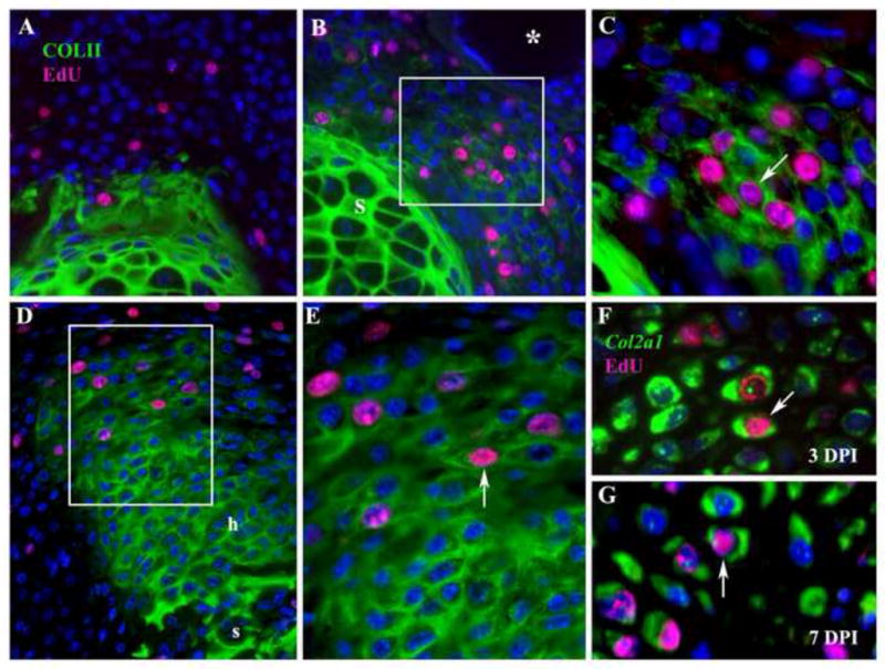
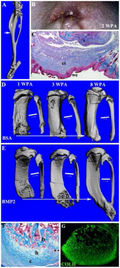
Bone morphogenetic proteins (BMPs) are required for bone development, the repair of damage skeletal tissue, and the regeneration of the mouse digit tip. Previously we showed that BMP treatment can induce a regeneration response in mouse digits amputated at a proximal level of the terminal phalangeal element (P3) (Yu et al., 2010). In this study, we show that the regeneration-inductive ability of BMP2 extends to amputations at the level of the second phalangeal element (P2) of neonatal digits, and the hindlimb of adult limbs. In these models the induced regenerative response is restricted in a segment-specific manner, thus amputated skeletal elements regenerate distally patterned skeletal structures but does not form joints or more distal skeletal elements. Studies on P2 amputations indicate that BMP2-induced regeneration is associated with a localized proliferative response and the transient expression of established digit blastema marker genes. This is followed by the formation of a new endochondral ossification center at the distal end of the bone stump. The endochondral ossification center contains proliferating chondrocytes that establish a distal proliferative zone and differentiate proximally into hypertrophic chondrocytes. Skeletal regeneration occurs from proximal to distal with the appearance of osteoblasts that differentiate in continuity with the amputated stump. Using the polarity of the endochondral ossification centers induced by BMP2 at two different amputation levels, we show that BMP2 activates a level-dependent regenerative response indicative of a positional information network. In summary, our studies provide evidence that BMP2 induces the regeneration of mammalian limb structures by stimulating a new endochondral ossification center that utilizes an existing network of positional information to regulate patterning during skeletal regeneration.


Figures

Figure 1
Figure 1. Induced regeneration of the second phalanx (P2)
Distal is toward the right in all images. A) Schematic diagram showing amputation level and the positioning of implanted bead following wound closure. PN3 digits are amputated through the middle of the P2 element (blue line). Four days after amputation (PN7), an Affigel blue bead (blue) is implanted into the mesenchymal tissue between wound epithelium and bone stump. B) Gross morphology of an regenerated digit (upper) with the BMP2 bead (arrow), and a similarly amputated control digit (lower) at 28 days post-implantation (DPI). C) Whole mount bone staining of a control amputated P2 digit (white line) treated with a BSA bead (*) at 14 DPI. D) Whole mount bone staining of a BMP2-treated amputated digit at 14 DPI showing skeletal outgrowth from the amputated stump (white line) induced by the BMP2 bead (*). E) Statistical comparison of the proximal-distal length of the P2 phalangeal bone at 14 DPI in BSA-treated and BMP2-treated digits in comparison to control unamputated digits (t-test, ± s.e.m., ***, P < 0.001). F) μCT reconstruction of a BMP2 induced regenerate at 14 DPI showing the smooth integration of the bone surface between the stump and regenerated skeletal tissues. G) Radiographic section of the digit displayed in F showing internal skeletal tissue irregularities (double arrow) identifying the amputation level and distinguishing the stump from the regenerated bone. Note that the marrow cavity is contiguous between the stump and regenerate. H) Whole mount bone staining (14 DPI) of a BMP2-induced regenerate showing bead (*) displacement associated with mis-directed skeletal outgrowth.
Figure 2
Figure 2. Endochondral ossification during BMP2 induced regeneration
Distal is toward the top in all images and microcarrrier beads are indicated by *. A–C) Histological analysis of control BSA-treated P2 amputations. A) At 3 DPI there is evidence of wound contraction based on differentiated epidermal structures along the periphery of the stump (s) and encroachment of a severed tendon (t) over the amputated skeletal stump. B) At 5 DPI the control amputation wound associated with a BSA bead is composed of vascularized (arrow) fibrous connective tissue covering the skeletal stump (s). C) At 7 DPI the amputation wound associated with the control BSA bead continues to accumulate fibrous connective tissue that covers the skeletal stump (s). D) Section in situ hybridization at 7 DPI showing cells expressing the osteoblast marker Osteocalcin forming a cap over the amputated skeletal stump (s). E–G) BMP2-treated P2 amputations. E) Histological section at 3 DPI showing the accumulation of mesenchymal cells (arrow) distal to the amputated skeletal stump (s) and associated with the BMP2 bead. The mesenchymal cell mass is flanked with connective tissue of the dermis (d) and is oriented in the direction of the BMP2 bead. E′) Section in situ hybridization at 3 DPI showing Col2a1 transcripts (arrowhead) associated with cells just distal to the skeletal stump (s) and not associated with the BMP2 bead. E″) Section in situ hybridization for Col10a1 transcripts showing the absence of hypertrophic chondrocytes distal to the amputated stump (s) at 3 DPI. F) Histological section at 5 DPI showing a reduced population of mesenchymal cells (m) associated with the BMP2 bead, and chondrocytic tissues (arrow) contiguous with, and distal to, the amputated skeletal stump (s). F′) Section in situ hybridization for Col2a1 transcripts (arrow) showing that chondrocytes associated with the skeletal stump (s) extend distally toward the BMP2 bead. F″) Section in situ hybridization for Col10a1 transcripts (arrow) showing that hypertrophic chondrocytes distal to the amputated stump (s) are present at 5 DPI. G) Histological section at 7 DPI showing few distal mesenchymal cells, and a large mass of chondrocytic tissue (c) contiguous with, and distal to, the amputated skeletal stump (s) that is associated with the BMP2 bead. H) Section in situ hybridization showing the initiation of Osteocalcin expression at the interface between the regenerating tissue (r) induced by the BMP2 bead and the amputated skeletal stump (s) at 7 DPI. I–J) Double color fluorescent section in situ hybridization to co-localize Col2a1 and Col10a1 transcripts in the proximal P2 growth plate at PN14 (I) and a BMP2-induced P2 regenerate at 7 DPI (J). I) Most growth plate chondrocytes either express Col2a1 (red) or Col10a1 (green) transcripts. However, some cells at the interface between proliferating chondrocytes and hypertrophic chondrocytes express both Col2a1 and Col10a1 transcripts (arrows), indicating a transitional state during the differentiation process. J) In BMP2-induced regenerates most cells either express Col2a1 (red) or Col10a1 (green) transcripts, but some co-expressing cells (arrows) are observed at the interface between Col2a1 expressing and Col10a1 expressing cells.
Figure 3
Figure 3. Patterning of BMP2-induced regeneration is level-dependent
Distal to toward the top in all images and microcarrrier beads are indicated by *. A) Section in situ hybridization at 7 DPI of a BMP2-induced regenerate from a proximal P3 amputation showing the localization of Col2a1 expressing chondrocytes relative to the BMP2 bead. Col2a1 expressing chondrocytes are not in direct contact with the BMP2 bead (double arrow) but are proximally localized. B) Section in situ hybridization at 7 DPI of the same BMP2-induced regenerate from a proximal P3 amputation shown in A, indicating that the Co10a1 expressing chondrocytes are in direct contact (arrow) with the BMP2 bead. Relative to the BMP2 bead, the Col2a1 expressing cells are proximal to the Col10a1 expressing cells. C) Section in situ hybridization at 7 DPI of a BMP2-induced P2 regenerate showing the localization of Col2a1 expressing chondrocytes relative the BMP2 bead. Col2a1 expressing chondrocytes are distally localized and in direct contact with the BMP2 bead (arrow). D) Section in situ hybridization at 7 DPI of the same BMP2-induced P2 regenerate shown in C, indicating that the Co10a1 expressing chondrocytes are proximally localized and not in direct contact (double arrows) with the BMP2 bead. Relative to the BMP2 bead, the Col2a1 expressing cells are distal to the Col10a1 expressing cells. E) Diagrammatic summary displaying the shift in polarity of the endochondral ossification centers induced by BMP2 (blue bead) from two different amputation levels (black arrows). The polarity of the endochondral ossification center is depicted by the proximal-distal positioning of the expression domains of Col2a1 (blue) and Col10a1 (red). In BMP2-induced regeneration of the proximal P3 amputation the Col2a1 expression domain is proximal to the Col10a1domain, whereas this orientation is reversed for BMP2-induced P2 regeneration. The polarity of the BMP2-induced endochondral ossification is shown by the red arrows. While BMP2 is capable of inducing regenerative responses from two distinct levels, the patterning of the induced regenerative response is level-specific and dictated by the amputation wound rather than by the inducer.
Figure 4
Figure 4. Blastema formation during induced regeneration
Distal is toward the top in all images and microcarrrier beads are indicated by *. A–E: BrdU incorporation in BMP2 treated and BSA control amputated digits. A) BSA treated amputations at 3 DPI display few BrdU labeled cells that are scattered within the healing dermis and skeletal stump, whereas the basal layer of the wound epithelium displays heavy BrdU labeling (arrowhead). B. BMP2 treated amputations at 3 DPI display numerous BrdU labeled cells within the distal mesenchyme (arrow) associated with the healing dermis with few labeled cells in the skeletal stump. There are also few BrdU labeled cells in the wound epithelium associated with the BMP2 bead (arrowhead). C. BSA treated amputations at 7 DPI display strong BrdU labeling within the wound epidermis and scattered labeling of cells within the wound dermis and stump tissues. D. BMP2 treated amputations at 7 DPI display a cluster of BrdU labeled cells associated with the microcarrier bead (arrowhead), whereas the regenerating stump is heavily labeled (arrow). E. Quantification of BrdU labeling by cell counts within the connective tissue and the stump in comparable regions of BSA and BMP2 treated digits at 3 and 7 DPI. The data show BMP2 enhanced proliferation in the connective tissue at 3 DPI, and in the skeletal stump at 7 DPI (t-test, ± s.e.m, *** p < 0.001). F,G: Proliferation studies of BMP2-induced P2 regeneration in the BRE-Gfp transgenic reporter mouse. EdU incorporation identified proliferating cells (magenta) and BMP responsive cells were identified based on GFP immunohistochemistry (green). F) Double labeled cells (arrows) distal to the amputated stump and associated with the BMP2 bead are present at 3 DPI. G) At 1 DPI double labeled mesenchymal cells (arrows) indicative of a proliferative response to BMP2 treatment are clustered between the BMP2 bead and the amputated stump. H–K: In situ hybridization to identify localized changes in expression of established digit blastema marker genes. H) In BMP2 treated digits at 1DPI, Msx1 transcripts are localized to cells directly associated with the BMP2 bead (arrow). I) In control BSA treated digits at 1 DPI, no Msx1 expression is induced. J) In BMP2 treated digits at 1DPI, Pedf expressing cells are scattered throughout the wound mesenchyme (arrows) and do not appear directly associated with the BMP2 bead. K) Pedf expressing cells are absent in the amputation wound of control BSA treated digits at 1 DPI.
Figure 5
Figure 5. Chondrocyte proliferation during BMP2-induced regeneration
Distal is toward the top in all images. A–E: Histological sections of control BSA-treated or BMP2-treated amputated P2 elements displaying Col II immunostaining (green) to identify chondrocytes in conjunction with EdU incorporation (magenta) to identify proliferating cells. A) Control digit treated with BSA bead at 3 DPI displays Col II expression restricted to the amputated skeletal stump, and few proliferating cells associated with the wound healing response following amputation. No proliferating chondrocytes are found in the amputation wound. B) P2 amputation treated with a BMP2 bead (*) at 3 DPI displays an aggregate of cells expressing Col II and positive for the incorporation of EdU distal to the stump (box). C) Higher magnification of cells in B (box) showing individual cells (arrow) with nuclei staining positive for EdU surrounded by Col II positive staining. D) P2 amputation treated with a BMP2 bead (top of the figure but not shown) at 7 DPI displaying an elongate skeletal outgrowth that is positive for Col II staining and contiguous with the stump (s). Cells staining positive for EdU incorporation are localized to the distal region of the outgrowth (box). Proximal hypertrophic chondrocytes (h) are not proliferating and correlate with regions of Col10a1 expressions (see Figure 3D). E) Higher magnification of cells in D (box) showing individual cells (arrow) that are labeled for EdU and surrounded by Col II positive staining. F–G: To definitively identity proliferating chondrocytes at 3 (F) and 7 (G) DPI, EdU incorporation (magenta) was used to identify proliferating cells in conjunction with FISH to identify Col2a1transcripts (green) localized in the cytoplasm. F) At 3 DPI double labeled cells (arrow) definitively identify proliferating chondrocytes induced by BMP2. G) At 7 DPI double labeled cells (arrow) identify proliferating chondrocytes localized to the growing apex of the BMP2 induced regenerate.
Figure 6
Figure 6. Regeneration response to Bmp2 after adult limb amputation
Distal is toward the bottom of all images. A) μCT image showing the skeleton of the mouse hind limb shank (consisting of the tibia (t) and fibula (f) that fuse distally), and the level of amputation (arrow). Simple amputation was made through the mid-shaft of the shank so as to transect both tibia and fibular proximal to the point of fusion. B) External examination indicated that wound closure is highly irregular due to the wound contraction response of mature skin and is complete by 2 WPA. A wound epithelium (*) is apparent by external examination. C) Mallory staining of histological section of an amputated limb after wound closure shows the formation of a wound epithelium (we) surrounded by mature epidermis, and underlain by connective tissue (ct) distal to the amputated skeletal stump (s). D–E: μCT scans of a BSA control (D) and a BMP2-treated limb (E) at 1, 3, and 8 WPA. Amputated limbs were treated after wound closure (2 WPA) by implantation of gelatin containing either BSA or BMP2 between the wound epithelium and the skeletal stump. The skeletal elements are aligned based on the proximal extent of the fibula and the arrows indicate the extent of the regenerative response. D) BSA control limbs showed a healing response that resulted in minor bone elongation. E) BMP2 treated amputations displayed organized distal bone growth resulting in skeletal elongation and distal bone fusion suggestive of a properly patterned regenerative response. Scale bars = 1 mm. F) Mallory staining of histological sections at 6 WPA of a BMP2 treated limb regenerate showing apical outgrowth associated with an endochondral ossification response involving distal chondrocytes (c), hypertrophic chondroctyes (h) and bone (b). G) Immunohistochemical staining for type II collagen of an adjacent section to the sample shown in F identifies an apical localization of chondrocytes associated with the BMP2-induced regenerative response. Following amputation in mice BMP2 induces a skeletal regenerative response BMP2 stimulated cell proliferation and an endochondral ossification center forms A distal growth plate like structure organizes the direction of skeletal outgrowth BMP2 induced regeneration is specific to the level of amputation BMP2 is a critical factor for successful limb regeneration in mammals

References

    1. Agrawal V, Johnson SA, Reing J, Zhang L, Tottey S, Wang G, Hirschi KK, Braunhut S, Gudas LJ, Badylak SF. Epimorphic regeneration approach to tissue replacement in adult mammals. Proc Natl Acad Sci U S A. 2010;107:3351–3355. - PMC - PubMed
    1. Bandyopadhyay A, Tsuji K, Cox K, Harfe BD, Rosen V, Tabin CJ. Genetic analysis of the roles of BMP2, BMP4, and BMP7 in limb patterning and skeletogenesis. PLoS Genet. 2006;2:e216. - PMC - PubMed
    1. Becker RO. Stimulation of partial limb regeneration in rats. Nature. 1972;235:109–111. - PubMed
    1. Brockes JP, Kumar A. Appendage regeneration in adult vertebrates and implications for regenerative medicine. Science. 2005;310:1919–1923. - PubMed
    1. Bryant SV, Gardiner DM. Retinoic acid, local cell-cell interactions, and pattern formation in vertebrate limbs. Dev Biol. 1992;152:1–25. - PubMed

Publication types

Substances