. 2012 Nov;122(11):4048-58.
doi: 10.1172/JCI64604. Epub 2012 Oct 8.

AFG3L2 supports mitochondrial protein synthesis and Purkinje cell survival

Affiliations

AFG3L2 supports mitochondrial protein synthesis and Purkinje cell survival

Eva R Almajan et al. J Clin Invest. 2012 Nov.

Abstract

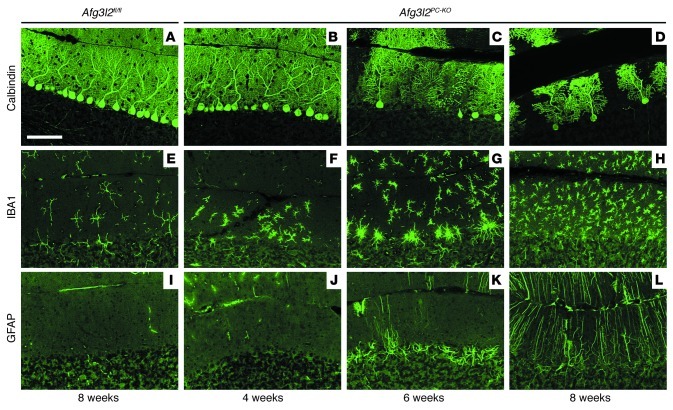
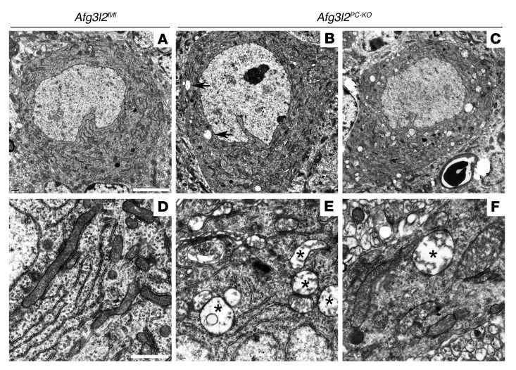
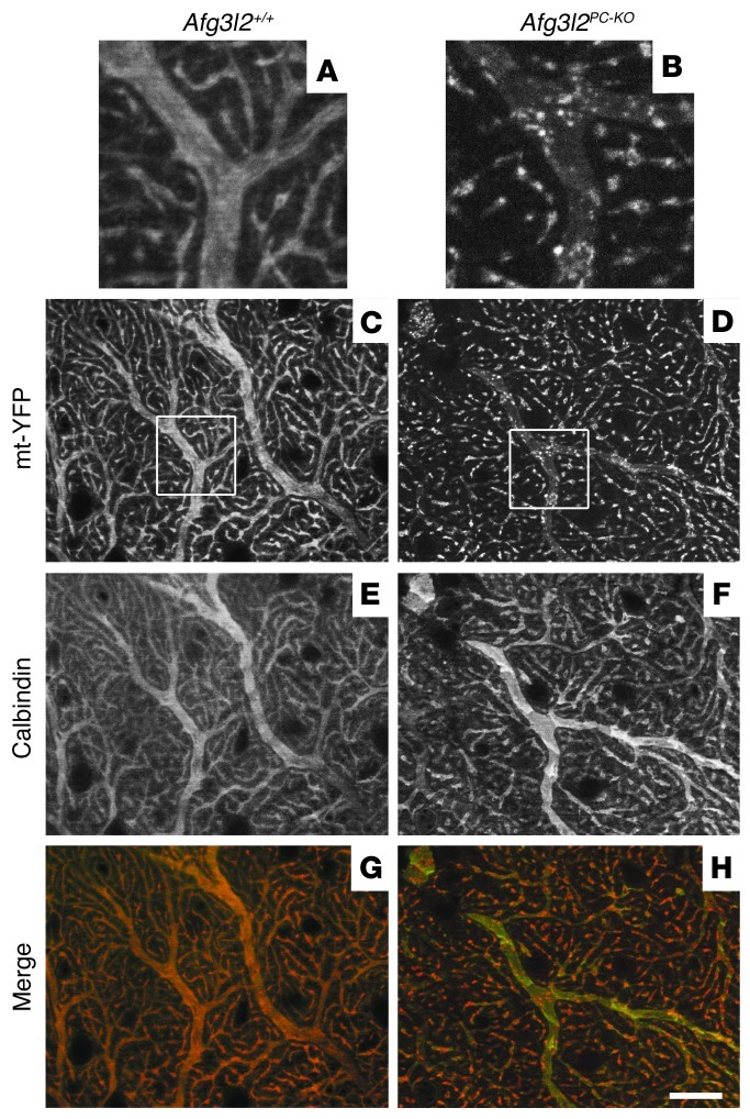
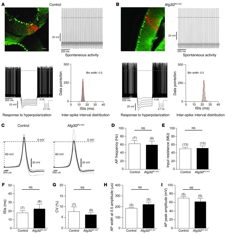
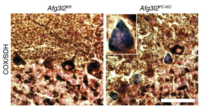
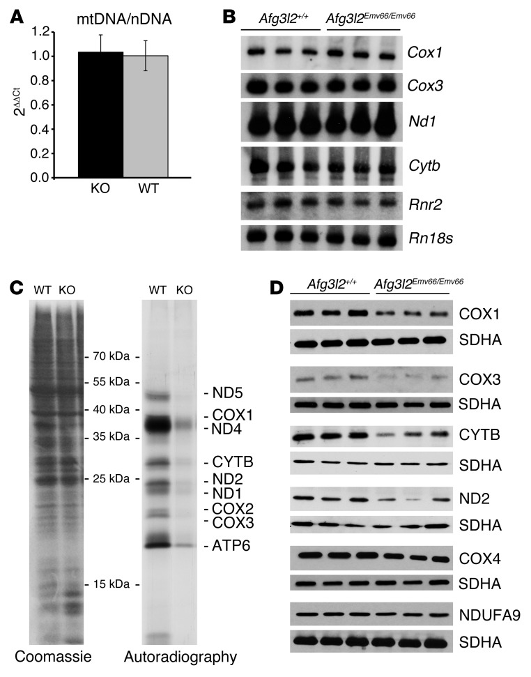
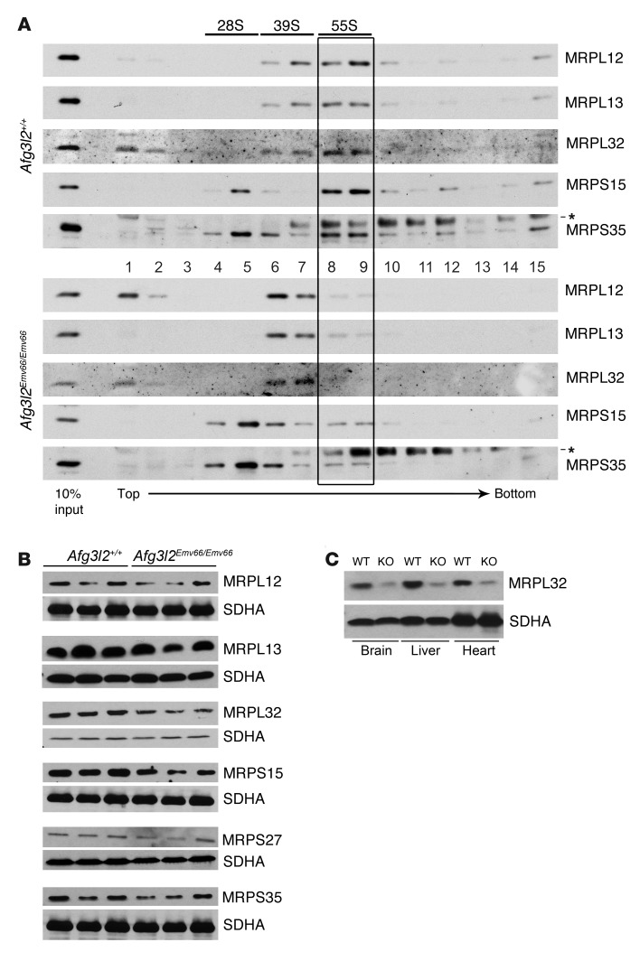
Mutations in the AFG3L2 gene have been linked to spinocerebellar ataxia type 28 and spastic ataxia-neuropathy syndrome in humans; however, the pathogenic mechanism is still unclear. AFG3L2 encodes a subunit of the mitochondrial m-AAA protease, previously implicated in quality control of misfolded inner mitochondrial membrane proteins and in regulatory functions via processing of specific substrates. Here, we used a conditional Afg3l2 mouse model that allows restricted deletion of the gene in Purkinje cells (PCs) to shed light on the pathogenic cascade in the neurons mainly affected in the human diseases. We demonstrate a cell-autonomous requirement of AFG3L2 for survival of PCs. Examination of PCs prior to neurodegeneration revealed fragmentation and altered distribution of mitochondria in the dendritic tree, indicating that abnormal mitochondrial dynamics is an early event in the pathogenic process. Moreover, PCs displayed features pointing to defects in mitochondrially encoded respiratory chain subunits at early stages. To unravel the underlying mechanism, we examined a constitutive knockout of Afg3l2, which revealed a decreased rate of mitochondrial protein synthesis associated with impaired mitochondrial ribosome assembly. We therefore propose that defective mitochondrial protein synthesis, leading to early-onset fragmentation of the mitochondrial network, is a central causative factor in AFG3L2-related neurodegeneration.


Figures

Figure 1
Figure 1. PCs degenerate over time in Afg3l2PC–KO mice.
Vibratome sections of the cerebellum were collected at various time points from Afg3l2fl/fl and Afg3l2PC–KO mice and stained for (AD) calbindin to show progressive PC loss, (EH) IBA1 to detect resting and activated microglia, and (IL) GFAP to mark reactive astrocytes. Scale bar: 100 μm.
Figure 2
Figure 2. Ultrastructural analysis of PCs in Afg3l2PC–KO mice.
(A) Electron micrographs of cerebella from 4-week-old mice show normal PCs in Afg3l2fl/fl mice. PCs in Afg3l2PC–KO mice appear grossly normal but display (B) a few (arrows) or (C) rarely many abnormal mitochondria in the cell body. Swollen mitochondria with disrupted cristae (labeled with asterisks) are present in (E) the cell body and (F) dendrites of PCs in Afg3l2PC–KO mice, (D) while normal mitochondria are present in the controls. White scale bars: 5 μm (AC); 1 μm (DF).
Figure 3
Figure 3. Abnormal mitochondrial network in Afg3l2PC–KO mice.
Analyses were performed in animals of the shown genotypes crossed with ROSA26+/SmY mice. (AD) The mitochondrial network is labeled with mt-YFP, (E and F) while PC dendrites are decorated using an antibody against calbindin. (G and H) In merged images, pseudocolors were used: calbindin is in green, while mt-YFP is in red. Images in A and B show enlarged views of the boxes in C and D, respectively. (C) mt-YFP expression in control mice shows an elongated network of mitochondria (E and G) that follows the pattern of PC dendrites labeled with calbindin, (A) with mitochondria filling the entire space of large caliber dendrites. (D) In Afg3l2PC–KO mice, the mitochondrial network shows initial fragmentation, with (B, F, and H) some large-caliber calbindin-stained dendrites depleted of mitochondria, which appear clustered. Scale bar: 20 μm.
Figure 4
Figure 4. Electrophysiological properties of PCs in 4- to 5-week-old Afg3l2PC–KO mice and control littermates.
PCs were recorded in the perforated patch clamp configuration and labeled with biocytin/streptavidin after the electrophysiological characterization. (A and B) Fluorescence images and electrophysiological data of (A) control and (B) Afg3l2PC–KO mice. Scale bars: 50 μm. The graphs show examples of the spontaneous activity and the inter-spike interval distributions during spontaneous activity and the membrane potential in response to hyperpolarizing current injections. Hyperpolarization induced a sag-like, slowly developing inward rectification. Dashed lines mark the 0-mV border. (C) PC action potential waveforms of control and Afg3l2PC–KO mice. The average of all neurons is shown in bold black. The average of individual neurons is shown in gray. (D) Membrane potential, (E) input resistance, (F) inter-spike interval (ISI), (G) coefficients of variance (CV) of the inter-spike interval distributions, (H) action potential (AP) width at 0.5 maximal amplitude, and (I) action potential maximal amplitude of control and Afg3l2PC–KO mice. Numbers over bars indicate the number of neurons. Error bars show SD.
Figure 5
Figure 5. Afg3l2PC–KO mice show COX-deficient PCs.
Combined COX-SDH enzymatic staining at 4 weeks of age reveals normal brown PCs in control mice, while several PCs stain blue in the Afg3l2PC–KO mice, indicating COX deficiency. Scale bar: 200 μm.
Figure 6
Figure 6. Impaired mitochondrial protein synthesis in brain mitochondria from Afg3l2Emv66/Emv66 mice.
(A) Real-time PCR quantification shows no statistically significant difference of the mtDNA content with respect to the nuclear DNA (nDNA) in the brains of Afg3l2+/+ (WT) and Afg3l2Emv66/Emv66 (KO) mice. Error bars show SD. (B) Northern blot analysis of a subset of mitochondrial transcripts reveals similar levels of expression in the brains of Afg3l2+/+ and Afg3l2Emv66/Emv66 mice. 18S RNA (Rn18s) was used as loading control. (C) In organello translation after labeling with 35S-met shows reduced de novo synthesized protein levels in Afg3l2Emv66/Emv66 mice when compared with those in Afg3l2+/+ mice. Coomassie brilliant blue staining of the gel shows equal loading of proteins. (D) The steady-state levels of mt-encoded COX1, COX3, CYTB, and ND2 are reduced in the brains of Afg3l2Emv66/Emv66 mice compared with those in Afg3l2+/+ mice, while levels of nuclear-encoded COX4 and NUDFA9 appear unaffected. The SDHA subunit of complex II was used as loading control.
Figure 7
Figure 7. Impaired mitoribosome assembly in Afg3l2Emv66/Emv66 mice.
(A) The mitoribosome profile shows a clear shift to less dense fractions in the pattern of large ribosomal subunits in brain mitochondria of Afg3l2Emv66/Emv66 mice when compared with those of the control Afg3l2+/+ mice and reduced levels of assembled mitoribosomes (55S, boxed area). Asterisks indicate unspecific signals. (B) Steady-state levels of mitochondrial ribosomal proteins in the brains of Afg3l2Emv66/Emv66 and Afg3l2+/+ mice. The levels of MRPL32 are reduced in the Afg3l2Emv66/Emv66 mice. The SDHA subunit of complex II was used as loading control. (C) MRPL32 levels are affected in several tissues from Afg3l2Emv66/Emv66 mice as compared with those in control Afg3l2+/+ mice. The SDHA subunit of complex II was used as loading control. Notably, the reduction in the levels of MRPL32 seems to be more or less pronounced depending on the mouse analyzed (compare B and C).

References

    1. Rugarli EI, Langer T. Mitochondrial quality control: a matter of life and death for neurons. EMBO J. 2012;31(6):1336–1349. doi: 10.1038/emboj.2012.38. - DOI - PMC - PubMed
    1. Arlt H, Tauer R, Feldmann H, Neupert W, Langer T. The YTA10-12 complex, an AAA protease with chaperone-like activity in the inner membrane of mitochondria. Cell. 1996;85(6):875–885. doi: 10.1016/S0092-8674(00)81271-4. - DOI - PubMed
    1. Lee S, Augustin S, Tatsuta T, Gerdes F, Langer T, Tsai FT. Electron cryomicroscopy structure of a membrane-anchored mitochondrial AAA protease. J Biol Chem. 2011;286(6):4404–4411. doi: 10.1074/jbc.M110.158741. - DOI - PMC - PubMed
    1. Leonhard K, Guiard B, Pellecchia G, Tzagoloff A, Neupert W, Langer T. Membrane protein degradation by AAA proteases in mitochondria: extraction of substrates from either membrane surface. Mol Cell. 2000;5(4):629–638. doi: 10.1016/S1097-2765(00)80242-7. - DOI - PubMed
    1. Hornig-Do HT, et al. Nonsense mutations in the COX1 subunit impair the stability of respiratory chain complexes rather than their assembly. EMBO J. 2012;31(5):1293–1307. doi: 10.1038/emboj.2011.477. - DOI - PMC - PubMed

Publication types

MeSH terms

Supplementary concepts