. 2012 Nov;122(11):3955-9.
doi: 10.1172/JCI63113. Epub 2012 Oct 8.

Progranulin deficiency promotes neuroinflammation and neuron loss following toxin-induced injury

Affiliations

Progranulin deficiency promotes neuroinflammation and neuron loss following toxin-induced injury

Lauren Herl Martens et al. J Clin Invest. 2012 Nov.

Erratum in

Abstract

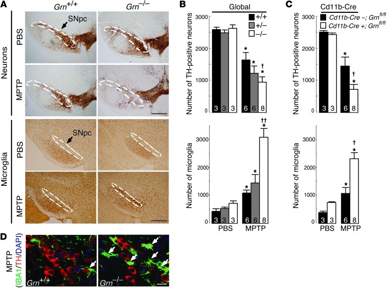
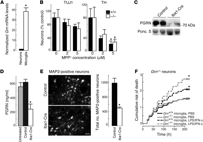
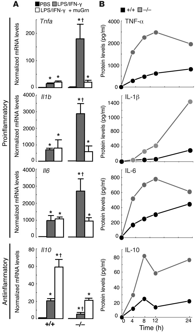
Progranulin (PGRN) is a widely expressed secreted protein that is linked to inflammation. In humans, PGRN haploinsufficiency is a major inherited cause of frontotemporal dementia (FTD), but how PGRN deficiency causes neurodegeneration is unknown. Here we show that loss of PGRN results in increased neuron loss in response to injury in the CNS. When exposed acutely to 1-methyl-4-(2'-methylphenyl)-1,2,3,6-tetrahydrophine (MPTP), mice lacking PGRN (Grn⁻/⁻) showed more neuron loss and increased microgliosis compared with wild-type mice. The exacerbated neuron loss was due not to selective vulnerability of Grn⁻/⁻ neurons to MPTP, but rather to an increased microglial inflammatory response. Consistent with this, conditional mutants lacking PGRN in microglia exhibited MPTP-induced phenotypes similar to Grn⁻/⁻ mice. Selective depletion of PGRN from microglia in mixed cortical cultures resulted in increased death of wild-type neurons in the absence of injury. Furthermore, Grn⁻/⁻ microglia treated with LPS/IFN-γ exhibited an amplified inflammatory response, and conditioned media from these microglia promoted death of cultured neurons. Our results indicate that PGRN deficiency leads to dysregulated microglial activation and thereby contributes to increased neuron loss with injury. These findings suggest that PGRN deficiency may cause increased neuron loss in other forms of CNS injury accompanied by neuroinflammation.


Figures

Figure 1
Figure 1. Increased neuron death and microglial activation caused by PGRN deficiency in a model of CNS injury.
(A) Representative SNpc sections showing decreased numbers of TH-positive neurons and increased numbers of IBA1-positive microglia following MPTP exposure in PGRN-deficient mice. Dashed lines indicate SNpc. Scale bars: 500 μm. (B) Quantification of TH-positive neurons and IBA1-positive microglia in the SNpc in the global PGRN-deficient mouse model. Numbers denote n per group. *P < 0.05, MPTP versus PBS; P < 0.05, Grn+/+ MPTP versus Grn–/– MPTP; ††P < 0.01, Grn–/– MPTP versus either Grn+/+ or Grn+/– MPTP. (C) Quantification of TH-positive and IBA1-positive microglia in the SNpc in the microglia-targeted PGRN-deficient mouse model. Numbers denote n per group. *P < 0.05, MPTP versus PBS; P < 0.05, Cd11b-Cre;Grnfl/fl MPTP versus Cd11b-Cre+;Grnfl/fl MPTP. (D) Representative confocal images showing that MPTP treatment decreased the number of TH-positive neurons (red) and increased the numbers of activated microglia (green) in the SNpc of Grn–/– mice. Arrows indicate activated microglia. Scale bar: 20 μm.
Figure 2
Figure 2. Microglial PGRN deficiency increases the death of cultured neurons.
(A) Grn mRNA levels in cultured primary neurons and microglia. *P < 0.001. (B) Similar numbers of surviving Grn+/+ and Grn–/– midbrain neurons (TUJ1 or TH immunolabeled) following MPP+ treatment. *P < 0.05, 5 μM versus untreated. (C) Immunoblot showing reduced levels of secreted PGRN in Grnfl/fl mixed cortical cultures infected with Iba1-Cre lentivirus. (D) Quantification of secreted PGRN in mixed cortical cultures. *P < 0.01. (E) Representative image showing decreased survival of MAP2-positive neurons in mixed cultures lacking microglial PGRN compared with control cultures. *P < 0.001. Scale bar: 20 μm. (F) Increased risk of death for wild-type neurons exposed to conditioned media from LPS/IFN-γ–treated PGRN-deficient microglia. *P < 0.0001 (log-rank test), LPS/IFN-γ–treated conditioned media compared with PBS-treated conditioned media.
Figure 3
Figure 3. PGRN-deficient microglia exhibit a hyperactivated inflammatory response.
(A) Increased inflammatory cytokine mRNA levels in LPS/IFN-γ–treated Grn–/– microglia, compared with Grn+/+ controls, that was rescued by lentiviral infection with murine PGRN (MuGrn). *P < 0.05 compared with PBS; P < 0.05 compared with Grn–/– LPS/IFN-γ. (B) Representative time plots revealing increased secretion of inflammatory cytokines by LPS/IFN-γ–treated Grn–/– microglia compared with similarly treated Grn+/+ cultures.

References

    1. He Z, Ismail A, Kriazhev L, Sadvakassova G, Bateman A. Progranulin (PC-cell-derived growth factor/acrogranin) regulates invasion and cell survival. Cancer Res. 2002;62(19):5590–5596. - PubMed
    1. He Z, Ong CH, Halper J, Bateman A. Progranulin is a mediator of the wound response. Nat Med. 2003;9(2):225–229. doi: 10.1038/nm816. - DOI - PubMed
    1. Tang W, et al. The growth factor progranulin binds to TNF receptors and is therapeutic against inflammatory arthritis in mice. Science. 2011;332(6028):478–484. doi: 10.1126/science.1199214. - DOI - PMC - PubMed
    1. Zhu J, et al. Conversion of proepithelin to epithelins: roles of SLPI and elastase in host defense and wound repair. Cell. 2002;111(6):867–878. doi: 10.1016/S0092-8674(02)01141-8. - DOI - PubMed
    1. Bateman A, Bennett HP. Granulins: the structure and function of an emerging family of growth factors. J Endocrinol. 1998;158(2):145–151. doi: 10.1677/joe.0.1580145. - DOI - PubMed

Publication types

MeSH terms