. 2012 Nov 29;491(7426):765-8.
doi: 10.1038/nature11463. Epub 2012 Oct 7.

Self-renewal of embryonic-stem-cell-derived progenitors by organ-matched mesenchyme

Affiliations

Self-renewal of embryonic-stem-cell-derived progenitors by organ-matched mesenchyme

Julie B Sneddon et al. Nature. .

Abstract

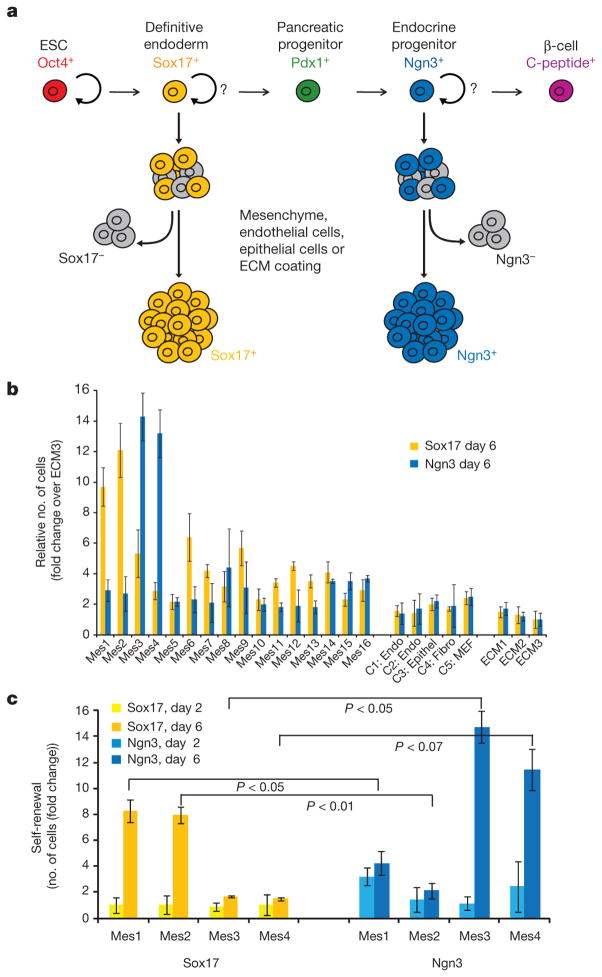
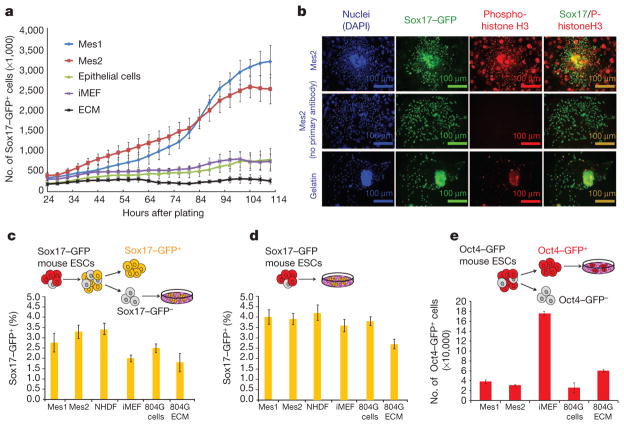
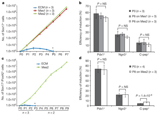
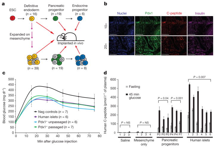
One goal of regenerative medicine, to use stem cells to replace cells lost by injury or disease, depends on producing an excess of the relevant cell for study or transplantation. To this end, the stepwise differentiation of stem cells into specialized derivatives has been successful for some cell types, but a major problem remains the inefficient conversion of cells from one stage of differentiation to the next. If specialized cells are to be produced in large numbers it will be necessary to expand progenitor cells, without differentiation, at some steps of the process. Using the pancreatic lineage as a model for embryonic-stem-cell differentiation, we demonstrate that this is a solvable problem. Co-culture with organ-matched mesenchyme permits proliferation and self-renewal of progenitors, without differentiation, and enables an expansion of more than a million-fold for human endodermal cells with full retention of their developmental potential. This effect is specific both to the mesenchymal cell and to the progenitor being amplified. Progenitors that have been serially expanded on mesenchyme give rise to glucose-sensing, insulin-secreting cells when transplanted in vivo. Theoretically, the identification of stage-specific renewal signals can be incorporated into any scheme for the efficient production of large numbers of differentiated cells from stem cells and may therefore have wide application in regenerative biology.


Conflict of interest statement

The authors declare no competing financial interests.

Figures

Figure 1
Figure 1. Screen for signals that expand definitive endoderm and endocrine progenitors
a, Schema for directed differentiation of β-cells and their progenitors. b, Number of Sox17–GFP+ cells or Ngn3–GFP+ cells after co-culture with primary mesenchyme lines (Mes1 through to Mes16), control endothelial cell lines (C1, C2), an epithelial cell line (C3), a fibroblast cell line (C4), MEFs (C5) or various ECM surfaces (ECM1, ECM2 and ECM3) for 6 days. c, The number of cells (Sox17+ and Ngn3+) after 2 or 6 days of co-culture. P values were calculated using Student’s t-test. Data represent the mean of two biological replicates ± s.d.
Figure 2
Figure 2. Effects of mesenchyme are due to proliferation, not induction, and are specific to the responding cell type
a, Number of Sox17–GFP+ cells over time during co-culture. b, Immunofluorescence staining of Sox17–GFP+ cells after 48 h co-culture with Mes2 or gelatin. c, Number of Sox17–GFP+ induced from sorted Sox17–GFP cells by 6 days of co-culture. d, Number of Sox17–GFP+ cells induced from mouse ESCs by 6 days of co-culture. e, Number of Oct4–GFP+ cells after co-culture. iMEF, irradiated MEFs; NHDF, normal human dermal fibroblasts. 804G ECM, extracellular matrix from 804G cells. Data represent the mean of two biological replicates ± s.d.
Figure 3
Figure 3. Long-term expansion of differentiation-competent mouse and human ESC-derived endoderm in the presence of mesenchyme
a, Number of mouse Sox17–GFP+ cells shown in relation to co-culture time. b, Efficiency of directed differentiation of unpassaged and passaged mouse Sox17–GFP+ cells. c, Number of human Sox17+ cells shown in relation to co-culture time. d, Efficiency of directed differentiation of unpassaged and passaged human Sox17+ cells. Data represent mean of biological replicates ± s.d. P values are based on two-tailed Student’s t-test. Px, passage number; 6 days (mouse) or 5 to 8 days (human) between passages. Asterisk denotes progressive loss of cells cultured on ECM alone.
Figure 4
Figure 4. Human ESC-derived cells expanded on mesenchyme give rise to insulin-expressing, glucose-responsive cells in vivo
a, Schematic depicting implantation of human ESC-derived progenitors. b, Immunofluorescence staining of human ESC-derived endoderm, passaged seven times on mesenchyme and engrafted for 3 months (top panel) or further differentiated to Pdx1+ stage and then engrafted for 2 months (bottom panel). c, Glucose-tolerance test of animals implanted with PBS or mesenchyme only, human islets or Pdx1+ pancreatic progenitors derived from unpassaged (P0), or passaged (P4 or P7) human endoderm. d, Fasting- and glucose-induced (45 min glucose) plasma human C-peptide levels. Pairs of bars represent two time points per animal; data represent mean of two technical replicates ± s.d.

References

    1. Kroon E, et al. Pancreatic endoderm derived from human embryonic stem cells generates glucose-responsive insulin-secreting cells in vivo. Nature Biotechnol. 2008;26:443–452. - PubMed
    1. Grigoriadis AE, et al. Directed differentiation of hematopoietic precursors and functional osteoclasts from human ES and iPS cells. Blood. 2010;115:2769–2776. - PMC - PubMed
    1. Perrier AL, et al. Derivation of midbrain dopamine neurons from human embryonic stem cells. Proc Natl Acad Sci USA. 2004;101:12543–12548. - PMC - PubMed
    1. Moore KA, Lemischka IR. Stem cells and their niches. Science. 2006;311:1880–1885. - PubMed
    1. Yamashita YM, Fuller MT. Asymmetric stem cell division and function of the niche in the Drosophila male germ line. Int J Hematol. 2005;82:377–380. - PubMed

Publication types

MeSH terms