

doi: 10.1038/nchembio.1087.
Epub 2012 Oct 7.
A RubisCO-like protein links SAM metabolism with isoprenoid biosynthesis
Affiliations
- PMID: 23042035
- PMCID: PMC3475740
- DOI: 10.1038/nchembio.1087
Item in Clipboard
A RubisCO-like protein links SAM metabolism with isoprenoid biosynthesis
Nat Chem Biol.
2012 Nov.
Abstract
Functional assignment of uncharacterized proteins is a challenge in the era of large-scale genome sequencing. Here, we combine in extracto NMR, proteomics and transcriptomics with a newly developed (knock-out) metabolomics platform to determine a potential physiological role for a ribulose-1,5-bisphosphate carboxylase/oxygenase (RubisCO)-like protein from Rhodospirillum rubrum. Our studies unraveled an unexpected link in bacterial central carbon metabolism between S-adenosylmethionine-dependent polyamine metabolism and isoprenoid biosynthesis and also provide an alternative approach to assign enzyme function at the organismic level.
Conflict of interest statement
The authors declare no competing financial interests.
Figures
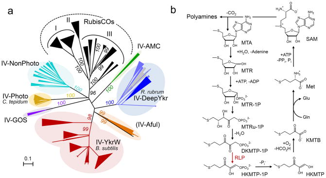
(A) Phylogenetic tree of the RubisCO/RLP superfamily. The unrooted phylogenetic tree is based on amino acid sequence analysis of 333 representative proteins (listed in Supplementary Table 6) that were restricted to a length of 409 amino acids and aligned with ClustalW. Tree topography and evolutionary distance are given by the neighbor joining method. Numbers at nodes represent the percentage bootstrap values for the clades of this group in 500 replications. Similar trees were obtained by using the minimum evolution and the maximum likelihood method. For extended views of each subtree, see Supplementary Fig. 12 and 13. The scale bar represents a difference of 0.1 substitutions per site. RubisCOs are classified into three well established subfamilies, (I, II and III). RLPs fall into six different subfamilies as described previously,: IV-AMC (metagenomic Leptospirillum sequences from an acid mine consortium); IV-DeepYkr; (R. rubrum group, including mainly alpha-, and gammaproteobacteria, some thermophilic species and Veillonellaceae); IV-YkrW (B. subtilis group, including many Bacilliales, Acidithiobacillales, and cyanobacteria); IV-GOS (metagenomic sequences from the global ocean sequencing program); IV-Photo (C. tepidum group, including many Chlorobiales and alphaproteobacteria,) and IV-NonPhoto (including many alpha-, and some beta proteobacteria). A seventh subgroup of RLPs, established in this extended phylogenetic analysis (IV-Aful, including Clostridiales and Archaeoglobus fulgidus) was described previously as a singleton (A. fulgidus DSM 4304) . (B) Function of the B. subtilis RLP in the classical methionine salvage pathway. Abbreviations: MTR, methylthioribose; DKMTP-1P, 2,3-diketo-5-methylthiopentyl-1-phosphate; HKMTP-1P, 2-hydroxy-3-keto-5-methylthiopentenyl-1-phosphate; HKMTP 1,2-dihydroxy-3-keto-5-methylthiopentene; KMTB, 2,4-keto-4-methylthiobutyrate.
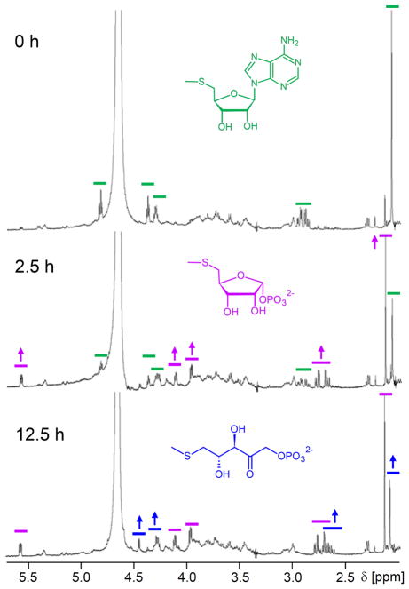
1H-NMR spectra of R. rubrum cell extract incubated with MTA that show the transformation of MTA into MTR-1P, and MTRu-1P over time. 0.78 mg cell extract protein of R. rubrum grown on MTA as sole sulfur source were incubated with 0.4 mM MTA at 30 °C. Spectra were recorded at different time points as indicated. Characteristic 1H-NMR signals for MTA (green), MTR-1P (purple), and MTRu-1P (blue) are indicated by colored lines. The full array experiment is shown in Supplementary Figure 1.

(A) Time-dependent formation of intermediates in the MTA-isoprenoid shunt upon MTA-feeding. Cell suspensions of R. rubrum (1 ml, OD578=6.0) were incubated with 0.4 mM MTA and analyzed after 2, 10, and 20 minutes respectively by LC-FTMS-metabolomics. (B) Time dependent formation of free thiols by R. rubrum upon MTA uptake. Cell suspensions of R. rubrum (1 ml, OD578=4.0) were incubated with 0.4 mM MTA. The supernatant was analyzed for consumption of MTA and formation of free thiols. Free thiols formed were identified as methanethiol by HPLC and FTMS (Supplementary Fig. 4). At least two independent cell batches were used in these assays. Data represent mean values ± standard deviation. (C) LC-FTMS-Metabolomics analysis of MTA-isoprenoid shunt mutants. Cell suspensions of R. rubrum wild type and different mutants were incubated with MTA (according to A) and analyzed after 10 minutes by LC-FTMS-metabolomics, see Supplementary Fig. 6 for the detailed analysis of the cupin mutant (D) Thiol release activities by R. rubrum wild type and different mutants. Cell suspensions of R. rubrum were incubated with MTA according to (B), and the formation of free thiols over time was quantified. At least two independent cell batches were used in these assays. Data represent mean value ± standard deviation.

The RLP is part of the central reaction sequence that involves the release of methanethiol from the molecule backbone. Whereas methanethiol can be recaptured as methionine via O-acetyl-L-homoserine sulfhydrylase (Rru_A0774), the rest of the molecule is converted into isoprenoid precursors. All intermediates of this proposed pathway that were identified by LC-FTMS-metabolomics are shown in boxed bar charts, with their individual increase in metabolite level after 0, 10 and 20 minutes feeding of MTA (+MTA, green bars) in comparison to control cells after 0, 10 and 20 min (control, black bars). Genes/proteins that were identified and characterized in this study are also shown and highlighted by colors.
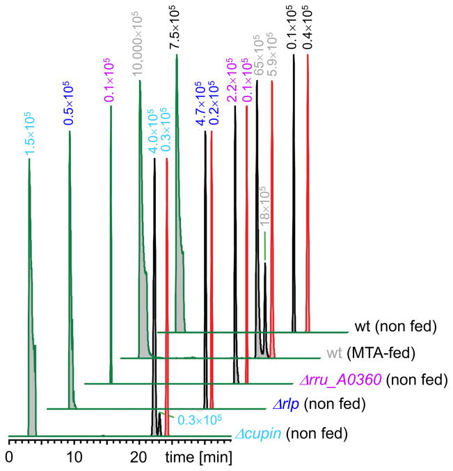
Metabolite extracts prepared from R. rubrum wildtype cells and mutants that had been cultivated on minimal medium with sulfate as sole sulfur source were analyzed for intermediates of the proposed MTA-isoprenoid shunt. The data are represented by extracted ion chromatograms (negative ion) at 2 ppm mass accuracy for 296.08228 (MTA; retention time 3.5 min; green traces), 259.00468 (methylthiopentose phosphates: MTR-1P, MTRu-1P, MTXu-5P, and MTRu-5P; retention time 22–23 min; black traces), and 213.01696 (DXP; retention time 24.5 min; red traces). The integrated intensities are inset and color coded for each sample. Note that the single traces are scaled to 100% relative intensity. For comparison, integrated intensities are also summarized in Supplementary Table 7.

(A) Differential induced gel electrophoresis (DIGE) analysis of changes in the proteome of R. rubrum. Proteins up-regulated in MTA-grown cells are shown in cyan, proteins up-regulated in sulfate-grown cells are shown in red, whereas proteins that are not changed under both conditions are shown in white. The calculated pI (first dimension) and the molecular mass standards (second dimension) of the DIGE gel are given. For more information on the five proteins highlighted in green that were selected for identification, see Supplementary Table 4. (B) RNA sequencing (RNAseq) analysis of changes in the transcriptome of R. rubrum. Genes annotated on the chromosome of R. rubrum, are represented by dots according to their physical location on the chromosome and their fold change in mRNA-level. Classical housekeeping genes are listed separately and highlighted by light gray dots in the graph. For more information on all transcripts up-regulated more than 30-fold, see Supplementary Table 5. (C) The “thiol cluster” that was identified by both DIGE and RNAseq analysis. The four proteins of the thiol cluster that were identified by DIGE are numbered and highlighted in green. Transcripts of the thiol-cluster that were identified by RNAseq. are highlighted in cyan, and the fold-upregulation of each gene is given separately.
References
-
- Hanson TE, Tabita FR. A ribulose-1,5-bisphosphate carboxylase/oxygenase (RubisCO)-like protein from Chlorobium tepidum that is involved with sulfur metabolism and the response to oxidative stress. Proceedings of the National Academy of Sciences of the United States of America. 2001;98:4397–4402. - PMC - PubMed
Publication types
MeSH terms
Substances
Grants and funding
LinkOut - more resources
Full Text Sources
Other Literature Sources
Molecular Biology Databases
