

Established breast cancer stem cell markers do not correlate with in vivo tumorigenicity of tumor-initiating cells
- PMID: 23042145
- PMCID: PMC3583871
- DOI: 10.3892/ijo.2012.1654
Established breast cancer stem cell markers do not correlate with in vivo tumorigenicity of tumor-initiating cells
Abstract
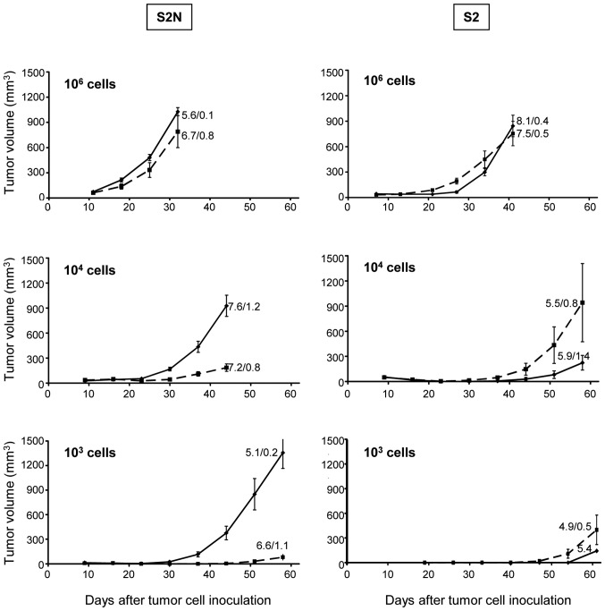
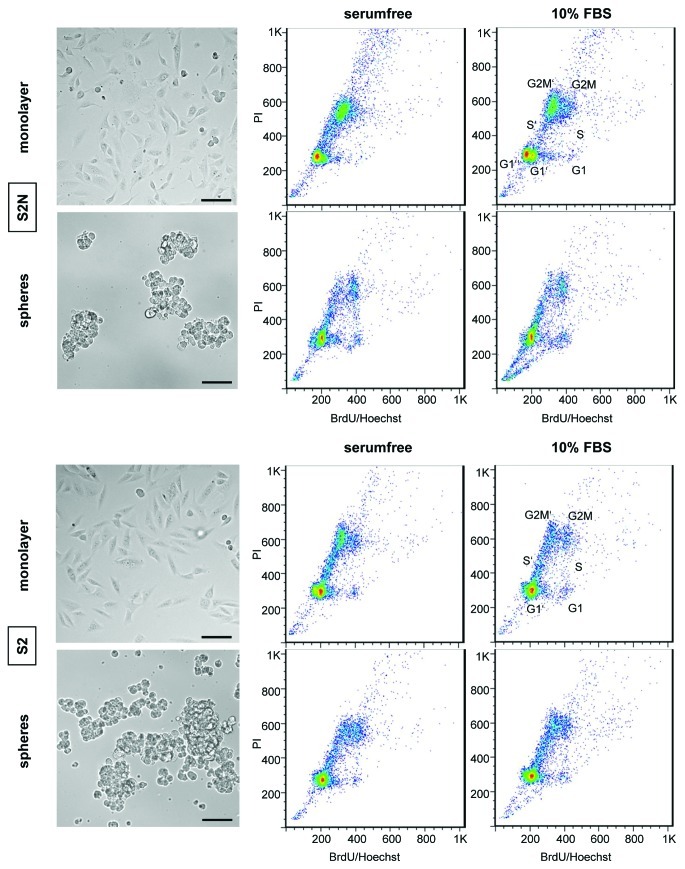
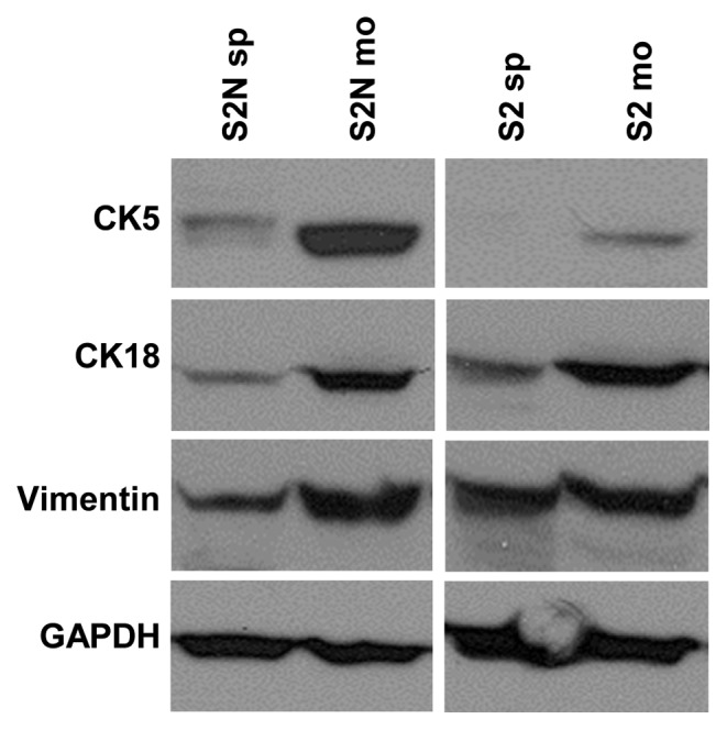
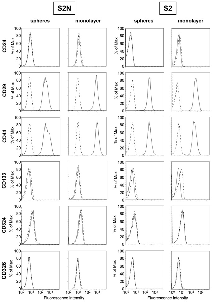
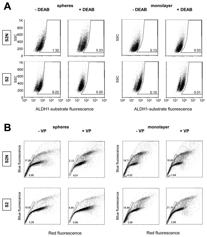
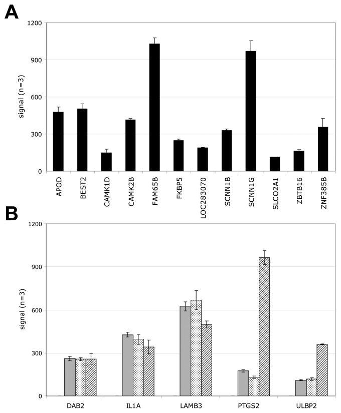
The tumor-initiating capacity of primary human breast cancer cells is maintained in vitro by culturing these cells as spheres/aggregates. Inoculation of small cell numbers derived from these non-adherent cultures leads to rapid xenograft tumor formation in mice. Accordingly, injection of more differentiated monolayer cells derived from spheres results in significantly decelerated tumor growth. For our study, two breast cancer cell lines were generated from primary tumors and cultured as mammospheres or as their adherent counterparts. We examined the in vivo tumorigenicity of these cells by injecting serial dilutions into immunodeficient mice. Inoculation of 106 cells per mouse led to rapid tumor formation, irrespective of cell line or culture conditions. However, after injection of only 103 cells, solely sphere cells were highly tumorigenic. In vitro, we investigated differentiation markers, established breast CSC markers and conducted mRNA profiling. Cytokeratin 5 and 18 were increased in both monolayer cell types, indicating a more differentiated phenotype. All cell lines were CD24(-)/CD44(+) and did not express CD133, CD326 or E-cadherin. ALDH1 activity was not detectable in any cell line. A verapamil‑sensitive Hoechst side population was present in sphere cells, but there was no correlation with tumorigenicity in vivo. mRNA profiling did not reveal upregulation of relevant transcription factors. In vitro cell cycle kinetics and in vivo tumor doubling times displayed no difference between sphere and monolayer cultures. Our data indicate that intrinsic genetic and functional markers investigated are not indicative of the in vivo tumori-genicity of putative breast tumor-initiating cells.
Figures

Similar articles
-
The role of CD133 in the identification and characterisation of tumour-initiating cells in non-small-cell lung cancer.Eur J Cardiothorac Surg. 2009 Sep;36(3):446-53. doi: 10.1016/j.ejcts.2009.03.063. Epub 2009 May 22. Eur J Cardiothorac Surg. 2009. PMID: 19464919
-
Phenotypic characterization of mammosphere-forming cells from the human MA-11 breast carcinoma cell line.Exp Cell Res. 2010 May 15;316(9):1576-86. doi: 10.1016/j.yexcr.2010.01.012. Epub 2010 Jan 11. Exp Cell Res. 2010. PMID: 20074564 Free PMC article.
-
Breast cancer stem cells expressing different stem cell markers exhibit distinct biological characteristics.Mol Med Rep. 2016 Dec;14(6):4991-4998. doi: 10.3892/mmr.2016.5899. Epub 2016 Oct 27. Mol Med Rep. 2016. PMID: 27840965 Free PMC article.
-
Functional sphere profiling reveals the complexity of neuroblastoma tumor-initiating cell model.Neoplasia. 2011 Oct;13(10):991-1004. doi: 10.1593/neo.11800. Neoplasia. 2011. PMID: 22028624 Free PMC article.
-
Breast cancer stem cells and intrinsic subtypes: controversies rage on.Curr Stem Cell Res Ther. 2009 Jan;4(1):50-60. doi: 10.2174/157488809787169110. Curr Stem Cell Res Ther. 2009. PMID: 19149630 Review.
Cited by
-
Distinct phenotypes of cancer cells on tissue matrix gel.Breast Cancer Res. 2020 Jul 31;22(1):82. doi: 10.1186/s13058-020-01321-7. Breast Cancer Res. 2020. PMID: 32736579 Free PMC article.
-
Tumorigenic and Metastatic Role of CD44-/low/CD24-/low Cells in Luminal Breast Cancer.Cancers (Basel). 2020 May 14;12(5):1239. doi: 10.3390/cancers12051239. Cancers (Basel). 2020. PMID: 32423137 Free PMC article.
-
The role of microRNAs in breast cancer stem cells.Int J Mol Sci. 2013 Jul 15;14(7):14712-23. doi: 10.3390/ijms140714712. Int J Mol Sci. 2013. PMID: 23860207 Free PMC article. Review.
-
Are breast cancer stem cells the key to resolving clinical issues in breast cancer therapy?Gland Surg. 2017 Feb;6(1):82-88. doi: 10.21037/gs.2016.08.03. Gland Surg. 2017. PMID: 28210556 Free PMC article. Review.
-
Sphere-derived tumor cells exhibit impaired metastasis by a host-mediated quiescent phenotype.Oncotarget. 2015 Sep 29;6(29):27288-303. doi: 10.18632/oncotarget.4803. Oncotarget. 2015. PMID: 26318423 Free PMC article.
References
-
- Wood LD, Parsons DW, Jones S, et al. The genomic landscapes of human breast and colorectal cancers. Science. 2007;318:1108–1113. - PubMed
-
- Stingl J, Caldas C. Molecular heterogeneity of breast carcinomas and the cancer stem cell hypothesis. Nat Rev Cancer. 2007;7:791–799. - PubMed
-
- Vermeulen L, Sprick MR, Kemper K, Stassi G, Medema JP. Cancer stem cells - old concepts, new insights. Cell Death Differ. 2008;15:947–958. - PubMed
-
- Polyak K, Weinberg RA. Transitions between epithelial and mesenchymal states: acquisition of malignant and stem cell traits. Nat Rev Cancer. 2009;9:265–273. - PubMed
-
- Hill RP. Identifying cancer stem cells in solid tumors: case not proven. Cancer Res. 2006;66:1891–1895. - PubMed
MeSH terms
Substances
LinkOut - more resources
Full Text Sources
Medical
Molecular Biology Databases
Research Materials
Miscellaneous
