

doi: 10.1038/nm.2924.
One path to understanding energy transduction in biological systems
Affiliations
- PMID: 23042356
- PMCID: PMC4799657
- DOI: 10.1038/nm.2924
Item in Clipboard
One path to understanding energy transduction in biological systems
Nat Med.
2012 Oct.
No abstract available
Figures
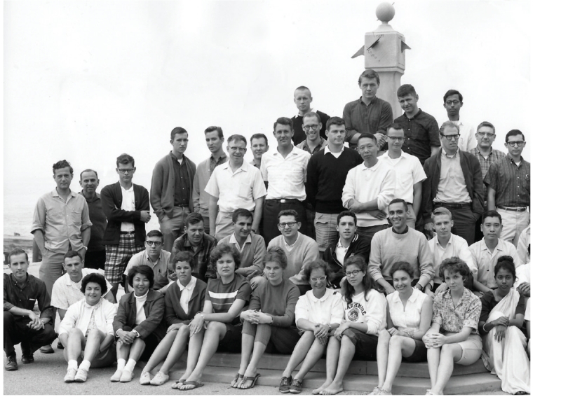
Woods Hole Physiology Course, 1963. Woody Hastings is in the top row, eighth from left.
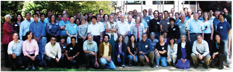
Many of the contributors to the discoveries regarding energy transduction by myosins and the roles of myosins in nonmuscle cells. This 2007 photo includes students, postdoctoral fellows, sabbatical visitors and two of my former mentors, Paul Berg and Charley Yanofsky, at a Spudich Symposium at Stanford that was organized by former members of my laboratory.
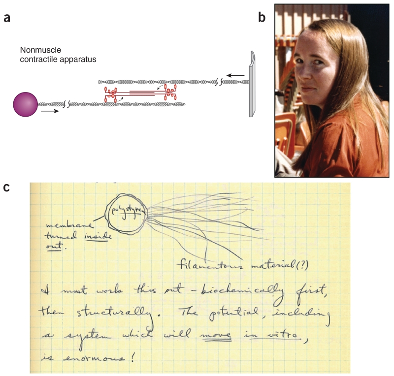
Dictyostelium has a muscle-like myosin and membrane-associated actin. (a) A possible scheme for pulling two membranes together (redrawn from ref. 6). (b) Margaret Clarke discovered myosin II in Dictyostelium and showed that it forms bipolar thick filaments, similar to muscle myosin. (c) Phagocytized polystyrene beads offered an opportunity to explore one version of an in vitro motility assay where the beads may be pulled along by myosin. Taken from my laboratory notebook, 21 January 1973.
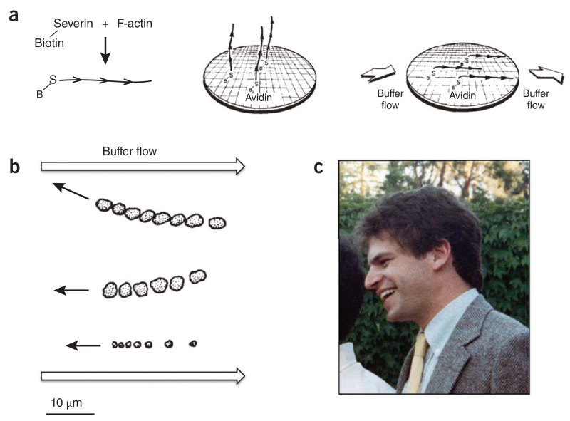
One approach to an in vitro motility assay from a totally defined system. (a) The concept was to observe myosin-coated beads moving along fixed actin filaments oriented by buffer flow. The actin filaments had biotinylated severin bound to their barbed ends; the barbed ends were attached to an avidin-coated surface by way of the tight avidin-biotin link. The filaments were oriented by buffer flow. B, biotin; S, severin. (b) Myosin-coated beads were observed by light microscopy to move upstream toward the barbed end of the surface-attached actin filaments. The position of each of the three bead aggregates is shown as a function of time. This was the first demonstration of quantitative, directed movement of myosin along actin with a totally defined system (taken from ref. 11). (c) Graduate student Steve Kron at a colleague’s wedding.
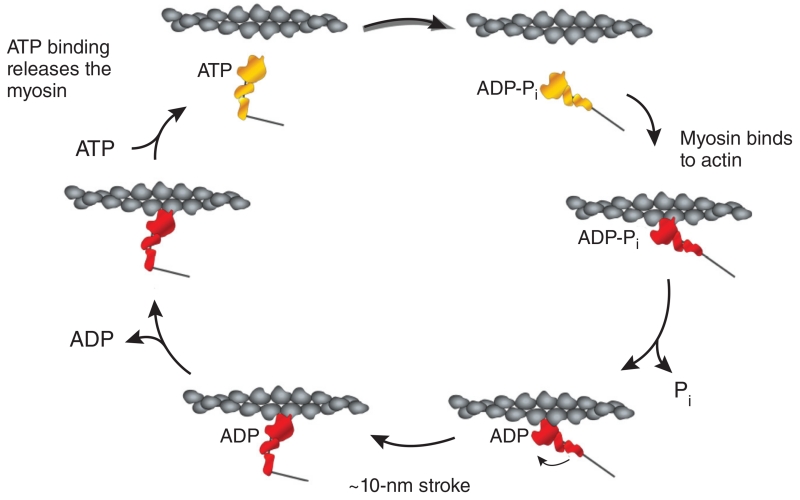
The actin-activated myosin chemomechanical cycle. This cycle, extensively studied by many researchers over several decades, was derived from kinetic studies of Lymn and Taylor. A mechanical stroke only occurs when the myosin is strongly bound to actin. Our mutational analyses of Dictyostelium myosin II probed each of the steps shown and provided structure-function analyses that helped define how the myosin motor works. ADP-Pi, ADP and inorganic phosphate, the products of ATP hydrolysis, remain bound to the active site until actin binds to the myosin.
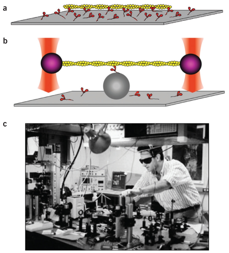
In vitro motility taken to the single-molecule level using the physics of laser trapping. (a) The Kron in vitro motility assay observing fluorescent actin filaments (yellow) moving on a myosin-coated (red) surface. (b) Two polystyrene beads attached to the ends of a single actin filament are trapped in space by laser beams. The filament is lowered onto a single myosin molecule on a bump on the surface (gray sphere). (c) Jeff Finer building the dual-beam laser trap in around 1990.
References
-
- Huxley HE. The mechanism of muscular contraction. Science. 1969;164:1356–1365. - PubMed
-
- Spudich JL. The multitalented microbial sensory rhodopsins. Trends Microbiol. 2006;14:480–487. - PubMed
-
- Spudich JA, Hastings JW. Inhibition of the bioluminescent oxidation of reduced flavin mononucleotide by 2-decenal. J. Biol. Chem. 1963;238:3106–3108. - PubMed
-
- Spudich JA, Huxley HE, Finch J. Regulation of skeletal muscle contraction. II. Structural studies of the interaction of the tropomyosin-troponin complex with actin. J. Mol. Biol. 1972;72:619–632. - PubMed
Publication types
MeSH terms
Substances
Personal name as subject
- Actions
Grants and funding
LinkOut - more resources
Full Text Sources
