Review
. 2012 Oct;7(10):623-9.
doi: 10.1038/nnano.2012.168.

Microfluidic technologies for accelerating the clinical translation of nanoparticles

Affiliations
Review

Microfluidic technologies for accelerating the clinical translation of nanoparticles

Pedro M Valencia et al. Nat Nanotechnol. 2012 Oct.

Abstract

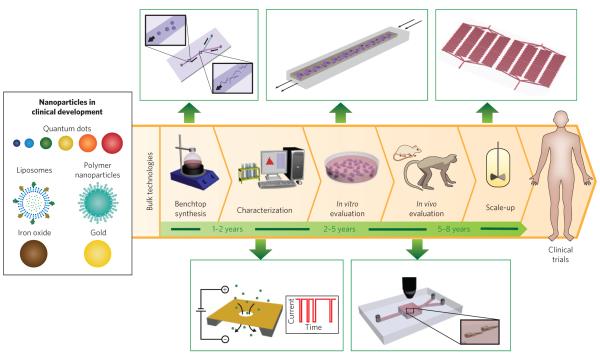
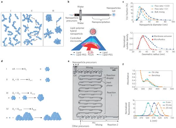
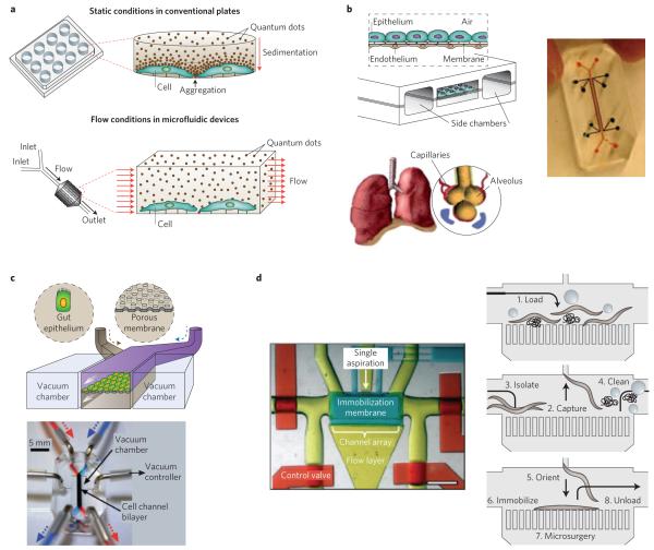
Using nanoparticles for therapy and imaging holds tremendous promise for the treatment of major diseases such as cancer. However, their translation into the clinic has been slow because it remains difficult to produce nanoparticles that are consistent 'batch-to-batch', and in sufficient quantities for clinical research. Moreover, platforms for rapid screening of nanoparticles are still lacking. Recent microfluidic technologies can tackle some of these issues, and offer a way to accelerate the clinical translation of nanoparticles. In this Progress Article, we highlight the advances in microfluidic systems that can synthesize libraries of nanoparticles in a well-controlled, reproducible and high-throughput manner. We also discuss the use of microfluidics for rapidly evaluating nanoparticles in vitro under microenvironments that mimic the in vivo conditions. Furthermore, we highlight some systems that can manipulate small organisms, which could be used for evaluating the in vivo toxicity of nanoparticles or for drug screening. We conclude with a critical assessment of the near- and long-term impact of microfluidics in the field of nanomedicine.


Figures

Figure 1
Figure 1. Nanoparticles in clinical development, steps for their translation (with average timescales) and microfluidic methods (green boxes) that could improve or complement current technologies
Synthesis is carried out in large reaction flasks, whereas microfluidic synthesis is carried out at micro and nano length scales that allow for improved control over reaction conditions. Characterization often involves taking a small sample of nanoparticles and measuring their properties offline, whereas nanopores embedded in microfluidic devices allow for real-time, in-line characterization. In vitro evaluation in plate wells produces a microenvironment far from that in vivo, whereas continuous flow in microfluidic systems result in conditions closer to those in vivo. In vivo evaluation in large animals is helpful for estimating the pharmacology of nanoparticles. To complement these studies microfluidic systems could enable real-time tracking of nanoparticles in large numbers of small organisms. Scale-up is generally carried out in reactor vessels several times larger than benchtop flasks, whereas parallelization of microfluidic channels can increase the production rate of nanoparticles with properties identical to the one at bench scale.
Figure 2
Figure 2. Microfluidic synthesis of nanoparticles
a, Schematic of the self-assembly mechanism of organic nanoparticles. On mixing with anti-solvent, polymers (or lipids) are brought to the vicinity of each other (I) then nucleate (II), subsequently aggregating into nanoparticles (III). b, Schematic of microfluidic synthesis of organic nanoparticles by rapid mixing through hydrodynamic flow focusing (top) and microvortices (bottom). Red and dark blue indicate organic and aqueous streams, respectively, while pink and light blue indicate their degree of mixing. PEG, polyethylene glycol; PLGA, poly(lactic-co-glycolic acid). c, Size distribution of polymeric nanoparticles (top) and liposomes (bottom) prepared in microfluidics compared with bulk synthesis. In both cases, narrower particle-size distributions are produced through microfluidics. d, Schematic of the self-assembly mechanism of inorganic nanoparticles. Individual molecules first nucleate (I and II), followed by aggregation of nuclei into nanoparticles (III). If the reaction is not quenched or stabilized, nanoparticles tend to agglomerate into bulk material (IV). A refers to individual molecules forming the nanoparticle, and An and Am refer to nuclei formed of n and m number of A molecules, respectively. e, Microfluidic synthesis of inorganic nanoparticles by rapid mixing through two-phase flow where reagents are embedded in fluid droplets carried by an inert fluid. f, Top: sharp versus broad absorption maximum of QDs synthesized in microchannels and bulk, respectively. Bottom: control of the absorption spectra of QDs as function of reaction time. Figure reproduced with permission from: a, ref. 16, © 2003 APS; b, Top: ref. 18, © 2008 ACS; Bottom: ref. 35, © 2012 ACS; c, Top: ref. 18, © 2008 ACS; Bottom: ref. 23, © 2008 Springer; d, ref. 24, © 2005 ACS; e, ref. 27, © 2004 RSC; f, Top: ref. 28, © 2010 Wiley; Bottom: ref. 27, © 2004 RSC.
Figure 3
Figure 3. Microfluidic systems for in vitro evaluation and screening of nanoparticles
a, Schematic of nanoparticle sedimentation in conventional plates, which could result in misinterpretation of results. In contrast, flow conditions in microfluidics provide a more-accurate method for evaluating nanoparticles in vitro. b, Left: schematic of the lung-on-a-chip that reconstitutes the critical functional alveolar-capillary interface of the human lung through a stretchable membrane containing an epithelium layer on one side and an endothelium layer on the other. Right: photograph of actual device. c, Top: schematic of the gut-on-a-chip made by flexible, porous, extracellular matrix-coated membrane lined by gut epithelial cells. The blue and brown arrows indicate two different streams of culture medium separated by a membrane, entering the channel from the top and bottom, respectively. Bottom: photograph of the gut-on-a-chip device made of polydimethylsiloxane elastomer. A syringe pump was used to perfuse dyes (red and blue) for channel visualization. d, Left: photograph of a dye-filled microfluidic system designed to handle C. elegans worms. Red, control valve layer; yellow, flow layer; blue, immobilization layer. Scale bar, 1 mm. Right: schematic showing load, capture, orient, immobilization and unload of the worm. Figure reproduced with permission from: a, ref. 39, © 2010 AIP; b, ref. 41, © 2010 AAAS; c, ref. 42, © 2012 RSC; d, ref. 48, © 2010 NAS.

References

    1. Petros RA, DeSimone JM. Strategies in the design of nanoparticles for therapeutic applications. Nature Rev. Drug. Discov. 2010;9:615–627. - PubMed
    1. Gregoriadis G. Drug entrapment in liposomes. FEBS Lett. 1973;36:292–296. - PubMed
    1. Hrkach J, et al. Preclinical development and clinical translation of a PSMA-targeted docetaxel nanoparticle with a differentiated pharmacological profile. Sci. Transl. Med. 2012;4:128ra39. This article describes the translation of the first targeted polymeric nanoparticle for drug delivery from discovery to clinical trials.

    1. Qiao R, Yang C, Gao M. Superparamagnetic iron oxide nanoparticles: from preparations to in vivo MRI applications. J. Mater. Chem. 2009;19:6274–6293.
    1. Haun JB, et al. Micro-NMR for rapid molecular analysis of human tumor samples. Sci. Transl. Med. 2011;3:71ra16. - PMC - PubMed

Publication types

MeSH terms