. 2013 May;228(5):1010-6.
doi: 10.1002/jcp.24247.

Mesenchymal stem cells contribute to endogenous FVIII:c production

Affiliations

Mesenchymal stem cells contribute to endogenous FVIII:c production

Chad Sanada et al. J Cell Physiol. 2013 May.

Abstract

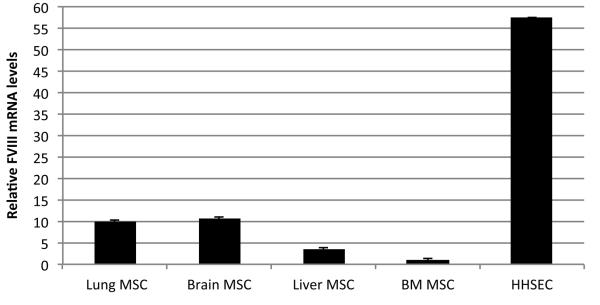
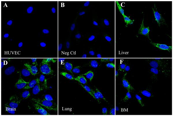
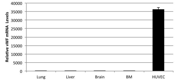
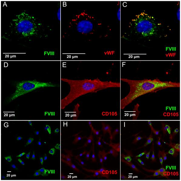
Besides the liver, it has been difficult to identify which organ(s) and/or cellular component(s) contribute significantly to the production of human FVIII:c (FVIII). Thus far, only endothelial cells have been shown to constitute a robust extrahepatic source of FVIII, possibly explaining both the diverse presence of FVIII mRNA in the body, and the observed increase in FVIII levels during liver failure. Here, we investigate whether human mesenchymal stem cells (MSC), ubiquitously present in different organs, could also contribute to FVIII production. MSC isolated from human lung, liver, brain, and bone marrow expressed FVIII message as determined by quantitative-RT-PCR. Using an antibody specific for FVIII, confocal microscopy, and umbilical cord-derived endothelial cells (HUVEC) as a negative control, we demonstrated that, in MSC, FVIII protein was not stored in granules; rather, it localized to the perinuclear region. Furthermore, functional FVIII was detected in MSC supernatants and cell lysates by aPTT and chromogenic assays. These results demonstrate that MSC can contribute at low levels to the functional FVIII pool, and advance the understanding of the physiology of FVIII production and secretion.


Figures

Figure 1
Figure 1. Relative FVIII mRNA levels in Human MSC
qRT-PCR data showing relative FVIII mRNA levels in different MSC populations in comparison to HHSEC. Values represent an average of 4 different independent experiments for each cell population.
Figure 2
Figure 2. Human MSC express FVIII protein
Cells were stained for FVIII (green) using an affinity purified FVIII:c antibody. Nuclei are counterstained with DAPI (blue). Images were taken on an Olympus Fluoview 1000 confocal microscope using a 40x objective. Representative images have an additional 3x digital zoom. HUVEC were used as a specificity control for the anti-FVIII:c antibody, showing that the antibody does not cross-react with VWF (A). Secondary antibody control (B). Panels C-F show different populations: Liver (C), Brain (D), Lung (E), BM (F).
Figure 3
Figure 3. Relative vWF mRNA levels in Human MSC
qRT-PCR data showing vWF mRNA levels in different MSC populations in comparison to HUVEC. Values represent an average of 3 different independent experiments for each cell population.
Figure 4
Figure 4. Human MSC secrete biologically active FVIII, but at lower levels than Hepatic Sinusoidal Endothelial Cells (HHSEC)
(A) Comparison between the different MSC populations and HHSEC of FVIII activity at 24 and 48h of culture, as determined by aPTT, expressed as a percentage of the activity present in normal human plasma. (B) Detail showing the FVIII activity of the different MSC populations as determined by aPTT, expressed as a percentage of that present in normal human plasma at 24 and 48h of culture.
Figure 5
Figure 5. Immunofluorescence Analysis Demonstrates Homogeneous Staining of FVIII in MSC
MSC and HUVEC were transduced with an expression/secretion optimized porcine FVIII in order to visualize intracellular FVIII localization. FVIII was detected by staining with an antibody against FVIII (green), while CD105 (red) staining reveals the MSC cell surface. All images were taken on a Fluoview 1000 confocal microscope with a 40x objective with various amounts of digital zoom factors as indicated by scale bars. (A-C) FVIII-transduced HUVEC co-store FVIII and VWF (red) in granules, and were used as a control. (D-F) High magnification of human MSC showing homogenous cytoplasmic staining of FVIII. (G-I) Low magnification of MSC labeled with an antibody against FVIII. Nuclei are counterstained with DAPI (blue).

References

    1. Airey JA, Almeida-Porada G, Colletti EJ, Porada CD, Chamberlain J, Movsesian M, Sutko JL, Zanjani ED. Human Mesenchymal Stem Cells Form Purkinje Fibers in Fetal Sheep Heart. Circulation. 2004;109(11):1401–1407. - PubMed
    1. Bianco P. Back to the future: moving beyond "mesenchymal stem cells". J Cell Biochem. 2011;112(7):1713–1721. - PMC - PubMed
    1. Bianco P, Robey PG, Simmons PJ. Mesenchymal Stem Cells: Revisiting History, Concepts, and Assays. Cell Stem Cell. 2008;2(4):313–319. - PMC - PubMed
    1. Bontempo FA, Lewis JH, Gorenc TJ, Spero JA, Ragni MV, Scott JP, Starzl TE. Liver transplantation in hemophilia A. Blood. 1987;69(6):1721–1724. - PMC - PubMed
    1. Caplan AI, Bruder SP. Mesenchymal stem cells: building blocks for molecular medicine in the 21st century. Trends Mol Med. 2001;7(6):259–264. - PubMed

Publication types