

Carotid artery plaque characterization using CT multienergy imaging
- PMID: 23042919
- PMCID: PMC7964496
- DOI: 10.3174/ajnr.A3285
Carotid artery plaque characterization using CT multienergy imaging
Abstract
Background and purpose: Carotid artery plaque types can be categorized with CT according to their HU values. The purpose of this work was to analyze carotid artery plaque characteristics by using multienergy imaging.
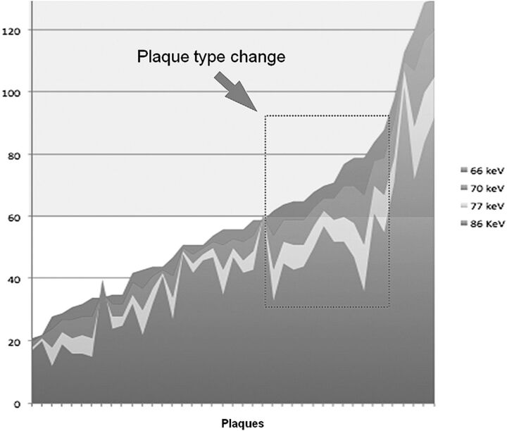
Methods and materials: Thirty-two consecutive patients (23 men; median age, 70 years) were retrospectively analyzed. Carotid arteries were studied with a multienergy CT scanner. All patients received a 15-mL timing bolus of contrast medium to synchronize the data acquisition followed by an injection of 60 mL of contrast medium at a 5-mL/s flow rate. Plaque analysis in 64 carotid arteries was performed, and datasets were reconstructed by using a dedicated workstation. For each plaque, the HU value was quantified with a 2-mm-square region of interest at monoenergy values of 66, 70, 77, and 86 keV. The Wilcoxon test was used to test the differences in HU values in the plaques at different kiloelectron volts.
Results: Four carotid arteries were excluded due to the absence of plaque, and another 7, because of the presence of calcified plaques. In the remaining 53 carotid arteries, Wilcoxon analysis showed a statistically significant difference in HU values among the monoenergy values of 66, 70, 77, and 86 keV (P=.0001). In particular, we found that with the increase in monochromatic kiloelectron volt values, there is a statistically significant reduction in the HU value of the plaque.
Conclusions: Results of this study suggest that the HU values of plaque may significantly change according to the selected kiloelectron volt; therefore, the HU-based plaque type (fatty, mixed, calcified) should be classified according to the energy level applied.
Figures


References
-
- Serfaty JM, Nonent M, Nighoghossian N, et al. , for the CARMEDAS Study Group. Plaque density on CT, a potential marker of ischemic stroke. Neurology 2006;66:118–20 - PubMed
-
- Naghavi M, Libby P, Falk E, et al. . From vulnerable plaque to vulnerable patient: a call for new definitions and risk assessment strategies: Part I. Circulation 2003;108:1664–72 - PubMed
-
- de Weert TT, Ouhlous M, Meijering E, et al. . In vivo characterization and quantification of atherosclerotic carotid plaque components with multidetector computed tomography and histopathological correlation. Arterioscler Thromb Vasc Biol 2006;26:2366–72 - PubMed
-
- Ajduk M, Pavić L, Bulimbasić S, et al. . Multidetector-row computed tomography in evaluation of atherosclerotic carotid plaques complicated with intraplaque hemorrhage. Ann Vasc Surg 2009;23:186–93 - PubMed
MeSH terms
Substances
LinkOut - more resources
Full Text Sources
Other Literature Sources
Medical