. 2012 Dec;194(23):6574-85.
doi: 10.1128/JB.01428-12. Epub 2012 Oct 5.

Polymorphisms in inc proteins and differential expression of inc genes among Chlamydia trachomatis strains correlate with invasiveness and tropism of lymphogranuloma venereum isolates

Affiliations

Polymorphisms in inc proteins and differential expression of inc genes among Chlamydia trachomatis strains correlate with invasiveness and tropism of lymphogranuloma venereum isolates

Filipe Almeida et al. J Bacteriol. 2012 Dec.

Abstract

Chlamydia trachomatis is a human bacterial pathogen that multiplies only within an intracellular membrane-bound vacuole, the inclusion. C. trachomatis includes ocular and urogenital strains, usually causing infections restricted to epithelial cells of the conjunctiva and genital mucosa, respectively, and lymphogranuloma venereum (LGV) strains, which can infect macrophages and spread into lymph nodes. However, C. trachomatis genomes display >98% identity at the DNA level. In this work, we studied whether C. trachomatis Inc proteins, which have a bilobed hydrophobic domain that may mediate their insertion in the inclusion membrane, could be a factor determining these different types of infection and tropisms. Analyses of polymorphisms and phylogeny of 48 Inc proteins from 51 strains encompassing the three disease groups showed significant amino acid differences that were mainly due to variations between Inc proteins from LGV and ocular or urogenital isolates. Studies of the evolutionary dynamics of inc genes suggested that 10 of them are likely under positive selection and indicated that most nonsilent mutations are LGV specific. Additionally, real-time quantitative PCR analyses in prototype and clinical strains covering the three disease groups identified three inc genes with LGV-specific expression. We determined the transcriptional start sites of these genes and found LGV-specific nucleotides within their promoters. Thus, subtle variations in the amino acids of a subset of Inc proteins and in the expression of inc genes may contribute to the unique tropism and invasiveness of C. trachomatis LGV strains.


Figures

Fig 1
Fig 1
Type III secretion (T3S) signals in C. trachomatis Inc proteins. Y. enterocolitica T3S-proficient (ΔHOPEMT) and T3S-defective (ΔHOPEMT ΔYscU) bacteria were used to analyze secretion of hybrid proteins comprising the first 20 amino acids of Inc proteins or of Y. enterocolitica SycT, fused to the mature form of TEM-1 β-lactamase (TEM-1). (A and B) Immunoblots show the result of representative assays in which proteins in culture supernatants (S, secreted proteins) and in bacterial pellets (P, nonsecreted proteins) from ∼5 × 10⁷ bacteria were loaded per lane. SycT and SycO are strictly cytosolic Yersinia T3S chaperones (28, 32). SycT20-TEM-1 was a negative control for the T3S assays. Immunodetection of SycO ensured that the presence of TEM-1 hybrid proteins in culture supernatants was not a result of bacterial lysis or contamination. (C) The percentage of secretion of each TEM-1 hybrid was calculated by densitometry as the ratio between the amount of secreted and total protein. The threshold to decide whether a protein was secreted was set to 5% (dashed line), based on the percentage of secretion of SycT20-TEM-1. Data are means ± SEM from at least three independent experiments. Please note that in this work we did not analyze all described putative Inc proteins (see Table 1) and that T3S signals were not analyzed for all known Inc proteins.
Fig 2
Fig 2
Polymorphisms in C. trachomatis Inc proteins. Polymorphic membrane proteins (Pmps) and housekeeping proteins (HKs) were analyzed as references. (A) Overall mean genetic distance (polymorphisms) based on the p distance between all possible pairs of amino acid (aa) sequences of Inc proteins, Pmps, and HKs among C. trachomatis strains. Proteins marked with an asterisk have a p distance that is equal to or higher than the average value for Inc proteins (0.017 [dashed line]). (B) Average mean genetic distance, based on the p distance between all possible pairs of amino acid sequences within (ocular [OC], urogenital [UROG], or LGV) or between (OC-UROG, OC-LGV, or UROG-LGV) C. trachomatis disease groups. (C) Venn diagrams showing the phylogenetic segregation of C. trachomatis disease groups based on neighbor-joining trees of Inc proteins, Pmps, or HKs and on pairwise p distances between and within disease groups for all possible pairs of Inc protein, PMP, or HK sequences from the C. trachomatis strains analyzed. All these analyses were performed by bootstrapping with 1,000 replicates. Error bars represent SEM.
Fig 3
Fig 3
Evolutionary dynamics of inc genes. Genes encoding polymorphic membrane proteins (pmp genes) or housekeeping (HK) proteins were used as reference. (A) Ratio of nonsynonymous (dN) to synonymous (dS) substitutions among C. trachomatis strains. The dashed line indicates neutrality (dN/dS = 1). The arrows specify genes likely under positive selection, according to the codon-based Z test of selection (see Materials and Methods). (B) Distribution of dN versus dS values for the 10 inc genes likely under positive selection, comparing the impact of artificially discarding ocular, urogenital, or LGV strains relative to the analysis with all C. trachomatis strains. The straight line in each graph indicates neutrality (dN/dS = 1). In all cases, inc genes likely under positive selection (codon-based Z test of selection) are depicted as black circles, while inc genes for which statistical support of likely positive selection can no longer be detected after discarding a particular group of strains are depicted as white circles. All these analyses were performed by bootstrapping with 1,000 replicates. For sake of clarity, SEMs for values in both panels are presented only in Table S5 in the supplemental material.
Fig 4
Fig 4
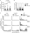
mRNA levels of inc genes during the developmental cycle of different C. trachomatis strains. The mRNA levels of 48 inc genes (A and B) and of ct058, ct192, and ct214 (C) were analyzed by RT-qPCR throughout the developmental cycle of the indicated prototype (B/Har36, C/TW3, E/Bour, L2/434, and L3/404) and clinical (F/CS465-95 and L2b/CS19-08) strains. (A) Peak of expression (highest mRNA levels during the developmental cycle) of each inc gene. The P values were calculated by two-tailed t tests. (B) Number of inc genes showing the indicated profiles of expression (variation of mRNA levels during the developmental cycle). (C) The expression values (mean ± SEM) resulted from raw RT-qPCR data (105) of each gene normalized to that of the 16S rRNA gene and are from at least two independent experiments. Complete data are shown in Table S6 in the supplemental material.
Fig 5
Fig 5
Identification of LGV-specific nucleotides in the promoter regions of ct192 and ct214 and within the ct059-ct058 transcript. (A) Genetic organization of ct058, ct192, and ct214 (the nomenclature of D/UW3 is used) depicting the fragments amplified in the transcriptional linkage analysis and the approximate locations of the transcriptional start sites (TSSs) determined by RACE in L2/434. (B) Transcriptional linkage analysis in L2/434: gDNA+, PCR from total DNA isolated from cells infected with L2/434; cDNA+, PCR from cDNA generated with reverse transcriptase (RT) from total RNA isolated from cells infected with L2/434; cDNA−, as for cDNA+ but without RT; gDNA−, PCR from DNA of uninfected cells. (C to E) Schematic view of the nucleotide sequences of the ct059 promoter region, ct059-ct058 intragenic region, and first codons of ct058 (C) and of the promoter regions of ct192 (D) and ct214 (E) in L2/434. The TSSs are labeled +1, and the predicted −10 and −35 σ66-like hexamers are underlined. The sequences of the three LGV strains used in RT-qPCR assays (Fig. 5) are 100% identical within the depicted regions (see Fig. S3 and S4 in the supplemental material). The identified LGV-specific nucleotides are indicated with an arrow, and letters below the sequence represent nucleotides present in those positions in the ocular and urogenital strains used in RT-qPCR assays. The full nucleotide sequences of these regions in the strains used in RT-qPCR assays are shown in Fig. S3 and S4.

References

    1. Abdelrahman YM, Belland RJ. 2005. The chlamydial developmental cycle. FEMS Microbiol. Rev. 29:949–959 - PubMed
    1. Albrecht M, Sharma CM, Reinhardt R, Vogel J, Rudel T. 2010. Deep sequencing-based discovery of the Chlamydia trachomatis transcriptome. Nucleic Acids Res. 38:868–877 - PMC - PubMed
    1. Alzhanov DT, Weeks SK, Burnett JR, Rockey DD. 2009. Cytokinesis is blocked in mammalian cells transfected with Chlamydia trachomatis gene CT223. BMC Microbiol. 9:2. - PMC - PubMed
    1. Bebear C, de Barbeyrac B. 2009. Genital Chlamydia trachomatis infections. Clin. Microbiol. Infect. 15:4–10 - PubMed
    1. Belland RJ, et al. 2003. Genomic transcriptional profiling of the developmental cycle of Chlamydia trachomatis. Proc. Natl. Acad. Sci. U. S. A. 100:8478–8483 - PMC - PubMed

Publication types

LinkOut - more resources