. 2012 Nov 15;186(10):1014-24.
doi: 10.1164/rccm.201207-1206OC. Epub 2012 Oct 4.

The alveolar epithelium determines susceptibility to lung fibrosis in Hermansky-Pudlak syndrome

Affiliations

The alveolar epithelium determines susceptibility to lung fibrosis in Hermansky-Pudlak syndrome

Lisa R Young et al. Am J Respir Crit Care Med. .

Abstract

Rationale: Hermansky-Pudlak syndrome (HPS) is a family of recessive disorders of intracellular trafficking defects that are associated with highly penetrant pulmonary fibrosis. Naturally occurring HPS mice reliably model important features of the human disease, including constitutive alveolar macrophage activation and susceptibility to profibrotic stimuli.

Objectives: To decipher which cell lineage(s) in the alveolar compartment is the predominant driver of fibrotic susceptibility in HPS.

Methods: We used five different HPS and Chediak-Higashi mouse models to evaluate genotype-specific fibrotic susceptibility. To determine whether intrinsic defects in HPS alveolar macrophages cause fibrotic susceptibility, we generated bone marrow chimeras in HPS and wild-type mice. To directly test the contribution of the pulmonary epithelium, we developed a transgenic model with epithelial-specific correction of the HPS2 defect in an HPS mouse model.

Measurements and main results: Bone marrow transplantation experiments demonstrated that both constitutive alveolar macrophage activation and increased susceptibility to bleomycin-induced fibrosis were conferred by the genotype of the lung epithelium, rather than that of the bone marrow-derived, cellular compartment. Furthermore, transgenic epithelial-specific correction of the HPS defect significantly attenuated bleomycin-induced alveolar epithelial apoptosis, fibrotic susceptibility, and macrophage activation. Type II cell apoptosis was genotype specific, caspase dependent, and correlated with the degree of fibrotic susceptibility.

Conclusions: We conclude that pulmonary fibrosis in naturally occurring HPS mice is driven by intracellular trafficking defects that lower the threshold for pulmonary epithelial apoptosis. Our findings demonstrate a pivotal role for the alveolar epithelium in the maintenance of alveolar homeostasis and regulation of alveolar macrophage activation.


Figures

Figure 1.
Figure 1.
Susceptibility to bleomycin-induced pulmonary fibrosis occurs only in those Hermansky-Pudlak syndrome (HPS) mouse models corresponding to genotypes linked to human lung fibrosis. HPS1mt, HPS2mt, HPS2ko, HPS3mt, CHSmt, and strain-matched C57BL/6J wild-type (WT) mice, aged 8 to 12 weeks, were challenged by intratracheal instillation of a single bleomycin dose of 0.025 units. (a) Survival of HPS1mt, HPS2mt, and HPS2ko mice was significantly different from WT, HPS3mt, and CHSmt when analyzed by log-rank test (n = 8 per group minimum; P < 0.01 for HPS1mt, HPS2mt, and HPS2ko versus other groups, with adjustment for multiple comparisons). (b) Representative lung histology images (hematoxylin and eosin, 10× magnification) obtained 7 days after mice were challenged with bleomycin. Light gray labels denote models corresponding to HPS genotypes associated with pulmonary fibrosis (HPS-1 and HPS-2) and dark gray labels denote models corresponding to human genotypes that have not been associated with pulmonary fibrosis (HPS-3 and Chediak-Higashi syndrome [CHS]). (c) Collagen quantitation using the Sircol Red assay from lungs harvested 7 days after bleomycin or controls. Values indicate totals for both lungs from each mouse; n = 12 per group for WT and HPS2mt, n = 8 per group for others. *P < 0.001 versus WT bleomycin challenged (analysis of variance with Tukey post-test).
Figure 2.
Figure 2.
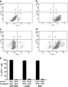
Generation of bone marrow chimeras. (a, b) Representative fluorescence-activated cell sorter (FACS) analysis of spleen cell populations 90 days after bone marrow transplantation. C57BL/6J mice that express green fluorescent protein (GFP) under the control of the ubiquitin promoter were used as wild-type (WT) mice in these experiments. (a) Results from a WT(GFP) recipient of HPS2mt marrow. (b) HPS2mt recipient of WT(GFP) marrow. (c, d) Representative FACS analysis of lung leukocyte populations 90 days after bone marrow transplantation. (c) Results from a WT(GFP) recipient of HPS2mt marrow. (d) HPS2mt recipient of WT(GFP) marrow. (e) Quantitation of the percentage of donor cell reconstitution in the spleen, whole lung, and bronchoalveolar lavage (BAL) 90 days after bone marrow transplantation. Results of FACS analysis and BAL cytospins were quantitated from three mice in each transplantation group.
Figure 3.
Figure 3.
Bone marrow transplantation of wild-type (WT) whole marrow fails to rescue the fibrotic susceptibility of HPS2mt and HPS1mt mice, and transplantation of HPS2mt or HPS1mt marrow into WT mice does not confer fibrotic susceptibility. Transplanted mice, age 20 to 26 weeks, were studied 90 days after bone marrow transplantation. Mice were challenged with a single intratracheal dose of bleomycin, 0.025 units. (a) Survival of bone marrow–transplanted HPS2mt and WT mice after bleomycin challenge. HPS2mt mice (gray) show increased mortality after bleomycin challenge despite transplantation with WT marrow. WT mice (black) had no mortality after bleomycin challenge despite transplantation with HPS2mt marrow. Chimeric mice (WT into WT and HPS2mt into HPS2mt) were used as experimental control groups. n = 6–10 per group; P < 0.001 for HPS2mt versus WT recipients by log-rank test. (b) Quantitation of lung collagen content demonstrates that increased pulmonary fibrosis occurs in the bleomycin-challenged HPS2mt mice despite transplantation with WT marrow. Lungs were harvested 7 days after bleomycin challenge and assayed using the Sircol Red assay. Values indicate totals for both lungs from each mouse; n = 3 per group for unchallenged, n = 10 for HPS2mt recipients of WT marrow, and n = 5 for other combinations. P = not significant among unchallenged mice at baseline, *P < 0.001 versus WT recipients after bleomycin challenge (analysis of variance with Tukey post-test). (c) Quantitation of lung collagen content in HPS1mt bone marrow transplant experiments. Mice were challenged with bleomycin 90 days after marrow transplantation, and lung collagen was evaluated 7 days later. n = 7 for HPS1mt recipients of WT marrow and n = 5 for WT recipients of HPS1mt marrow, *P < 0.01. (d) Representative lung histology images with Trichrome staining (20× magnification), 7 days after bleomycin challenge, are shown from each of the transplanted groups. Note that only minimal fibrosis is present in WT recipients, even after transplantation with HPS2mt or HPS1mt marrow. In contrast, severe fibrosis occurs in HPS2mt and HPS1mt recipients, despite transplantation of WT marrow. (eh) Impact of bone marrow transplantation on alveolar macrophage activation phenotype of constitutive cytokine secretion. Alveolar macrophages were isolated by bronchoalveolar lavage and cultured, and tumor necrosis factor (TNF)-α (e, f) and macrophage inflammatory protein (MIP)-1α (g, h) were assayed from the cell culture media supernatant by ELISA. For e and g, n = 4–8 per group, *P < 0.05 for both HPS2mt recipients versus WT recipients. For f and h, n = 6 for WT recipients of HPS1mt marrow, n = 10 for HPS1mt recipients of WT marrow, *P < 0.05.
Figure 4.
Figure 4.
Transgenic correction of the defective β3 subunit stabilizes the AP-3 complex as evidenced by detection of the mu and delta subunits of the heterooligomer. (a) Construct used for generation of hSPC-AP3 transgenic mice. The cDNA for murine AP-3/β3A was generated by polymerase chain reaction (PCR) amplification, fully sequenced, and inserted into a vector containing the human SP-C promoter and β-globin sequences as follows: hSPC promoter (3.7 kb)–intronic β globin sequence–AP3 (3.4 kb)–β globin–poly A. This construct was linearized and injected into the pronuclei of fertilized mouse C57BL/6J embryos and implanted into pseudopregnant C57BL/6J dams. Progeny were screened by PCR to identify founders, which were then crossed back into HPS2mt or HPS2ko mice. (b) PCR detection of a sequence spanning the hSPC promoter to rabbit β-globin. HPS2mt and HPS2ko denote the background HPS2 mice, and (+) represents the plasmid for the positive control. The PCR strategy distinguishes transgene-positive mice from two different founder lines (TGA+ and TGB+) from wild-type (WT) and transgene-negative mice (TGA− and TGB−). (c) Quantitative PCR analysis of AP3b1 from alveolar type II cells from transgene-positive, transgene-negative, and WT mice. Data were normalized to Actb. Note that AP3b1 was not detected from HPS2ko mice. *P < 0.05 versus WT, #P < 0.05 versus HPS2mt, HPS2ko, TGA− and TGB−, and P = not significant, versus WT, **P < 0.01 versus all others. (d) Rescue of the AP-3 complex stability as demonstrated by presence of μ3 in transgene-positive mice. Western blots of cells isolated from HPS2mt mice show stabilization of the AP-3 μ3 subunit in alveolar type II cells in transgene-positive mice (TG+), but not in the spleen, nor in transgene-negative (TGneg) mice. β-actin expression is shown as the loading control. (e) Rescue of the AP-3 complex as demonstrated by delta (δ) subunit detection in transgene-positive mice. Example Western blot using a monoclonal antibody to the delta subunit confirms functional expression of the β3 subunit in transgene-positive (TG+) mice. The delta subunit is not detected in the HPS2mt TGneg mice. β-actin expression is shown as the loading control. (f, g) Epithelial-specific transgenic correction of AP-3 reduces excess monocyte chemotactic protein (MCP)-1 and chemokine (C-X-C motif) ligand 1 (CXCL1) production from HPS2mt alveolar type II cells. MCP-1 and CXCL1 levels were assayed from conditioned media of unchallenged primary murine alveolar type II cells. For f, *P < 0.01 versus WT and TG+, **P < 0.05 versus WT. For g, *P < 0.01 versus WT and #P < 0.05 versus WT and TGneg, n = 9 to 12 per group. (h) Epithelial-specific transgenic correction of AP-3 also reduces the size of enlarged lamellar bodies in HPS2mt alveolar type II cells. Images of lamellar bodies were captured by a technician blinded to the genotype of samples, and the area of lamellar bodies was quantitated on 10,000× images of the ultrastructural examination using Olympus Soft Imaging Solutions. *P < 0.01 versus WT and **P < 0.05 versus WT and TGneg.
Figure 5.
Figure 5.
Epithelial-specific transgenic correction of AP-3 protects Hermansky-Pudlak syndrome (HPS) mice from bleomycin-induced mortality and pulmonary fibrosis and also dampens constitutive alveolar macrophage activation. (a) Increased survival of transgene-positive HPS mice after intratracheal bleomycin challenge. Transgene-positive (open squares) and transgene-negative (circles) HPS2mt mice, aged 6 to 8 weeks, were challenged by intratracheal instillation of a single bleomycin dose of 0.025 units per mouse. Survival was significantly increased in the transgene-positive HPS2mt mice based on analysis using the log-rank test (n = 7 per group, P < 0.01). (b) Trichrome staining (40×) of representative lung histology images from Day 7 bleomycin-challenged transgene-positive mice and littermate control mice, for HPS2 transgene-negative (HPS2 TGneg), both HPS transgene-positive lines (HPS2mt-TGA+ and HPS2mt-TGB+), and HPS2ko transgene-positive and -negative mice. (c) Quantitation of the total lung collagen content demonstrates that the transgene-positive HPS2mt and HPS2ko mice are relatively resistant to bleomycin-induced fibrosis compared with transgene-negative littermate control mice. Mice were challenged with intratracheal bleomycin, and lungs were harvested for Sircol collagen assay on Day 7. n = 6 each for all groups except n = 9 each for HPS2mt TGA+ and TGneg. *P < 0.01 versus TGneg mice and P < 0.05 versus wild-type mice. (d, e) Epithelial-specific transgenic correction of AP-3 also corrects the alveolar macrophage activation phenotype. Primary alveolar macrophages were isolated by bronchoalveolar lavage and cultured, and tumor necrosis factor (TNF)-α (d) and macrophage inflammatory protein (MIP)-1α (e) were assayed in the alveolar macrophage cell culture media supernatant by ELISA. *P < 0.01 versus WT and TGneg, **P < 0.05 versus WT, n = 6 to 12 per group.
Figure 6.
Figure 6.
Alveolar epithelial apoptosis correlates with fibrotic susceptibility in Hermansky-Pudlak syndrome (HPS) mice. (a) Evaluation of early alveolar apoptosis in HPS mouse models. Lungs were inflation-fixed 5 hours after intratracheal bleomycin challenge, terminal deoxynucleotidyl transferase-mediated dUTP nick end labeling (TUNEL) assay, and pro-SP-C immunofluorescence were performed, and the mean number of dual-positive cells (TUNEL-positive, pro-SP-C positive type II cells) were determined from a minimum of 10 fields counted by two independent observers blind to mouse genotype. n = 3–6 per group, P = not significant (n.s.) for unchallenged mice (not shown). P = n.s. for wild-type (WT) versus CHSmt and HPS3mt 5 hours after bleomycin, and *P < 0.01 for HPS1mt, HPS2mt, and HPS2ko versus WT, CHSmt, or HPS3mt (analysis of variance [ANOVA]). (b) Quantitation of TUNEL-positive type II cells from WT, HPS2mt-TGneg, and HPS2mt-TG+ mice. n = 4 mice per group, *P < 0.001 for TGneg versus WT, **P < 0.01 for TG+ versus TGneg littermate control mice, and P = n.s. for TG+ versus WT. (c) Bleomycin-induced cell death in primary murine type II cells in vitro, measured by lactate dehydrogenase (LDH) cytotoxicity assay. Primary murine type II cells were isolated and cultured in vitro, and LDH was expressed as LDH% (LDH media/LDH cells + media) compared with WT control. n = 4 mice per group; P < 0.0001 for both mouse strain and bleomycin dose effect by two-way ANOVA. (d) Pan-caspase inhibition protects HPS mice from excess bleomycin-induced pulmonary fibrosis. HPS2mt or WT mice, aged 8 to 10 weeks, were challenged with intratracheal bleomycin 24 hours after initiation of intraperitoneal caspase inhibitor Q-VD-Oph or vehicle control (dimethyl sulfoxide), and the total lung collagen content was measured by Sircol assay 7 days after the bleomycin challenge. n = 10 for WT and HPS2mt with caspase inhibitor, n = 10 for WT with vehicle, and n = 7 for HPS2mt with vehicle control. *P < 0.01 versus all other groups. P = n.s. for HPS2mt with caspase inhibitor versus all WT.

Comment in

References

    1. King TE, Jr, Pardo A, Selman M. Idiopathic pulmonary fibrosis. Lancet 2011;378:1949–1961 - PubMed
    1. Gahl WA, Brantly M, Kaiser-Kupfer MI, Iwata F, Hazelwood S, Shotelersuk V, Duffy LF, Kuehl EM, Troendle J, Bernardini I. Genetic defects and clinical characteristics of patients with a form of oculocutaneous albinism (Hermansky-Pudlak syndrome). N Engl J Med 1998;338:1258–1264 - PubMed
    1. Cullinane AR, Curry JA, Carmona-Rivera C, Summers CG, Ciccone C, Cardillo ND, Dorward H, Hess RA, White JG, Adams D, et al. A BLOC-1 mutation screen reveals that PLDN is mutated in Hermansky-Pudlak syndrome type 9. Am J Hum Genet 2011;88:778–787 - PMC - PubMed
    1. Gochuico BR, Huizing M, Golas GA, Scher CD, Tsokos M, Denver SD, Frei-Jones MJ, Gahl WA. Interstitial lung disease and pulmonary fibrosis in Hermansky-Pudlak syndrome type 2, an adaptor protein-3 complex disease. Mol Med 2012;18:56–64 - PMC - PubMed
    1. Brantly M, Avila NA, Shotelersuk V, Lucero C, Huizing M, Gahl WA. Pulmonary function and high-resolution CT findings in patients with an inherited form of pulmonary fibrosis, Hermansky-Pudlak syndrome, due to mutations in HPS-1. Chest 2000;117:129–136 - PubMed

Publication types

MeSH terms