. 2012 Nov 16;287(47):39275-90.
doi: 10.1074/jbc.M112.378109. Epub 2012 Oct 5.

ATG8 family proteins act as scaffolds for assembly of the ULK complex: sequence requirements for LC3-interacting region (LIR) motifs

Affiliations

ATG8 family proteins act as scaffolds for assembly of the ULK complex: sequence requirements for LC3-interacting region (LIR) motifs

Endalkachew Ashenafi Alemu et al. J Biol Chem. .

Abstract

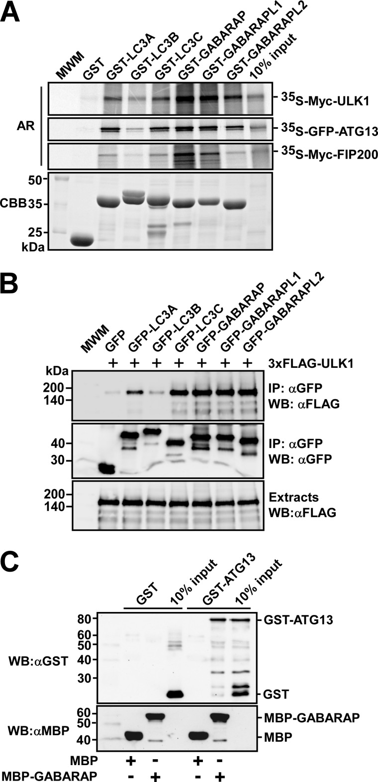
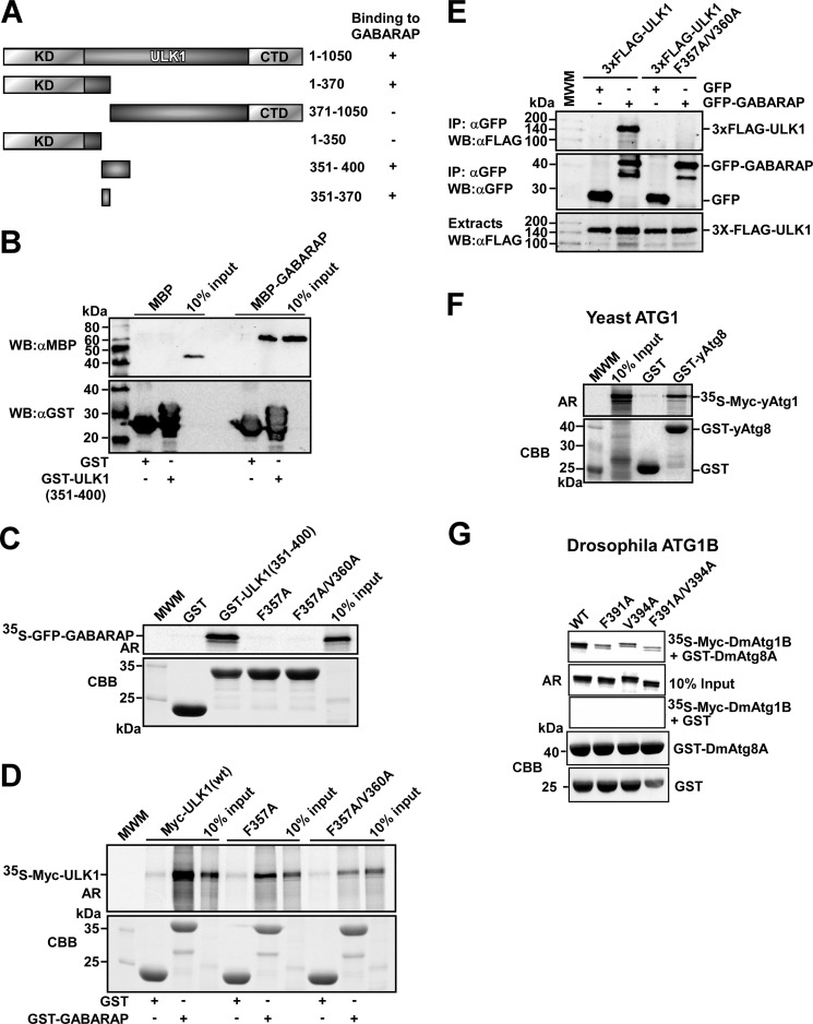
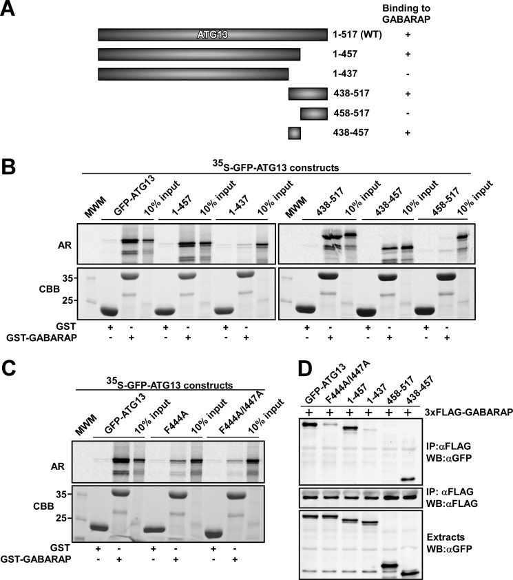
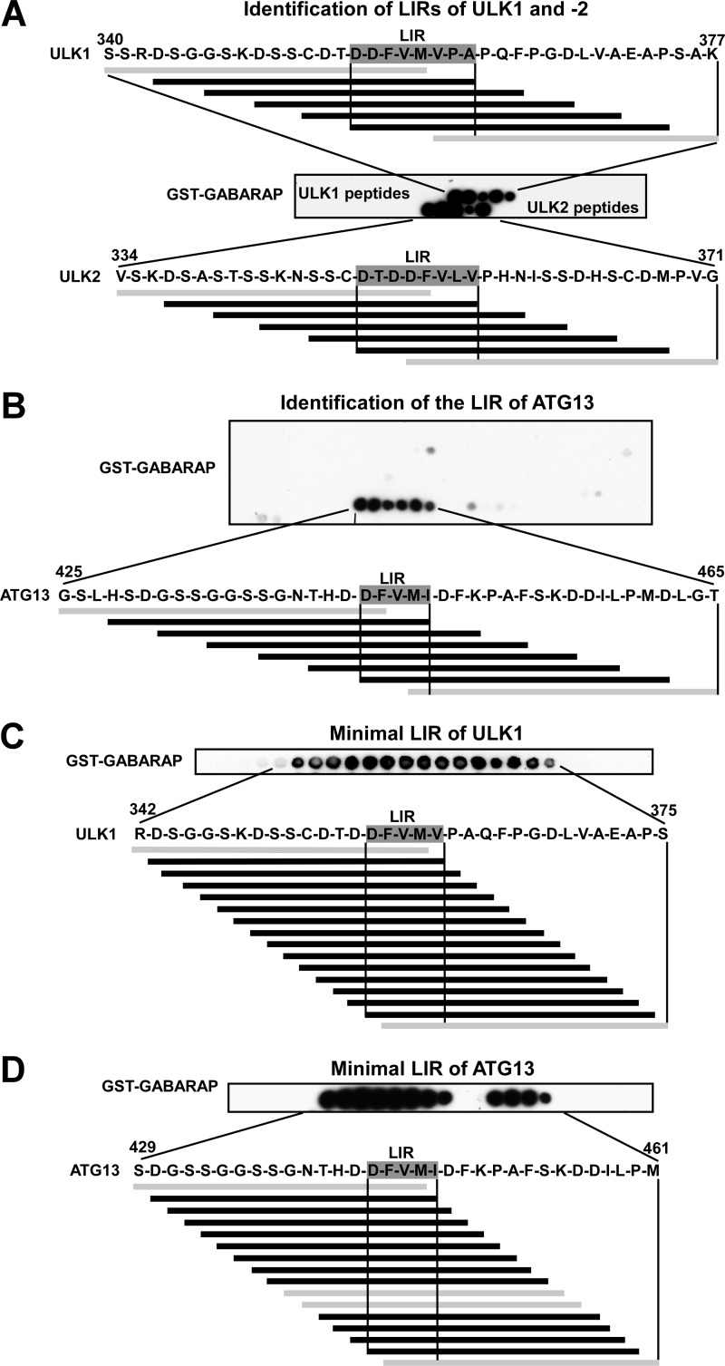
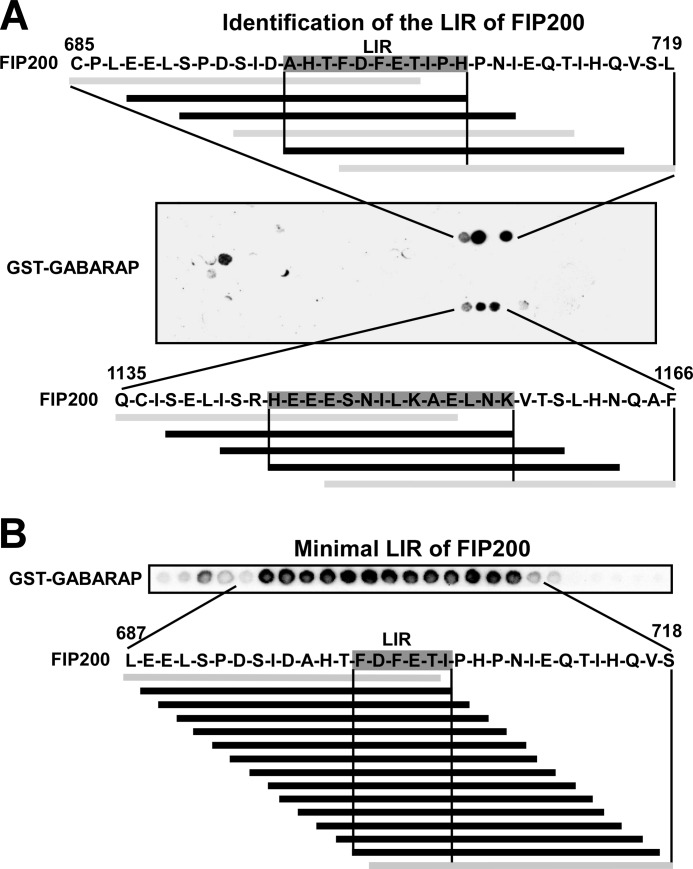
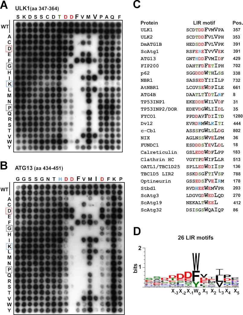
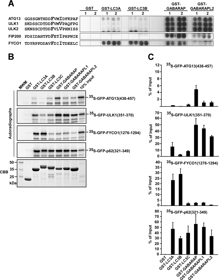
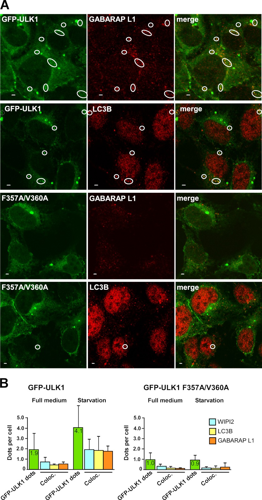
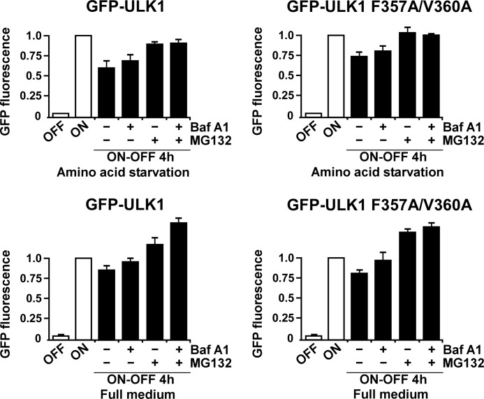
Autophagy is a lysosome-dependent degradation system conserved among eukaryotes. The mammalian Atg1 homologues, Unc-51 like kinase (ULK) 1 and 2, are multifunctional proteins with roles in autophagy, neurite outgrowth, and vesicle transport. The mammalian ULK complex involved in autophagy consists of ULK1, ULK2, ATG13, FIP200, and ATG101. We have used pulldown and peptide array overlay assays to study interactions between the ULK complex and six different ATG8 family proteins. Strikingly, in addition to ULK1 and ULK2, ATG13 and FIP200 interacted with human ATG8 proteins, all with strong preference for the GABARAP subfamily. Similarly, yeast and Drosophila Atg1 interacted with their respective Atg8 proteins, demonstrating the evolutionary conservation of the interaction. Use of peptide arrays allowed precise mapping of the functional LIR motifs, and two-dimensional scans of the ULK1 and ATG13 LIR motifs revealed which substitutions that were tolerated. This information, combined with an analysis of known LIR motifs, provides us with a clearer picture of sequence requirements for LIR motifs. In addition to the known requirements of the aromatic and hydrophobic residues of the core motif, we found the interactions to depend strongly on acidic residues surrounding the central core LIR motifs. A preference for either a hydrophobic residue or an acidic residue following the aromatic residue in the LIR motif is also evident. Importantly, the LIR motif is required for starvation-induced association of ULK1 with autophagosomes. Our data suggest that ATG8 proteins act as scaffolds for assembly of the ULK complex at the phagophore.


Figures

FIGURE 1.
FIGURE 1.
ULK1, ATG13, and FIP200 interact with human ATG8 family proteins. A, GST pulldown assays showing that ULK1, ATG13, and FIP200 all preferentially bind to the GABARAP subfamily proteins. The indicated proteins were in vitro translated in the presence of [35S]methionine and tested in GST pulldown assays for interaction with the indicated GST-fused recombinant proteins. Autoradiograph (AR, top panels) and Coomassie stained immobilized GST or GST fusion proteins (CBB, bottom panel) are shown. B, co-precipitation of FLAG-ULK1 with GFP-tagged ATG8 homologues from HEK293 cell extracts. Cells were co-transfected with the indicated constructs, and GFP or GFP fusion proteins were immunoprecipitated with GFP antibodies. Precipitated GFP-tagged proteins (middle panel), co-precipitated ULK1 (upper panel), and inputs of ULK1 (lower panel) were analyzed by Western blotting using the indicated antibodies. C, MBP pulldown assay showing direct interaction between ATG13 and GABARAP. All proteins were produced in E. coli and GST and GST-ATG13 were eluted from the beads and tested for interaction with immobilized MBP or MBP-GABARAP. The proteins were detected by Western blotting (WB) using the indicated antibodies. Anti-GST staining (upper panel) and anti-MBP staining (lower panel) are shown.
FIGURE 2.
FIGURE 2.
Identification of a GABARAP interacting LIR motif in ULK1. A, schematic representation of the domain organization of human ULK1. The ULK1 fragments used to map the GABARAP-interacting region are indicated. (+) and (−) indicate the presence or absence of GABARAP binding, respectively. B, GST pulldown assay showing a direct interaction between GABARAP and a deletion fragment of ULK1 encompassing amino acids 351–400. All proteins were produced in E. coli, and MBP or MBP-GABARAP was eluted from the beads and tested for interaction with immobilized GST or GST-ULK1(351–400). Precipitated GST or GST-ULK1(351–400) and co-precipitated MBP or MBP-GABARAP were analyzed by Western blotting using the indicated antibodies. C, GST pulldown assay demonstrating loss of binding of the F357A and F357A/V360A point mutations of ULK1(351–400) to in vitro translated GFP-GABARAP. D, full-length ULK1 with the F357A and F357A/V360A point mutations that lost binding affinity for GABARAP. The GST pulldown assay was carried out using in vitro translated full-length ULK1 constructs and bacterially expressed and immobilized GST or GST-GABARAP. E, the F357A/V360A point mutations prevent co-precipitation of ULK1 with GFP-GABARAP from HEK293 cell extracts. Cells were co-transfected with the indicated constructs, and GFP or GFP-GABARAP were immunoprecipitated (IP) using GFP antibodies. The inputs of wild type or mutated 3xFLAG-ULK1 (lower panel), precipitated GFP or GFP-GABARAP (middle panel), and co-precipitated FLAG-ULK1 (upper panel) were analyzed by Western blotting (WB) using the indicated antibodies. F, yeast Atg1 interacts with yeast Atg8. In vitro translated yAtg1 was tested in a GST pulldown assay for interaction with GST or GST-yAtg8. Autoradiograph (AR) and Coomassie staining of immobilized GST or GST-yAtg8 (CBB) are shown. G, Drosophila Atg1B interacts with Drosophila Atg8A via a LIR motif. In vitro translated DmAtg1B, wild-type, and LIR mutant, were tested in a GST pulldown assay for interaction with GST or GST-DmAtg8A. Autoradiograph and Coomassie staining of immobilized GST or GST-DmAtg8A (CBB) are shown.
FIGURE 3.
FIGURE 3.
Identification of a GABARAP interacting LIR motif in ATG13. A, schematic representation of full-length ATG13 and its deletion constructs used to map the GABARAP-interacting region. (+) and (−) indicate the presence or absence of GABARAP binding, respectively. B, amino acids 438–457 of ATG13 are needed for its binding to GABARAP. The indicated GFP-tagged ATG13 constructs were in vitro translated in the presence of [35S]methionine and tested in GST pulldown assays for binding to recombinant GST or GST-GABARAP. C, the F444A and F444A/I447A point mutations strongly reduce the affinity of ATG13 for GABARAP. The GST pulldown assays were conducted using in vitro translated GFP-ATG13 constructs and bacterially expressed GST or GST-GABARAP. For B and C, autoradiograph (AR, upper panels) and Coomassie staining of immobilized proteins (CBB, lower panels) are shown. D, GFP fusions of wild type or the indicated point or deletion mutants of ATG13 were co-transfected with FLAG-GABARAP in HEK293 cells. FLAG-GABARAP was immunoprecipitated (IP) with FLAG antibodies. The inputs of the various GFP-ATG13 constructs (upper panel), precipitated FLAG-GABARAP (middle panel), and co-precipitated GFP-ATG13 constructs (upper panel) were analyzed by Western blotting (WB) using the indicated antibodies.
FIGURE 4.
FIGURE 4.
Identification of the LIR motifs in ULK1, ULK2, and ATG13 using peptide arrays. A and B, identification of GABARAP-interacting LIR motifs in ULK1, ULK2 (A), and ATG13 (B). Arrays of 20-mer peptides covering the interacting region of ULK1, ULK2, and full-length ATG13 were synthesized on cellulose membranes. Each peptide was shifted three amino acids relative to the previous peptide. C and D, minimal LIR motifs of ULK1 (C) and ATG13 (D). Arrays of 18-mer peptides with single amino acid increments, covering a 42-amino acid region with the LIR motif in the center, were synthesized on cellulose membranes. The arrays were probed with 1 μg/ml of GST or GST-GABARAP for 2 h, and binding to GST (not shown) or GST-GABARAP was detected with anti-GST antibodies. Shown are sequence data for the GABARAP interacting peptides. Interacting peptides are in black, whereas noninteracting peptides are in gray.
FIGURE 5.
FIGURE 5.
Identification of LIR motifs in FIP200 using peptide arrays. A, an array of 20-mer peptides covering the full-length of FIP200 was performed as described in the legend to Fig. 4. B, an array of 18-mer peptides with single amino acid increments was performed as described for ULK1 and ATG13 (see legend to Fig. 4).
FIGURE 6.
FIGURE 6.
Characterization of the LIR motifs of ULK1 and ATG13. A, two-dimensional peptide arrays scans analyzing the effect of single amino acid substitutions at all positions of the indicated 18-mer peptides from ULK1 (amino acids 347–364); B, or ATG13 (amino acids 434–451). Each position of the 18-mer peptides was replaced with all 20 amino acids and GST-GABARAP was detected with anti-GST antibodies. C, alignment of published LIR motifs verified by binding assays. The amino acid position (Pos.) of the conserved aromatic residue in the LIR motifs is indicated for each protein. All sequences are from the human proteins except for the four yeast proteins Atg1, -3, -19, and -32, plant NBR1, and Drosophila ATG1B. The sequences and references are the following: p62 (16), NBR1 (19), AtNBR1 (43), ATG4B (44), TP53INP1 and TP53INP2/DOR (45), FYCO1 (21), Dvl2 (46), NIX (47), FUNDC1 (48), Calreticulin (49), clathrin heavy chain (50), OATL1/TBC1D25 (22), TBC1D5 (23), Optineurin (20), Stbd1 (51), yeast Atg3 (52), yeast Atg19 (35) and Atg32 (53). D, sequence logo of the LIR motifs aligned in C made using WebLogo 3 (54).
FIGURE 7.
FIGURE 7.
Specificity of isolated LIR motifs analyzed by use of peptide arrays and GST pulldown assays. A, the indicated 20-mer peptides from ATG13, ULK1, ULK2, FIP200, and FYCO1 were synthesized on cellulose membranes in parallels (indicated as 1 and 2). The arrays were probed with 1 μg/ml of GST, GST-LC3A, GST-LC3B, GST-GABARAP, or GST-GABARAPL2 for 2 h, as indicated. Binding was detected with anti-GST antibodies. B, GST pulldown assays testing binding of isolated LIR motifs to GST or various ATG8 homologues fused to GST. The indicated fragments of ULK1, ATG13, FYCO1, and p62 were in vitro translated as GFP fusion proteins and tested in GST pulldown assays for binding to GST or the indicated GST fusion proteins. Autoradiographs (upper panels) and Coomassie staining of the immobilized GST or GST fusion proteins used in the assay (CBB, lower panel) are shown. C, quantitative representations of the experiments depicted in B. Quantifications of the mean % binding with S.D. from three independent experiments are shown.
FIGURE 8.
FIGURE 8.
The LIR motif in ULK1 is needed for amino acid starvation-induced co-localization of ULK1 with endogenous LC3B, GABARAP L1, and WIPI2 on autophagosomes. A, co-localization of wild type GFP-ULK1 with endogenous GABARAP L1 and LC3B in punctuated structures in HEK293 Flp-In T-Rex cells. Expression of GFP-ULK1 (wild type or mutated) was induced for 24 h with tetracycline. Cells were then incubated for 1 h in starvation medium without amino acids, fixed, and stained with anti-LC3B or anti-GABARAP L1 antibodies as indicated. Representative images are shown, the circles indicate dots with co-localization. Bars, 2 μm. B, quantitative analysis of intracellular punctuated structures in HEK293 Flp-In T-Rex cells expressing GFP-ULK1 (wild type or LIR-mutated). Expression of GFP-ULK1 was induced as in A, and cells were then incubated for 1 h in full medium or starvation medium. Cells were fixed and stained with anti-WIPI2, anti-LC3B, or anti-GABARAP L1 antibodies before being analyzed by confocal microscopy. Each bar indicating the number of GFP-ULK1 positive dots per cell (green color) is based on an analysis of 490–570 cells, whereas 100–180 cells were analyzed for co-localization with endogenous WIPI2, LC3B, and GABARAP L1. Under starvation conditions, the average co-localization of WIPI2, LC3B, and GABARAP L1 with wild type ULK1 dots was 47, 45, or 43%, respectively, and for the LIR mutant ULK1 the corresponding numbers were 25, 15, and 22%.
FIGURE 9.
FIGURE 9.
Only a minor fraction of GFP-ULK1 is degraded by autophagy. HEK293 Flp-In T-Rex cells stably expressing GFP-ULK1 (wild type or LIR-mutated) from an inducible promoter were grown overnight in full medium supplemented with tetracycline, resulting in accumulation of GFP-ULK1 (ON). Tetracycline was removed (promoter shut-off, ON-OFF), and the cells were incubated for 4 h in full medium or starvation medium, both supplemented or not with bafilomycin A1 and/or MG132 as indicated. Degradation of GFP-ULK1 (wild type or LIR-mutated) was measured by flow cytometry, using loss of GFP fluorescence as readout. For each condition, median GFP fluorescence was determined from a population of at least 10,000 cells. Bars show the average GFP fluorescence from three independent experiments, relative to the ON condition.

References

    1. Tomoda T., Bhatt R. S., Kuroyanagi H., Shirasawa T., Hatten M. E. (1999) A mouse serine/threonine kinase homologous to C. elegans UNC51 functions in parallel fiber formation of cerebellar granule neurons. Neuron 24, 833–846 - PubMed
    1. Young A. R., Chan E. Y., Hu X. W., Köchl R., Crawshaw S. G., High S., Hailey D. W., Lippincott-Schwartz J., Tooze S. A. (2006) Starvation and ULK1-dependent cycling of mammalian Atg9 between the TGN and endosomes. J. Cell Sci. 119, 3888–3900 - PubMed
    1. Chan E. Y. (2012) Regulation and function of uncoordinated-51 like kinase proteins. Antioxidants Redox Signal. 17, 775–785 - PubMed
    1. Weidberg H., Shvets E., Elazar Z. (2011) Biogenesis and cargo selectivity of autophagosomes. Annu. Rev. Biochem. 80, 125–156 - PubMed
    1. Nakatogawa H., Suzuki K., Kamada Y., Ohsumi Y. (2009) Dynamics and diversity in autophagy mechanisms. Lessons from yeast. Nat. Rev. Mol. Cell Biol. 10, 458–467 - PubMed

Publication types

MeSH terms

Substances