Review
. 2013:53:187-210.
doi: 10.1146/annurev-pharmtox-011112-140204. Epub 2012 Oct 8.

Creating order from chaos: cellular regulation by kinase anchoring

Affiliations
Review

Creating order from chaos: cellular regulation by kinase anchoring

John D Scott et al. Annu Rev Pharmacol Toxicol. 2013.

Abstract

Second messenger responses rely on where and when the enzymes that propagate these signals become active. Spatial and temporal organization of certain signaling enzymes is controlled in part by A-kinase anchoring proteins (AKAPs). This family of regulatory proteins was originally classified on the basis of their ability to compartmentalize the cyclic adenosine monophosphate (cAMP)-dependent protein kinase (also known as protein kinase A, or PKA). However, it is now recognized that AKAPs position G protein-coupled receptors, adenylyl cyclases, G proteins, and their effector proteins in relation to protein kinases and signal termination enzymes such as phosphodiesterases and protein phosphatases. This arrangement offers a simple and efficient means to limit the scope, duration, and directional flow of information to sites deep within the cell. This review focuses on the pros and cons of reagents that define the biological role of kinase anchoring inside cells and discusses recent advances in our understanding of anchored second messenger signaling in the cardiovascular and immune systems.


Figures

Figure 1
Figure 1
The evolution of protein kinase A (PKA) anchoring models. (a) Schematic diagram (circa 1990) (18) presenting a model of the anchored RII complex. (b) Contemporary model based on the crystal structure of RIIα1–45 dimer in complex with the A-kinase anchoring protein (AKAP)–in silico binding helix (36, 48).
Figure 2
Figure 2
Combinatorial assembly of cardiac A-kinase anchoring protein (AKAP) signaling complexes. The common name (first column) and alternate name(s) (second column) of each anchoring protein are indicated. Schematic representations of cardiac AKAPs highlight enzyme-binding sites and functional domains (third column). Binding partners are indicated (fourth column). Abbreviations: AC, adenylyl cyclase; AMP, adenosine monophosphate; βAR, β-adrenergic receptor; DH, Dbl homology domain; ERK, extracellular signal-regulated kinase; HIF-1α, hypoxia-inducible factor 1α; IP3-R, inositol 3,4,5-phosphate receptor; KCNQ, KvLQT potassium channel subunit; KSR-1, kinase suppressor of Ras1; Lfc, Lbc first cousin; MEK, mitogen-activated protein kinase kinase; MT, mitochondrial transit peptide; NCX1, sodium-calcium exchanger 1; NFATc, nuclear factor of activated T cells; NMDA-R, N-methyl-d-aspartate receptor; PDE, phosphodiesterase; PDK1, phosphoinositide-dependent kinase-1; PH, pleckstrin homology domain; PKA/C/D/N, protein kinase A/C/D/N; PP1/2A/2B, protein phosphatase 1/2A/2B; PTPD1, protein tyrosine phosphatase D1; RGS, regulator of G protein signaling; RSK, ribosomal S6 kinase; RyR2, ryanodine receptor 2; SAP97, synapse-associated protein 97; Siah2, seven in absentia homolog 2; Trek-1, two pore-domain potassium channel; TrpV1, transient receptor potential cation channel V1; VHL, von Hippel–Lindau protein.
Figure 3
Figure 3
The A-kinase anchoring protein (AKAP) terrain of cardiomyocytes. The subcellular distribution of AKAPs in cardiomyocytes is depicted. Subcellular organelles, anchoring proteins, and key effector proteins are indicated. Abbreviations: KCNQ, IKs potassium channel subunit; KSR, kinase suppressor of Ras; PLN, phospholamban; RyR, ryanodine receptor; SERCA, sarcoplasmic/endoplasmic reticulum Ca2+ pump; SKIP, sphingosine kinase interacting protein.
Figure 4
Figure 4
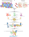
The immunoregulatory role of anchored cAMP signaling in T cells. (a) Tumor-infiltrating lymphocytes are inhibited by peripherally induced Tregs (orange cells) exposed to chronic antigenic stimulation. Persistent infections, such as HIV, lead to chronic inflammation and immunosuppression, both of which involve production of PGE2 and other inflammatory mediators. (b) Generation of peripherally induced or adaptive Tregs. These cells express COX-2, produce PGE2, and stimulate FOXP3 expression. In contrast, naturally occurring Tregs can transfer cAMP to responder T cells through gap junctions. Pericellular accumulation of adenosine also elicits immunosuppressive responses through a pathway whereby CD39 and CD73 ectoenzymes metabolize ATP to generate adenosine. T cell inhibition and PGE2-responsive induction of FOXP3 can occur as a result of the secretion of PGE2 by LPS-activated monocytes. (c) cAMP inhibits TCR-mediated immunoregulatory functions at membranes. This occurs in lipid rafts through a receptor–G protein–AC–cAMP–PKA type I–Csk pathway that acts on the Src family tyrosine kinase Lck. (d) Ezrin links transmembrane receptors such as CD43 to the actin cytoskeleton via its N-terminal FERM domain and to F-actin via its C terminus. Ezrin functions as an AKAP, bringing type I PKA in proximity to its substrate Csk via a supramolecular signaling complex consisting of PKA, ezrin, EBP50, Cbp/PAG, and Csk. Abbreviations: AC, adenylyl cyclase; AKAP, A-kinase anchoring protein; cAMP, cyclic adenosine monophosphate; COX-2, cyclooxygenase 2; Csk, C-terminal Src kinase; EPB50, ERM binding protein 50; EP-R, receptor for E series of prostaglandins; FERM, band 4.1 protein and ezrin, radixin, and moesin (ERM) protein domain; Lck, lymphocyte-specific protein tyrosine kinase; LPS, lipopolysaccharide; PAG, phosphoprotein associated with glycosphingolipid-enriched microdomains; PGE2, prostaglandin E2; PKA, protein kinase A; TCR, T cell receptor; Treg, regulatory T cell.

References

    1. Rall TW, Sutherland EW, Berthet J. The relationships of epinephrine and glucagon to liver phosphorylase. J. Biol. Chem. 1957;224:463–475. - PubMed
    1. Sutherland EW. Studies on the mechanism of hormone action. Science. 1972;171:401–408. - PubMed
    1. Fischer EH, Krebs EG. Conversion of phosphorylase b to phosphorylase a in muscle extracts. J. Biol. Chem. 1955;216:121–132. - PubMed
    1. Corbin JD, Soderling TR, Park CR. Regulation of adenosine 3′,5′-monophosphate-dependent protein kinase. J. Biol. Chem. 1973;248:1813–1821. - PubMed
    1. Potter RL, Stafford PH, Taylor S. Regulatory subunit of cyclic AMP-dependent protein kinase I from porcine skeletal muscle: purification and proteolysis. Arch. Biochem. Biophys. 1978;190:174–180. - PubMed

Publication types