. 2012 Oct 8:11:337.
doi: 10.1186/1475-2875-11-337.

Neutral lipids associated with haemozoin mediate efficient and rapid β-haematin formation at physiological pH, temperature and ionic composition

Affiliations

Neutral lipids associated with haemozoin mediate efficient and rapid β-haematin formation at physiological pH, temperature and ionic composition

Melvin A Ambele et al. Malar J. .

Abstract

Background: The malaria parasite disposes of host-derived ferrihaem (iron(III)protoporphyrin IX, Fe(III)PPIX) by conversion to crystalline haemozoin in close association with neutral lipids. Lipids mediate synthetic haemozoin (β-haematin) formation very efficiently. However, the effect on reaction rates of concentrations of lipid, Fe(III)PPIX and physiologically relevant ions and biomolecules are unknown.

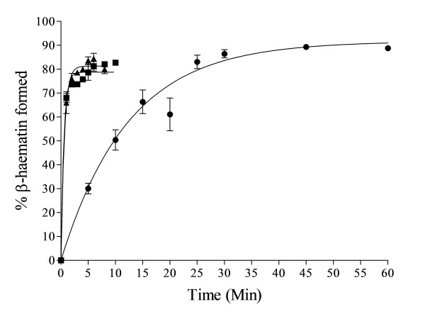
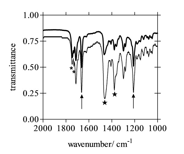
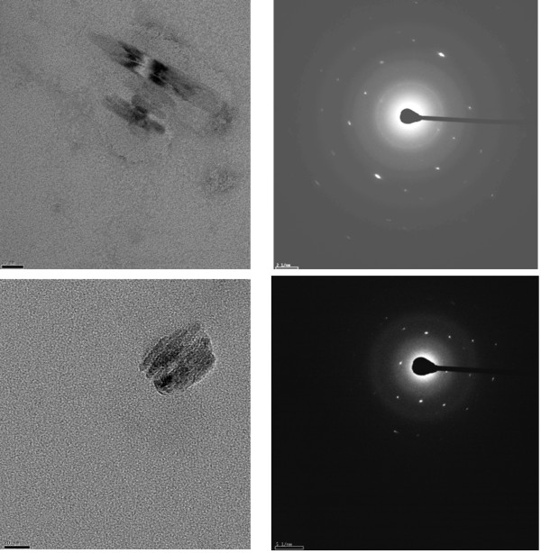
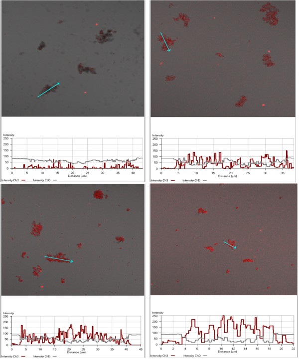
Methods: Lipid emulsions containing Fe(III)PPIX were prepared in aqueous medium (pH 4.8, 37°C) to mediate β-haematin formation. The reaction was quenched at various times and free Fe(III)PPIX measured colorimetrically as a pyridine complex and the kinetics and yields analysed. Products were also characterized by FTIR, TEM and electron diffraction. Autofluorescence was also used to monitor β-haematin formation by confocal microscopy.

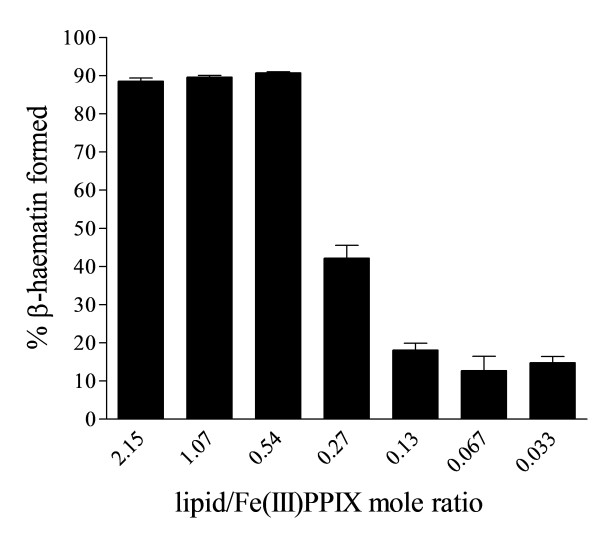
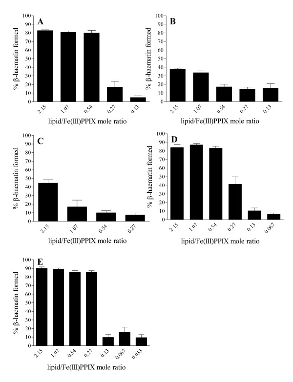
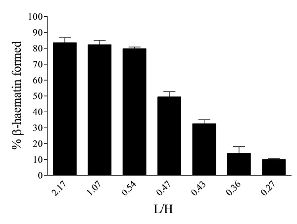
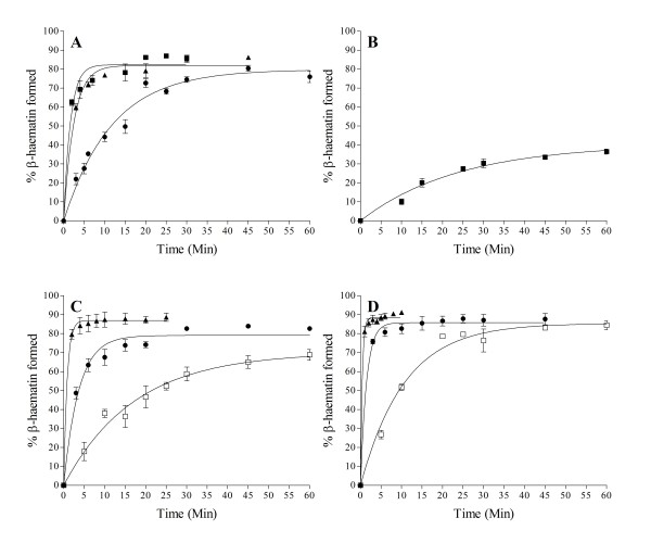
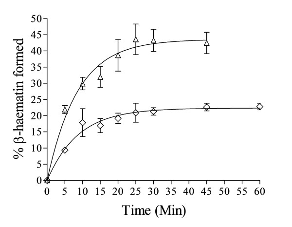
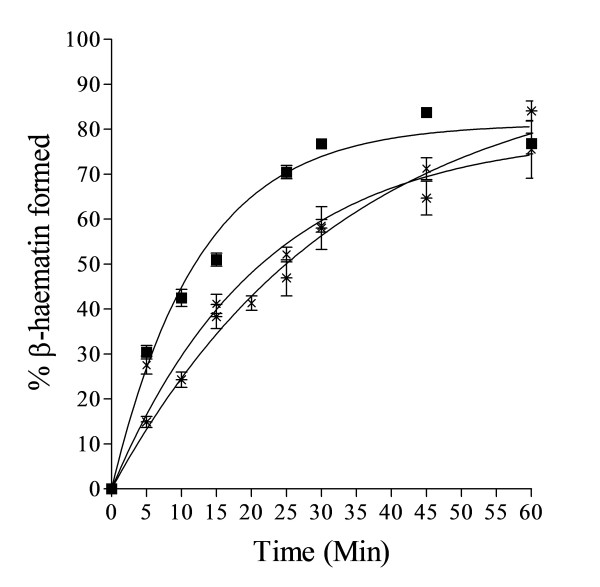
Results: At fixed Fe(III)PPIX concentration, β-haematin yields remained constant with decreasing lipid concentration until a cut-off ratio was reached whereupon efficiency decreased dramatically. For the haemozoin-associated neutral lipid blend (NLB) and monopalmitoylglycerol (MPG), this occurred below a lipid/Fe(III)PPIX (L/H) ratio of 0.54. Rate constants were found to increase with L/H ratio above the cut-off. At 16 μM MPG, Fe(III)PPIX concentration could be raised until the L/H ratio reached the same ratio before a sudden decline in yield was observed. MPG-mediated β-haematin formation was relatively insensitive to biologically relevant cations (Na(+), K(+), Mg(2+), Ca(2+)), or anions (H(2)PO(4)(-), HCO(3)(-), ATP, 2,3-diphosphoglycerate, glutathione). Confocal microscopy demonstrated β-haematin formation occurs in association with the lipid particles.

Conclusions: Kinetics of β-haematin formation have shown that haemozoin-associated neutral lipids alone are capable of mediating β-haematin formation at adequate rates under physiologically realistic conditions of ion concentrations to account for haemozoin formation.


Figures

Figure 1
Figure 1
β-haematin formation with NLB gave a high yield at higher lipid/Fe(III)PPIX mol ratios. The high yield stays relatively constant from 2.15 L/H (64.3 μM total lipid) to 0.54 L/H after which the yield suddenly drops to a value of about 10% below 0.13 L/H. Citric buffer (50 mM, pH 4.8), 37°C, 30 min incubation. Constant total Fe(III)PPIX concentration of 30 μM. Error bars represent SEM (L/H = 2.15 and 1.07, n = 6; L/H = 0.54 and 0.27, n = 8; L/H = 0.13 and 0.067, n = 4; L/H = 0.033, n = 2).
Figure 2
Figure 2
Yields of β-haematin formed in the presence of individual constituent lipids of NLB vary with the identity of the lipid. High yields of about 60–80% were obtained with MPG at L/H ratios from 2.15 to 0.54 with a sharp decrease in yield to less than 10% below 0.27 L/H (A). MSG and DPG at 2.15 L/H gave lower yields of about 40%, which decrease sharply to about 10–20% at L/H ratios of 0.54 and 1.07 respectively (B and C). DLG gave high yields around 80–90% at higher mol ratios (2.15–0.54) which decreases sharply to about 40% at 0.27 L/H mol ratio and drops further to about 10% and lower at 0.13 L/H (D). DOG appears to be the most efficient lipid with a high yield of almost 90% between 2.15 L/H and 0.27 L/H before decreasing sharply between 0.27 and 0.13 L/H (E). Citric buffer (50 mM, pH 4.8), 37°C, incubated for 30 min. Constant total Fe(III)PPIX concentration of 30 μM. Error bars represent SEM (A, C – E, n = 3; B, n = 4).
Figure 3
Figure 3
Yields of about 80% β-haematin were obtained with 7.4 to 30 μM Fe(III)PPIX and 16.1 μM MPG (2.17 to 0.54 L/H ratio). The yield started to decline from 34 to 37 μM Fe(III)PPIX (0.47–0.43 L/H) to about 30–50% and levels out to a lower yield of about 10% at 45 μM (0.36 L/H) and below. Incubation time was 30 min, citric buffer (50 mM, pH 4.8), 37°C. Error bars represent SEM (n = 3).
Figure 4
Figure 4
Fast kinetics were observed with NLB at higher mol ratios of 2.15 (■) and 1.07 L/H (▲). These reactions were complete within 10 min with yields of about 80% and conform to first-order kinetics. The rate constants were 1.9 ± 0.2 and 1.6 ± 0.1 min−1 respectively. Relatively slower kinetics were obtained at 0.54 L/H mol ratio (●), with k = 0.082 ± 0.006 min−1. Citric buffer (50 mM, pH 4.8), 37°C. Constant total Fe(III)PPIX concentration of 30 μM. Error bars represent SEM (n = 4).
Figure 5
Figure 5
MPG exhibited fast kinetics at 2.15 L/H (■) and 1.07 L/H mol ratio (▲) withk= 0.61 ± 0.07 and 0.40 ± 0.03 min−1respectively, while at 0.54 L/H mol ratio (●), the reaction is much slower withk= 0.085 ± 0.006 min−1, similar to that of NLB (A). The reaction with MSG at the highest lipid concentration (2.15 L/H) is slower than NLB and MPG at 0.54 L/H mol ratio, with k = 0.044 ± 0.007 min−1 (B). Kinetics with DLG at 1.07 L/H (▲) and 0.54 L/H (●) appear to be faster than that of NLB at same mol ratio with k = 1.2 ± 0.2 and 0.27 ± 0.02 min−1 respectively, while at 0.27 L/H mol ratio (□) the reaction was slower with k = 0.058 ± 0.009 min−1 (C). DOG mediated exceptionally fast kinetics at 1.07 L/H (▲) and 0.54 L/H mol ratio (●) with k = 2.4 ± 0.2 and 0.69 ± 0.08 min−1 respectively, while the kinetics at 0.27 L/H (□) were slower with k = 0.094 ± 0.008 min−1, comparable to NLB and MPG at the mol ratio of 0.54 L/H (D). Citric buffer (50 mM, pH 4.8), 37°C. Constant total Fe(III)PPIX concentration of 30 μM. Error bars represent SEM (n = 4, except D for which n = 3).
Figure 6
Figure 6
Kinetics of β-haematin formation at ratios of 0.46 L/H (Δ) withk= 0.12 ± 0.02 and 0.36 L/H () withk= 0.11 ± 0.02 min−1respectively, are comparable to that at 0.54 L/H (wherek= 0.085 ± 0.006 min−1). Citric buffer (50 mM, pH 4.8), 37°C, MPG. Total Fe(III)PPIX concentrations were 35 and 45 μM respectively. Error bars represent SEM (n = 4).
Figure 7
Figure 7
FTIR spectra of dried product obtained as Nujol mulls. Products were those formed using NLB (thick line) or MPG (thin line) and show characteristic β-haematin peaks at 1662 cm-1 and 1210 cm-1 (arrows). Both products gave an identical infrared spectrum. Nujol peaks (★) and contaminating lipid peaks (*) are marked. Reaction conditions: 2.15 L/H ratio (64.3 μM total lipid), citric buffer (50 mM, pH 4.8), 37°C, 10 min incubation.
Figure 8
Figure 8
TEM (left panels) and electron diffraction (right panels) of β-haematin obtained from an MPG mediated process (L/H = 0.54). The particles are very small (scale bar = 20 nm), but show well defined diffraction spots conclusively confirming that the product is crystalline.
Figure 9
Figure 9
Confocal laser microscopy images of MPG lipid aggregates in extracts taken at various time points during lipid mediated β-haematin formation. Reaction times were 10 min (top left), 20 min (top right), 30 min (bottom left) and 60 min (bottom right). The dark non-fluorescent areas are unconverted Fe(III)PPIX, either precipitated, or lipid associated. Fluorescence profiles taken along cross-sections indicated by an arrow in each image are given below the images in red. The grey trace (ChD line) represents the transmitted light channel). Steady formation of β-haematin crystals is indicated by increasing fluorescence and is in qualitative agreement with kinetic traces, indicating about 60 min required to reach completion. Excitation was at 516 nm, emission in the range 575–630 nm. Reaction mediated by MPG, 0.54 L/H ratio, citric buffer (50 mM, pH 4.8), 37°C. Confocal microscopy was performed on wet samples. Acquisition conditions: λex = 561 nm; λem = 575–630 nm; pinhole diameter 444 μm; output power 20% transmission; scan zoom 1.0; objective LD C-Apochromat 40×/1.1 W. No z-stacking.
Figure 10
Figure 10
Effect of buffer on the kinetics of β-haematin formation. Rate constants obtained with citric (■), acetic (×) and MES buffers (*) were 0.080 ± 0.005, 0.047 ± 0.006 and 0.030 ± 0.004 min-1 respectively. Reaction mediated by MPG (0.54 L/H), pH 4.8 (50 mM buffer), 37°C. Constant total Fe(III)PPIX concentration of 30 μM. Error bars represent SEM (n = 4).

References

    1. Noland GS, Briones N, Sullivan DJ. The shape and size of hemozoin crystals distinguishes diverse Plasmodium species. Mol Biochem Parasitol. 2003;130:91–99. doi: 10.1016/S0166-6851(03)00163-4. - DOI - PubMed
    1. Oliveira M, d'Avila JCP, Torres CR, Oliveira PL, Tempone AJ, Rumjanek FD, Braga CMS, Silva JR, Dansa-Petretski M, Oliveira MA, De Souza W, Ferreira ST. Haemozoin in Schistosoma mansoni. Mol Biochem Parasitol. 2000;111:217–221. doi: 10.1016/S0166-6851(00)00299-1. - DOI - PubMed
    1. Oliveira MF, Silva JR, Dansa-Petretski M, de Souza W, Lins U, Braga CMS, Masuda H, Oliveira PL. Haem detoxification by an insect. Nature. 1999;400:517–518. doi: 10.1038/22910. - DOI - PubMed
    1. Tekwani BL, Walker LA. Targeting the hemozoin synthesis pathway for new antimalarial drug discovery: technologies for in vitro β-hematin formation assay. Combinatorial Chem High Througput Scr. 2005;8:61–77. - PubMed
    1. Hänscheid T, Egan TJ, Grobusch MP. Haemozoin: from melatonin pigment to drug target, diagnostic tool, and immune modulator. Lancet Infect Dis. 2007;7:675–685. doi: 10.1016/S1473-3099(07)70238-4. - DOI - PubMed

Publication types

LinkOut - more resources