. 2013 Jan;1827(1):38-49.
doi: 10.1016/j.bbabio.2012.09.016. Epub 2012 Oct 5.

Sirtuin-4 modulates sensitivity to induction of the mitochondrial permeability transition pore

Affiliations

Sirtuin-4 modulates sensitivity to induction of the mitochondrial permeability transition pore

Manish Verma et al. Biochim Biophys Acta. 2013 Jan.

Retraction in

Abstract

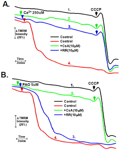
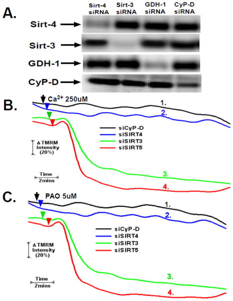
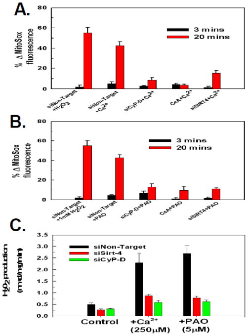
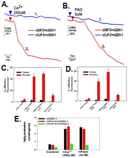
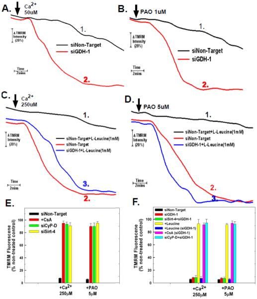
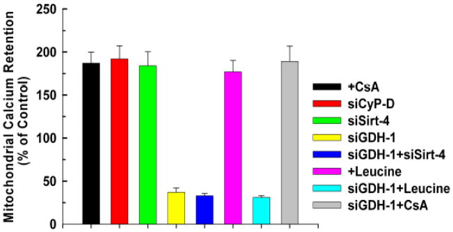
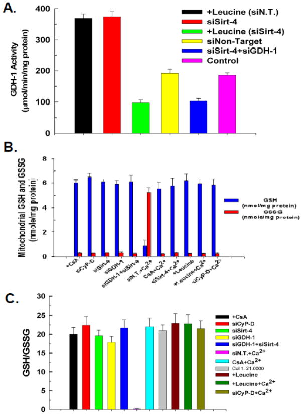
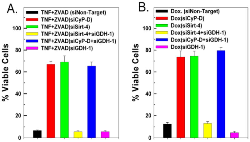
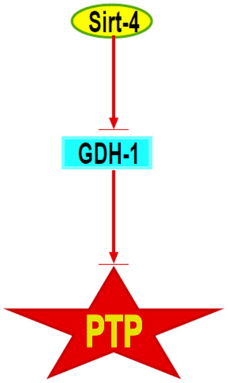
The sustained opening of the mitochondrial permeability transition pore (PTP) is a decisive event in the onset of irreversible cell injury. The PTP is modulated by numerous exogenous and endogenous effectors, including mitochondrial membrane potential, ions and metabolites. Mitochondrial sirtuins have recently emerged as pivotal mediators of mitochondrial metabolism. In the present study, we demonstrate that sirt-4 modulates sensitivity to PTP onset induced by calcium and the oxidative cross linking reagent phenylarsine oxide, and PTP dependent cytotoxicity brought about by TNF or doxorubicin. Moreover, the ability of sirt-4 to modulate onset of the PTP is dependent on the expression of glutamate dehydrogenase-1.


Figures

Figure 1
Figure 1. Measurement and control of PTP induction in permeabilized cells
A and B. HeLa cells were loaded with 200nM TMRM in DMEM for 30 minutes. The cells were then washed twice with PBS and incubated further for 5 minutes in respiratory buffer containing 20nM of TMRM. Digitonin at 2.5μg/ml was then added to permeabilize the plasma membrane and the cells were mounted on a heated stage kept at 37°C. Where indicated, cells were pretreated with 10μM of cyclosporin A or ruthenium red for five minutes. Phenylarsine oxide (PAO) or Ca2+ were added at final concentrations of 5μM and 250μM, respectively. TMRM fluorescence was monitored over a 20 minute time course. 5μM of CCCP was added at the 18 minute time point. The result is the average of three independent experiments.
Figure 2
Figure 2. Sirt-4 controls sensitivity to PTP induction by Ca2+ and PAO
A. HeLa cells were plated and then transfected with siRNAs targeting sirt-4, sirt-3, sirt-5, cyclophilin-D or a non-targeting control. Following 48 hours, the cells were harvested. Mitochondrial extracts were prepared and separated on 12% SDS-PAGE gels followed by blotting to PVDF membranes. The western blots were developed with antibodies against sirt-4, CyP-D, GDH-1, sirt-3 or sirt-5. The results are representative of three independent experiments. B and C. HeLa cells were plated and then transfected with siRNAs targeting sirt-4, sirt-3, sirt-5 or cyclophilin-D. Following 48 hours, the cells were loaded with 200nM TMRM in DMEM for 30 minutes. The cells were then washed twice with PBS and incubated further for 5 minutes in respiratory buffer containing 20nM of TMRM. Digitonin at 2.5μg/ml was then added to permeabilize the plasma membrane and the cells were mounted on a heated stage kept at 37°C.the cells were loaded with TMRM and mounted on a heated microscopy stage. Following permeabilization with digitonin, Ca2+ or PAO were added at final concentrations of 250μM and 5μM, respectively. Time-lapse microscopy was conducted over a 20 minute time course with TMRM fluorescence intensity assessed as described in materials and methods. The result is the average of three independent experiments.
Figure 3
Figure 3. Down-regulation of sirt-4 prevents PTP dependent ROS production
A and B. HeLa cells were plated and then transfected with siRNAs targeting sirt-4, cyclophilin-D or a non-targeting control. Following 48 hours the HeLa cells were loaded with 200nM MitoSOX in DMEM for 30 minutes. The cells were then washed twice with PBS and incubated further for 5 minutes in respiratory buffer containing 20nM of MitoSOX. Digitonin at 2.5μg/ml was then added to permeabilize the plasma membrane and the cells were mounted on a heated stage kept at 37°C. Where indicated, cells were pretreated with 10μM of cyclosporin A for five minutes. Ca2+ and PAO were added at final concentrations of 250μM and 5μM, respectively. Time-lapse microscopy was conducted over a 20 minute time course with MitoSox fluorescence intensity assessed as described in materials and methods. The results are the mean of three independent experiments ± the standard deviation. C. HeLa cells were plated and then transfected with siRNAs targeting sirt-4 or CyP-D. Following 48 hours, the cells were placed in respiratory buffer and permeabilized with digitonin (2.5μg/ml). Amplex-red reagent was added at 5μM along with 10U/ml of horseradish peroxidase. Following addition of Ca2+ or PAO, fluorescence intensity was measured using a Synergy HT microplate reader with an excitation of 570nm and emission at 585nm at 37°C at 1 minute intervals. The results are the mean of three independent experiments ± the standard deviation.
Figure 4
Figure 4. Glutamate dehydrogenase-1 expression is necessary for sirt-4 depletion to protect against the PTP
A and B. HeLa cells were plated and then transfected with an siRNA targeting GDH-1 simultaneously with siRNAs targeting sirt-4 or cyclophilin-D. Following 48 hours, HeLa cells were loaded with 200nM TMRM in DMEM for 30 minutes. The cells were then washed twice with PBS and incubated further for 5 minutes in respiratory buffer containing 20nM of TMRM. Digitonin at 2.5μg/ml was then added to permeabilize the plasma membrane and the cells were mounted on a heated stage kept at 37°C. Following permeabilization with digitonin, Ca2+ and PAO were added at final concentrations of 250μM and 5μM, respectively. Time-lapse microscopy was conducted over a 20 minute time course with TMRM fluorescence intensity assessed as described in materials and methods. The result is the average of three independent experiments. C and D. HeLa cells were plated and then transfected with an siRNA targeting GDH-1 simultaneously with siRNAs targeting sirt-4 or cyclophilin-D. Following 48 hours, the cells were loaded with 200nM of MitoSOX in DMEM for 30 minutes. The cells were then washed twice with PBS and incubated further for 5 minutes in respiratory buffer containing 20nM of MitoSOX. Digitonin at 2.5μg/ml was then added to permeabilize the plasma membrane and the cells were mounted on a heated stage kept at 37°C. Following permeabilization with digitonin, Ca2+ and PAO were added at final concentrations of 250μM and 5μM, respectively. Time-lapse microscopy was conducted over a 20 minute time course with MitoSOX fluorescence intensity assessed as described in materials and methods. The results are the mean of three independent experiments ± the standard deviation. E. HeLa cells were plated and then transfected with siRNAs targeting sirt-4, CyP-D individually or in tandem with siRNA targeting GDH-1. Following 48 hours, the cells were placed in respiratory buffer and permeabilized with digitonin (2.5μg/ml). Amplex-red reagent was added at 5μM along with 10U/ml of horseradish peroxidase. Following addition of Ca2+ or PAO, fluorescence intensity was measured using Synergy HT microplate reader with an excitation of 570nm and emission at 585nm at 37°C at 1 minute intervals. The results are the mean of three independent experiments ± the standard deviation.
Figure 5
Figure 5. Glutamate Dehydrogenase Regulates PTP Sensitivity
A and B. HeLa cells were plated and then transfected with siRNAs targeting GDH-1 or a non-targeting control. Following 48 hours, HeLa cells were loaded with 200nM TMRM in DMEM for 30 minutes. The cells were then washed twice with PBS and incubated further for 5 minutes in respiratory buffer containing 20nM of TMRM. Digitonin at 2.5μg/ml was then added to permeabilize the plasma membrane and the cells were mounted on a heated stage kept at 37°C. Following permeabilization with digitonin, Ca2+ or PAO were added at final concentrations of 50μM and 2.5μM, respectively. Time-lapse microscopy was conducted over a 20 minute time course with TMRM fluorescence intensity assessed as described in materials and methods. The result is the average of three independent experiments. C and D. HeLa cells were plated and then transfected with siRNAs targeting GDH-1 or a non-targeting control. Following 48 hours, HeLa cells were loaded with 200nM TMRM in DMEM for 30 minutes. The cells were then washed twice with PBS and incubated further for 5 minutes in respiratory buffer containing 20nM of TMRM. Digitonin at 2.5μg/ml was then added to permeabilize the plasma membrane and the cells were mounted on a heated stage kept at 37°C. Following permeabilization with digitonin, leucine at a final concentration of 1mM was added. Following 5 minutes of pre-incubation with 1mM leucine, Ca2+ or PAO were added at final concentrations of 250μM and 5μM, respectively. Time-lapse microscopy was conducted over a 20 minute time course with TMRM fluorescence intensity assessed as described in materials and methods. The results are the mean of three independent experiments ± the standard deviation. E and F. HeLa cells were plated at 50,000 cells/well in 24 well plates and transfected with the indicated siRNA. After forty eight hours, the cells were loaded with 200nM TMRM in DMEM for 30 minutes. The cells were incubated further for 5 minutes in respiratory buffer containing 2.5μg/ml digitonin and 20nM of TMRM. Fluorescence intensity was measured using a Synergy HT microplate reader with an excitation of 550nm and emission at 573nm at 37°C at 1 minute intervals. After 4 minutes, either phenylarsine oxide or Ca2+ was added at the indicated concentrations, with fluorescence measured over 20 minutes. The results are presented as % of TMRM retained in non-treated cells transfected with non target siRNA at the 20 minute time point. The results are the mean of three independent experiments ± the standard deviation.
Figure 6
Figure 6. Depletion of Sirt-4 enhances mitochondrial calcium retention capacity
HeLa cells were plated at 50,000 cells/well in 24 well plates and transfected with the indicated siRNA. After forty eight hours, the cells were incubated for 5 minutes in respiratory buffer containing 2.5μg/ml digitonin and 1μM of the Ca2+ indicator, Calcium Green-5N (excitation 505 nm; emission: 535 nm; Molecular Probes). Calcium was added in pulses of 10μM and uptake measured as a decrease of Calcium Green-5N fluorescence. The results are the mean of three independent experiments ± the standard deviation.
Figure 7
Figure 7. GDH-1 activity and mitochondrial GSH
A. HeLa cells were plated at 50,000 cells/well in 24 well plates and transfected with the indicated siRNA. Forty eight hours after transfection, cells from 4 wells were harvested by trypsinization and washed twice with ice cold PBS. Mitochondria were isolated and lysates prepared. Optical density at 450 nm was measured with Synergy HT microplate reader (BioTek, Winooski, VT) at 37°C at 3 minute intervals for 1 hour. The results are expressed in percentage increase or decrease in activity compared to non-treated cells transfected with non-targeting control siRNA. The results are the mean of three independent experiments ± the standard deviation. B. HeLa cells were plated at 50,000 cells/well in 24 well plates and transfected with the indicated siRNA. Forty eight hours after transfection, cells from 4 wells were harvested by trypsinization and washed twice with ice cold PBS. Mitochondria were isolated and lysates prepared. The supernatant was used to determine the GSH content. Fluorescence was measured at excitation of 308nm and emission of 460nm using a Synergy HT microplate reader. The results are presented as percentage of GSH content compared to non-treated and non-target transfected cells. The results are the mean of three independent experiments ± the standard deviation. C. The GSH/GSSG ratio was calculated from the measured concentrations of GSH and GSSG.
Figure 8
Figure 8. Down-regulation of sirt-4 prevents TNF and doxorubicin cytotoxicity
A and B. HeLa cells were transfected with 50nM of a non-targeting control siRNA or siRNAs targeting sirt-4 or CyP-D, separately or in tandem with siRNA targeting GDH-1 or siGDH-1 alone. Following 48 hours incubation, the cells were incubated with 20ng/ml of TNFα in the presence of 20μM of ZVAD, or treated with 10μM of doxorubicin. After 24 hours, the cells were harvested and cell viability assessed utilizing propidium iodide as described in materials and methods. Values are the means of three independent experiments with the error bars indicating standard deviations.
Figure 9
Figure 9. Control of mitochondrial permeability transition pore sensitivity by Sirt-4
Proposed pathway by which modulation of sirt-4 activity can stimulate GDH-1, which in turn inhibits PTP opening and loss of cell viability.

Comment in

  • Findings of research misconduct.
    [No authors listed] [No authors listed] NIH Guide Grants Contracts (Bethesda). 2016 Jun 10:NOT-OD-16-104. NIH Guide Grants Contracts (Bethesda). 2016. PMID: 27311138 Free PMC article. No abstract available.
  • Findings of Research Misconduct.
    [No authors listed] [No authors listed] Fed Regist. 2016 May 13;81(93):29871. Fed Regist. 2016. PMID: 27737277 Free PMC article. No abstract available.

References

    1. Di Lisa F, Menabo R, Canton M, Barile M, Bernardi P. Opening of the mitochondrial permeability transition pore causes depletion of mitochondrial and cytosolic NAD+ and is a causative event in the death of myocytes in postischemic reperfusion of the heart. J Biol Chem. 2001;276:2571–2575. - PubMed
    1. Halestrap AP, Connern CP, Griffiths EJ, Kerr PM. Cyclosporin A binding to mitochondrial cyclophilin inhibits the permeability transition pore and protects hearts from ischaemia/reperfusion injury. Molecular & Cellular Biochemistry. 1997;174:167–172. - PubMed
    1. Sharov VG, Todor A, Khanal S, Imai M, Sabbah HN. Cyclosporine A attenuates mitochondrial permeability transition and improves mitochondrial respiratory function in cardiomyocytes isolated from dogs with heart failure. J Mol Cell Cardiol. 2007;42:150–158. - PMC - PubMed
    1. Baines CP, Kaiser RA, Purcell NH, Blair NS, Osinska H, Hambleton MA, Brunskill EW, Sayen MR, Gottlieb RA, Dorn GW, Robbins J, Molkentin JD. Loss of cyclophilin D reveals a critical role for mitochondrial permeability transition in cell death. Nature. 2005;434:658–662. - PubMed
    1. Nakagawa T, Shimizu S, Watanabe T, Yamaguchi O, Otsu K, Yamagata H, Inohara H, Kubo T, Tsujimoto Y. Cyclophilin D-dependent mitochondrial permeability transition regulates some necrotic but not apoptotic cell death. Nature. 2005;434:652–658. - PubMed

Publication types

MeSH terms

LinkOut - more resources