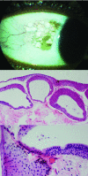
Cystic benign melanosis of the conjunctiva
- PMID: 23044615
- PMCID: PMC3467456
- DOI: 10.1097/ICO.0b013e31823d1ec4
Cystic benign melanosis of the conjunctiva
Abstract
Purpose: To report the clinical and histologic features of cystic benign melanosis.
Methods: This case series reports on the clinical and histopathologic features of 3 patients with enlarging, cystic, brown, pigmented, conjunctival lesions.
Results: Slit-lamp examination showed cystic melanotic lesions of bulbar conjunctiva. Histopathologic examination of the biopsy specimens showed epithelial lined cysts in the substantia propria, goblet cells, and secondary pigmentation of basilar keratinocytes.
Conclusions: Cystic benign melanosis, a unique conjunctival lesion, should be differentiated from cystic nevus and primary acquired melanosis.
Figures


References
-
- Shields CL, Demirci H, Karatza E, et al. Clinical survey of 1643 melanocytic and nonmelanocytic conjunctival tumors. Ophthalmology. 2004;111:1747–54. - PubMed
-
- Grossniklaus HE, Green WR, Luckenbach M, et al. Conjunctival lesions in adults. A clinical and histopathologic review. Cornea. 1987;6:78–116. - PubMed
-
- Henkind P, editor. Conjunctival melanocytic lesions: natural history. Aesculapius Publishing Co; Birmingham, AL: 1978. - PubMed
-
- Folberg R, Jakobiec FA, Bernardino VB, et al. Benign conjunctival melanocytic lesions. Clinicopathologic features. Ophthalmology. 1989;96:436–61. - PubMed
-
- Novais GA, Fernandes BF, Belfort RN, et al. Incidence of melanocytic lesions of the conjunctiva in a review of 10 675 ophthalmic specimens. Int J Surg Pathol. 2010;18:60–3. - PubMed
Publication types
MeSH terms
Grants and funding
LinkOut - more resources
Full Text Sources

