Review
. 2012 Nov;13(11):795-806.
doi: 10.1038/nrg3317. Epub 2012 Oct 9.

Evolution of the cancer genome

Affiliations
Review

Evolution of the cancer genome

Lucy R Yates et al. Nat Rev Genet. 2012 Nov.

Abstract

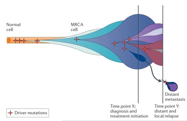
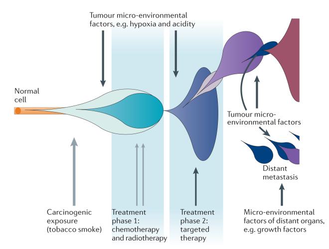
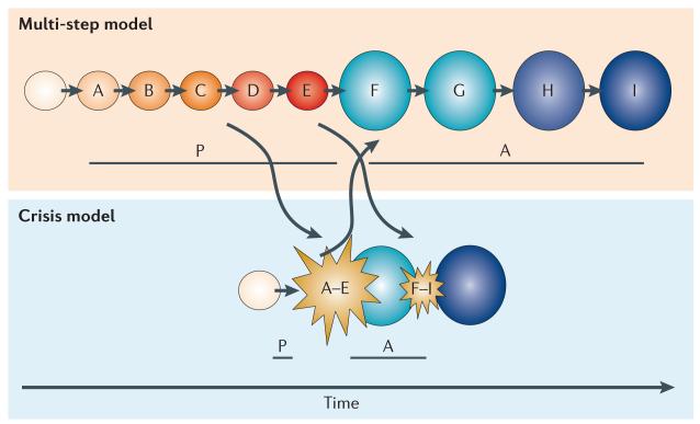
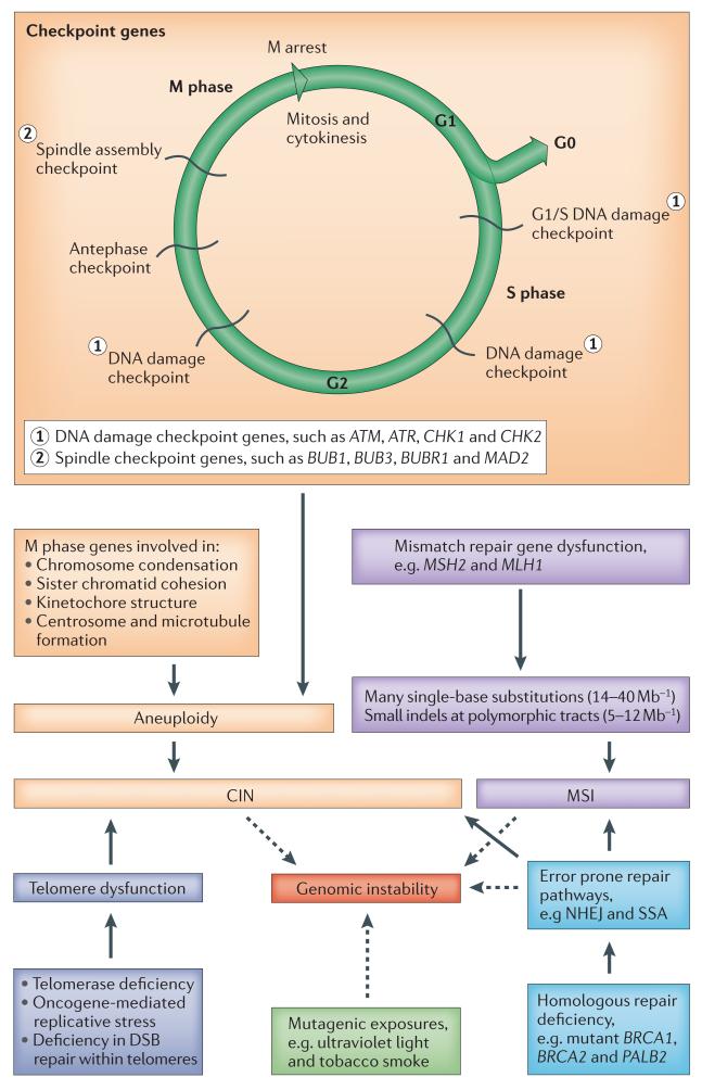
The advent of massively parallel sequencing technologies has allowed the characterization of cancer genomes at an unprecedented resolution. Investigation of the mutational landscape of tumours is providing new insights into cancer genome evolution, laying bare the interplay of somatic mutation, adaptation of clones to their environment and natural selection. These studies have demonstrated the extent of the heterogeneity of cancer genomes, have allowed inferences to be made about the forces that act on nascent cancer clones as they evolve and have shown insight into the mutational processes that generate genetic variation. Here we review our emerging understanding of the dynamic evolution of the cancer genome and of the implications for basic cancer biology and the development of antitumour therapy.


Figures

Figure 1
Figure 1. The evolution of clonal populations
Cancers are genomically diverse and dynamic entities. Unique clones (represented by different coloured bubbles) emerge as a consequence of accumulating driver mutations in the progeny of a single most recent common ancestor (MRCA) cell. Ongoing linear and branching evolution results in multiple simultaneous subclones that may individually be capable of giving rise to episodes of disease relapse and metastasis. The dynamic clonal architecture is shaped by mutation and competition between subclones in light of environmental selection pressures, including those that are exerted by cancer treatments.
Figure 2
Figure 2. The role of the environment in evolutionary adaptation
A multitude of environmental factors may shape the evolutionary processes within a single cancer. Blue and purple bubbles represent successive cancer clones, the expansion of which is altered by directly mutagenic factors (grey arrows) and non-mutagenic factors (black arrows).
Figure 3
Figure 3. Stepwise versus crisis-driven mutation accumulation
Multi-step and crisis event models of carcinogenesis are represented. It is thought that these pathways are not necessarily mutually exclusive but that they may coincide and overlap. In this example, mutations A–E (orange to red circles) are those that are required to initiate clonal expansion and malignant transformation, whereas mutations F–H (blue circles) drive ongoing evolution and the acquisition of aggressive clinical characteristics. The pre-malignant phase (P) and the time from malignancy onset to acquisition of an aggressive phenotype (A) are reduced in the crisis event model compared to the multi-step model. This indicates that standard screening techniques that aim to detect pre-invasive and early malignancies may be inadequate in cancers that develop through crisis events.
Figure 4
Figure 4. ‘Mutator mutations’ drive genomic instability in cancers
There are two major recognized routes by which genomic instability may arise. Chromosomal instability (CIN) is common across all types of cancer and may be numeric (aneuploidy) or structural. CIN may arise through mutations in a wide range of genes involved in cell cycling and division (orange boxes) or through other diverse mechanisms, such as telomeric dysfunction or as a consequence of failure in homologous repair. Microsatellite instability (MSI) is less common and occurs as a result of mutations in the mismatch repair genes (purple boxes). Instability may also directly arise as a consequence of defects in homologous repair, necessitating the use of alternative error prone pathways, such as non-homologous end joining (NHEJ) and single-strand annealing (SSA). Error-prone pathways may result in both chromosomal instability and genomic instability through frequent small deletions or substitutions. Mutagenic exposures may also contribute to genomic instability. ATM, ataxia telangiectasia mutated; ATR, ataxia telangiectasia and Rad3-related; BUB1, budding uninhibited by benzimidazoles 1; BUBR1, budding uninhibited by benzimidazoles 1 beta; BRCA1, breast cancer 1, early onset; BRCA2, breast cancer 2, early onset; DSB, double-strand break; indel, insertion or deletion mutation; MAD2, MAD2 mitotic arrest deficient-like 1; MSH2, mutS homologue 2, colon cancer, nonpolyposis type 1; MLH1, mutL homologue 1, colon cancer nonpolyposis type 2; PALB2, partner and localizer of BRCA2.

References

    1. Nowell PC. The clonal evolution of tumor cell populations. Science. 1976;194:23–28. - PubMed
    1. Hanahan D, Weinberg RA. Hallmarks of cancer: the next generation. Cell. 2011;144:646–674. - PubMed
    1. Vogelstein B, Kinzler KW. The multistep nature of cancer. Trends Genet. 1993;9:138–141. - PubMed
    1. Stratton MR, Campbell PJ, Futreal PA. The cancer genome. Nature. 2009;458:719–724. - PMC - PubMed
    1. Campbell PJ, et al. Subclonal phylogenetic structures in cancer revealed by ultra-deep sequencing. Proc. Natl Acad. Sci. USA. 2008;105:13081–13086. - PMC - PubMed

Publication types