. 2012 Dec;92(12):1749-59.
doi: 10.1038/labinvest.2012.141. Epub 2012 Oct 8.

Lack of MMP10 exacerbates experimental colitis and promotes development of inflammation-associated colonic dysplasia

Affiliations

Lack of MMP10 exacerbates experimental colitis and promotes development of inflammation-associated colonic dysplasia

Felicitas L Koller et al. Lab Invest. 2012 Dec.

Abstract

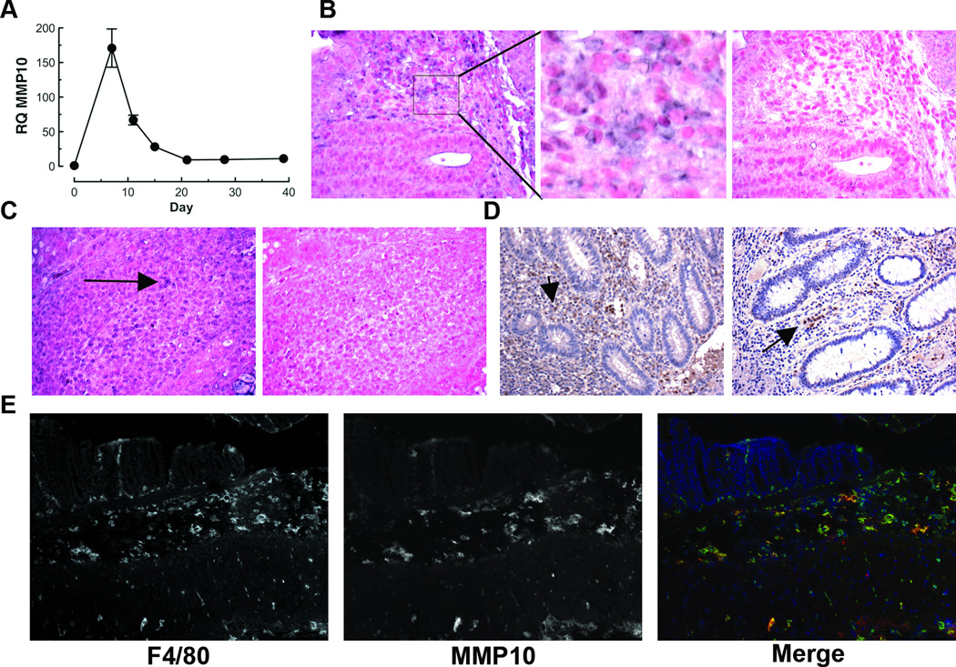
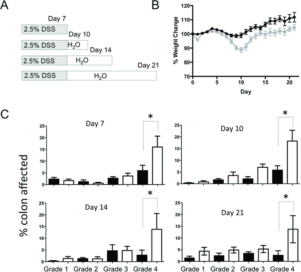
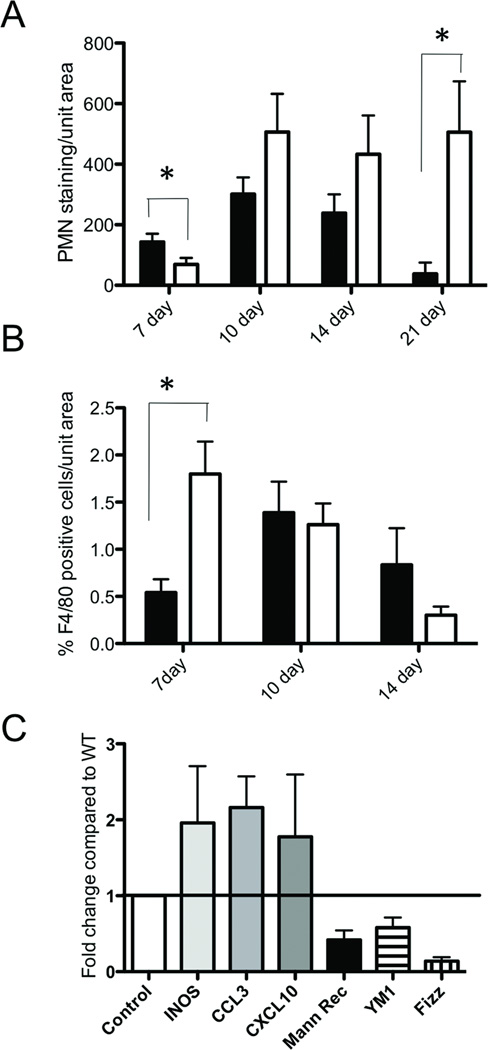
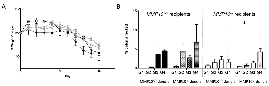
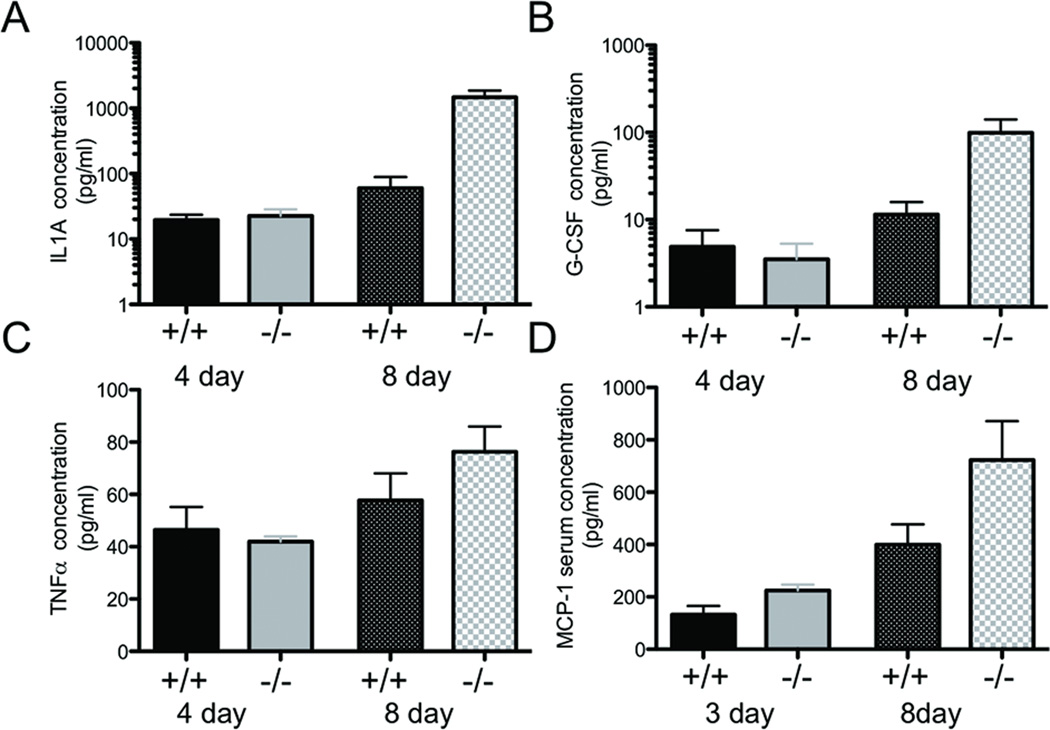
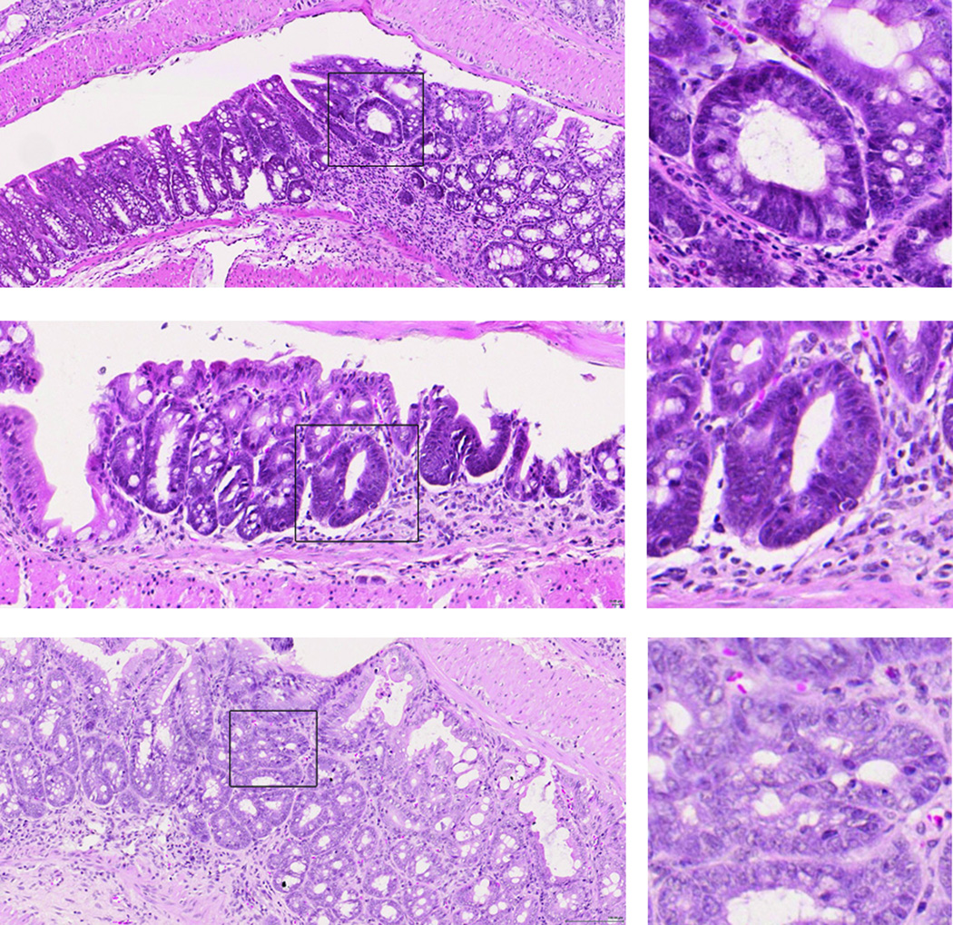
Inflammatory bowel diseases (IBD) such as ulcerative colitis (UC) represent serious health burdens because of both the tissue-damaging disease itself and an elevated risk of colon cancer. The increased expression of many members of the matrix metalloproteinase (MMP) family of enzymes that occurs in colitis has long been associated with the destructive nature of the disease. Recent findings in cancer and other MMP-associated diseases, however, led us to question whether MMPs are indeed detrimental in the setting of colitis. Here, we focus on a single MMP family member, MMP10, and assess its role in a murine model of colonic tissue damage induced by dextran sulfate sodium (DSS) treatment. Using mice genetically deficient for MMP10, we find that absence of this enzyme leads to significantly worse disease scores and failure to resolve inflammation even after extended recovery periods. We show that MMP10 is produced predominantly by infiltrating myeloid cells in both murine and human colitis. Through bone marrow transplant experiments, we confirm that bone marrow-derived MMP10 contributes to colitis severity. Mice lacking MMP10 have a significantly higher propensity for development of dysplastic lesions in the colon after two rounds of DSS exposure. Thus, we conclude that MMP10 is required for resolution of DSS-induced colonic damage, and in its absence, chronic inflammation and ultimately dysplasia occurs.


Figures

Fig 1
Fig 1. MMP10 is induced by inflammation in murine and human colonic tissue
(A) Real-time PCR analysis of MMP10 expression in colon tissue of C57BL/6 mice at different timepoints before, during and after a 7-day treatment with DSS. (B, C) In situ hybridization for Mmp10 transcript in colon tissue specimens from DSS-treated (B) or Il10−/− (C) C57BL/6 mice. Antisense and sense probes were both labeled with digoxigenin and applied at the same concentration. Positive signal is purple (e.g. arrows); nuclei are counterstained with Fast Red. A higher magnification of the area bounded by a rectangle in the DSS anti-sense image is shown in the adjacent panel. (D) Immunohistochemical detection of MMP10 protein in specimens of colon tissue from 2 different IBD patients. Positive signal is brown; nuclei are counterstained blue with hematoxylin. (E) Immunofluorescent co-staining of a frozen section of colon from a mouse after a 7-day treatment with DSS. The murine macrophage marker F4/80 is shown on the left, MMP10 is shown in the center and merged image showing F4/80 (green), MMP10 (red) and nuclear stain (blue) is on the right.
Fig 2
Fig 2. MMP10-deficient mice are significantly more injured by DSS treatment than wildtype mice
(A) Scheme showing the four timepoints after a 7-day exposure to 2.5% DSS at which cohorts of mice were analyzed. (B) Average weight change, relative to each individual starting weight, for all mice on the DSS protocol. Wildtype mice are shown in black, whereas Mmp10−/− mice are in grey. (C) Histological scores for wildtype (black bars) and MMP10−/− (white bars) colon sections from mice assessed at the 4 different timepoints outlined in (A). The description for each of the grades is given in the Materials & Methods. Shown is the average percentage of the colon length assessed at each grade for each group of mice.
Fig 3
Fig 3. Leukocyte recruitment is altered in DSS-treated MMP10-deficient mice
(A) The average level of PMN immunostaining per unit area of colonic tissue in widltype (black bars) or MMP10−/− (white bars) mice at each of the 4 timepoints after DSS treatment. (B) The average number of F4/80-positive macrophages per unit area of colon tissue from wild-type (black bars) or MMP10−/− (white bars) mice at 3 timepoints after DSS treatment. Macrophages were undetectable at 21 days. (C) Whole colon lysates from 7-day DSS-treated mice were analyzed by realtime PCR for markers of macrophage activation status. Shown are the levels of markers of M1 macrophages (INOS, CXCL3 and CCL10) and M2 macrophages (mannose receptor, Fizz and Ym1) in MMP10-null colons compared to levels in wild-type colons after normalization using levels of GAPDH.
Fig 4
Fig 4. Bone marrow transplant experiments suggest colitis severity is largely controlled by bone marrow-derived MMP10
(A) Weight changes over the course of DSS treatment and 3-day recovery. Black symbols indicate wildtype recipients, while grey indicates MMP10−/− recipients. Filled circles indicate wildtype donors, while empty circles indicate MMP10−/− donors. (B) Histologic grades of colonic tissue specimens from bone marrow transplant mice. The dark bars on the left indicate wildtype recipients, while the light bars on the right are MMP10−/− recipients. Solid bars are wildtype donors and hashed bars are MMP10−/− donors. The grades (assigned as described in Materials & Methods) are indicated by G1 through G4, with the bars representing the average percentage of colon length calculated for each grade in each group of mice.
Fig 5
Fig 5. Levels of cytokines and chemokines detected by ELISA in fluids from DSS-treated mice
Levels of IL1a (A), G-CSF (B), and TNFα (C) measured in colonic lavage samples collected from mice on day 4 or day 8 of a 7-day DSS treatment protocol. Day 8 mice had one recovery day before collection. (D) Levels of the chemokine MCP-1 detected in serum from mice on day 3 or day 8 of a 7-day DSS treatment protocol.
Fig 6
Fig 6. Dysplastic lesions are present in the colons of MMP10−/− mice after two rounds of DSS treatment
Shown are examples of hematoxylin and eosin-stained lesions from 3 different MMP10−/− mice. Boxed regions showing examples of dysplasic lesions are shown in higher magnification on the right. Scale bar = 100 µm.

References

    1. Osterman MT, Lichtenstein GR. Chapter 112 - Ulcerative Colitis. In: Feldman M, Friedman LS, Brandt LJ, editors. Sleisenger's and Fordtran's Gastrointestinal and Liver Disease. 9th ed. Philadelphia, PA: Saunders; 2010. p. 1982.
    1. Itzkowitz SH, Yio X. Inflammation and cancer IV. Colorectal cancer in inflammatory bowel disease: the role of inflammation. Am J Physiol Gastrointest Liver Physiol. 2004;287:G7–G17. - PubMed
    1. Sandborn WJ, Loftus EV. Balancing the risks and benefits of infliximab in the treatment of inflammatory bowel disease. Gut. 2004;53:780–782. - PMC - PubMed
    1. Ng SC, Kamm MA. Therapeutic strategies for the management of ulcerative colitis. Inflamm Bowel Dis. 2009;15:935–950. - PubMed
    1. Baugh MD, Perry MJ, Hollander AP, et al. Matrix metalloproteinase levels are elevated in inflammatory bowel disease. Gastroenterology. 1999;117:814–822. - PubMed

Publication types

MeSH terms