

Identification of a novel mutation in the CDHR1 gene in a family with recessive retinal degeneration
- PMID: 23044944
- PMCID: PMC3799916
- DOI: 10.1001/archophthalmol.2012.1906
Identification of a novel mutation in the CDHR1 gene in a family with recessive retinal degeneration
Abstract
Objectives: To describe the clinical phenotype and identify the molecular basis of disease in a consanguineous family of Palestinian origin with autosomal recessive retinal degeneration.
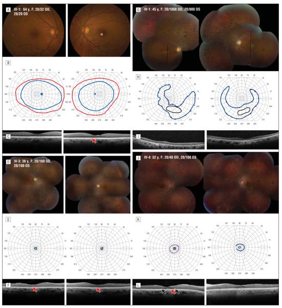
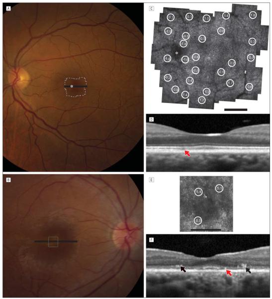
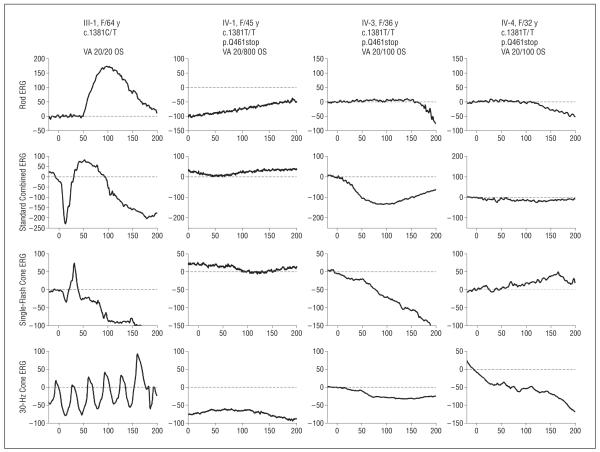
Methods: Eight family members were evaluated with visual acuity and perimetry tests, color fundus photographs, full-field electroretinography, and optical coherence tomography. Cone photoreceptors surrounding the fovea were imaged in 2 members, using adaptive optics scanning laser ophthalmoscopy. Exome was captured using probes and sequenced. Readings were mapped to reference hg19. Variant calls and annotations were performed, using published protocols. Confirmation of variants and segregation analysis was performed using dideoxy sequencing.
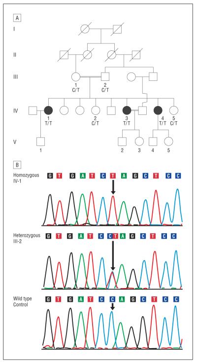
Results: Analysis detected 24 037 single-nucleotide variants in one affected family member, of which 3622 were rare and potentially damaging to encoded proteins. Further analysis revealed a novel homozygous nonsense change, c.1381 C>T, p.Gln461X in exon 13 of the CDHR1 gene, which segregated with retinal degeneration in this family. Affected members had night blindness beginning during adolescence with progressive visual acuity and field loss and unmeasurable electroretinographic responses, as well as macular outer retinal loss, although residual cones with increased cone spacing were observed in the youngest individual.
Conclusions: Exome analysis revealed a novel CDHR1 nonsense mutation segregating with progressive retinal degeneration causing severe central vision loss by the fourth decade of life. High-resolution retinal imaging revealed outer retinal changes suggesting that CDHR1 is important for normal photoreceptor structure and survival.
Clinical relevance: Exome sequencing is a powerful technique that may identify causative genetic variants in families with autosomal recessive retinal degeneration.
Figures
Similar articles
-
Ocular Phenotype of a Family with FAM161A-associated Retinal Degeneration.Ophthalmic Genet. 2016;37(1):44-52. doi: 10.3109/13816810.2014.929716. Epub 2014 Jul 9. Ophthalmic Genet. 2016. PMID: 25007332 Free PMC article.
-
A novel splice site mutation of CDHR1 in a consanguineous Israeli Christian Arab family segregating autosomal recessive cone-rod dystrophy.Mol Vis. 2012;18:2915-21. Epub 2012 Dec 1. Mol Vis. 2012. PMID: 23233793 Free PMC article.
-
Clinical characteristics of recessive retinal degeneration due to mutations in the CDHR1 gene and a review of the literature.Ophthalmic Genet. 2018 Jan-Feb;39(1):51-55. doi: 10.1080/13816810.2017.1363244. Epub 2017 Sep 8. Ophthalmic Genet. 2018. PMID: 28885867 Review.
-
A Specific Macula-Predominant Retinal Phenotype Is Associated With the CDHR1 Variant c.783G>A, a Silent Mutation Leading to In-Frame Exon Skipping.Invest Ophthalmol Vis Sci. 2019 Aug 1;60(10):3388-3397. doi: 10.1167/iovs.18-26415. Invest Ophthalmol Vis Sci. 2019. PMID: 31387115
-
Multimodal fundus imaging in fundus albipunctatus with RDH5 mutation: a newly identified compound heterozygous mutation and review of the literature.Doc Ophthalmol. 2012 Aug;125(1):51-62. doi: 10.1007/s10633-012-9336-z. Epub 2012 Jun 6. Doc Ophthalmol. 2012. PMID: 22669287 Review.
Cited by
-
A mutation in IFT43 causes non-syndromic recessive retinal degeneration.Hum Mol Genet. 2017 Dec 1;26(23):4741-4751. doi: 10.1093/hmg/ddx356. Hum Mol Genet. 2017. PMID: 28973684 Free PMC article.
-
High-resolution Imaging in Male Germ Cell-Associated Kinase (MAK)-related Retinal Degeneration.Am J Ophthalmol. 2018 Jan;185:32-42. doi: 10.1016/j.ajo.2017.10.023. Epub 2017 Nov 16. Am J Ophthalmol. 2018. PMID: 29103961 Free PMC article.
-
Identification of the genetic determinants responsible for retinal degeneration in families of Mexican descent.Ophthalmic Genet. 2018 Jan-Feb;39(1):73-79. doi: 10.1080/13816810.2017.1373830. Epub 2017 Sep 25. Ophthalmic Genet. 2018. PMID: 28945494 Free PMC article.
-
Whole Genome Sequencing Revealed Mutations in Two Independent Genes as the Underlying Cause of Retinal Degeneration in an Ashkenazi Jewish Pedigree.Genes (Basel). 2017 Aug 24;8(9):210. doi: 10.3390/genes8090210. Genes (Basel). 2017. PMID: 28837078 Free PMC article.
-
Rescue of cone and rod photoreceptor function in a CDHR1-model of age-related retinal degeneration.Mol Ther. 2024 May 1;32(5):1445-1460. doi: 10.1016/j.ymthe.2024.03.026. Epub 2024 Mar 19. Mol Ther. 2024. PMID: 38504520 Free PMC article.
References
-
- [Accessed January 8, 2012];RetNet: summary of genes and loci causing retinal diseases. https://sph.uth.tmc.edu/Retnet/sum-dis.htm.
-
- Briggs CE, Rucinski D, Rosenfeld PJ, Hirose T, Berson EL, Dryja TP. Mutations in ABCR (ABCA4) in patients with Stargardt macular degeneration or cone-rod degeneration. Invest Ophthalmol Vis Sci. 2001;42(10):2229–2236. - PubMed
-
- Wells J, Wroblewski J, Keen J, et al. Mutations in the human retinal degeneration slow (RDS) gene can cause either retinitis pigmentosa or macular dystrophy. Nat Genet. 1993;3(3):213–218. - PubMed
-
- Boon CJ, Klevering BJ, Leroy BP, Hoyng CB, Keunen JE, den Hollander AI. The spectrum of ocular phenotypes caused by mutations in the BEST1 gene. Prog Retin Eye Res. 2009;28(3):187–205. - PubMed
Publication types
MeSH terms
Substances
Grants and funding
LinkOut - more resources
Full Text Sources
Molecular Biology Databases
