. 2013 Sep 5;32(36):4214-21.
doi: 10.1038/onc.2012.439. Epub 2012 Oct 8.

Anti-tumorigenic effects of Type 1 interferon are subdued by integrated stress responses

Affiliations

Anti-tumorigenic effects of Type 1 interferon are subdued by integrated stress responses

S Bhattacharya et al. Oncogene. .

Abstract

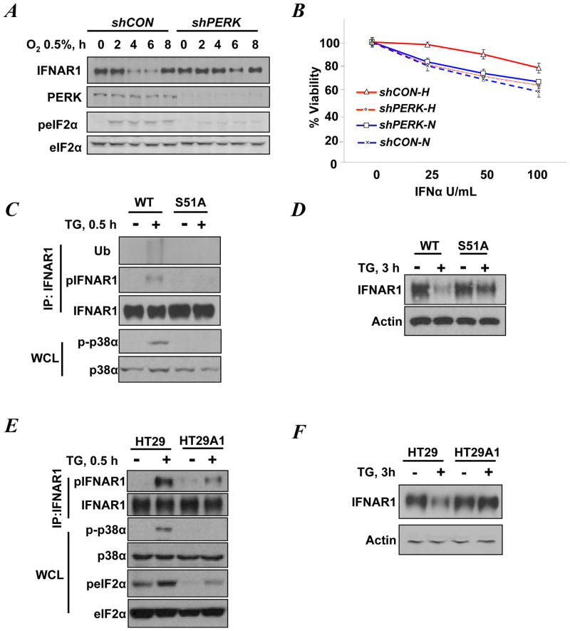
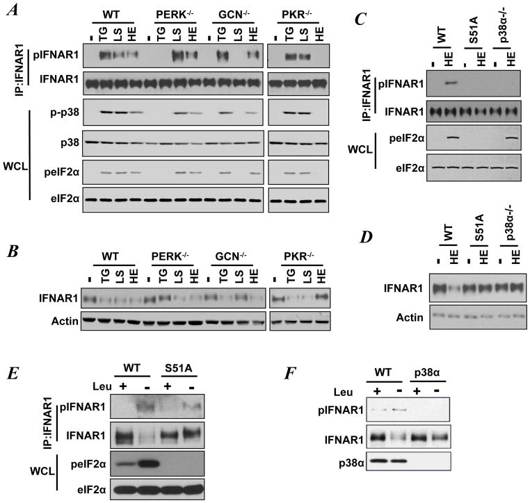
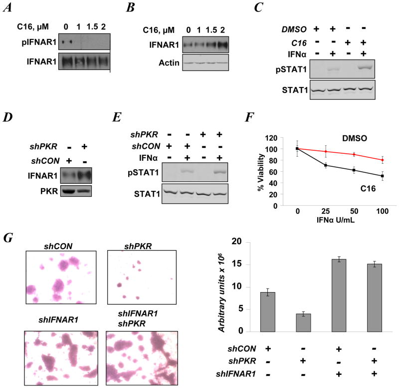
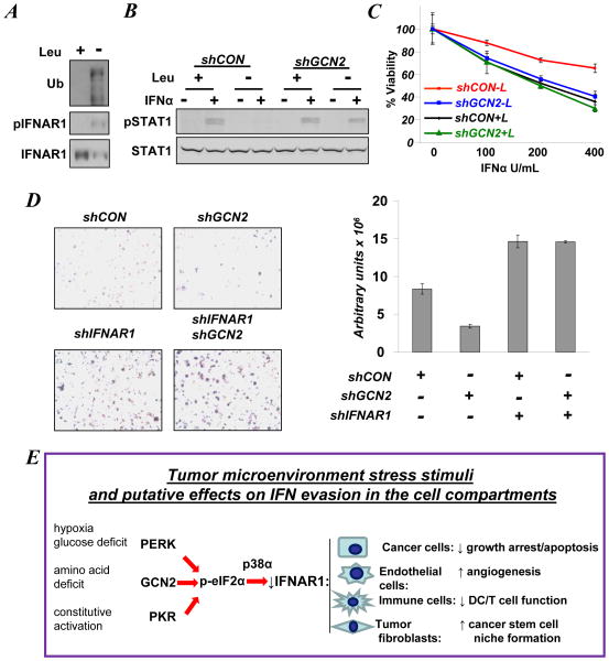
Viral and pharmacological inducers of protein kinase RNA-activated (PKR)-like ER kinase (PERK) were shown to accelerate the phosphorylation-dependent degradation of the IFNAR1 chain of the Type 1 interferon (IFN) receptor and to limit cell sensitivity to IFN. Here we report that hypoxia can elicit these effects in a PERK-dependent manner. The altered fate of IFNAR1 affected by signaling downstream of PERK depends on phosphorylation of eIF2α (eukaryotic translational initiation factor 2-α) and ensuing activation of p38α kinase. Activators of other eIF2α kinases such as PKR or GCN2 (general control nonrepressed-2) are also capable of eliminating IFNAR1 and blunting IFN responses. Modulation of constitutive PKR activity in human breast cancer cells stabilizes IFNAR1 and sensitizes these cells to IFNAR1-dependent anti-tumorigenic effects. Although downregulation of IFNAR1 and impaired IFNAR1 signaling can be elicited in response to amino-acid deficit, the knockdown of GCN2 in melanoma cells reverses these phenotypes. We propose that, in cancer cells and the tumor microenvironment, activation of diverse eIF2α kinases followed by IFNAR1 downregulation enables multiple cellular components of tumor tissue to evade the direct and indirect anti-tumorigenic effects of Type 1 IFN.


Conflict of interest statement

CONFLICT OF INTEREST:

The authors declare no conflict of interest except for Dr. D.P. Baker who is an employee of BiogenIDEC, Inc and owns stock of this company.

Figures

Figure 1
Figure 1. PERK inducers trigger phosphorylation and downregulation of IFNAR1 in a manner that depends on phosphorylation of eIF2α
A. WM266-4 cells that received shRNA against PERK or GFP (control, shCON) were exposed to 0.5% O2 for the indicated times. Levels of IFNAR1, PERK, eIF2α, and the phosphorylation of eIF2α on S51 were detected by immunoblotting. B. WM266-4 cells transduced with the indicated shRNA and incubated under normoxic (N) or hypoxic (H) conditions for 4h were treated with the indicated doses of IFNα. The extent of PERK knockdown in these cells is shown in panel A. Cell viability was assessed 24 h later. C. Phosphorylation, ubiquitination, and levels of IFNAR1 immunopurified from untreated or TG-treated (0.5 h) MEFs were analyzed by immunoblotting using the indicated antibodies. Levels and activation of p38α in the whole cell lysates were also analyzed. D. Immunoblotting analyses of the levels of IFNAR1 and β-actin (used as a loading control) in untreated or TG-treated (3 h) MEFs. E. Immunoblotting analysis of phosphorylation and levels of IFNAR1, p38α, and eIF2α in untreated or TG-treated (0.5 h) HT29-derived cells treated. F. Immunoblotting analyses of the levels of IFNAR1 and β-actin (used as a loading control) in untreated or TG-treated (3 h) HT29-derived cells.
Figure 2
Figure 2. Diverse stress stimuli induce eIF2α phosphorylation to mediate phosphorylation and downregulation of IFNAR1
A. Immunoblotting analyses of phosphorylation and levels of IFNAR1, p38α, and eIF2α in MEFs exposed to TG (for 30 min) or heparin (HE, for 40 min), or leucine starvation (LS, for 24 h). B. Immunoblotting analyses of the levels of IFNAR1 and β-actin in MEFs treated with TG (for 3 h), or HE (for 7 h), or incubated with media without leucine (LS, 48 h). C. Immunoblotting analyses of phosphorylation and levels of IFNAR1 and eIF2α in MEFs exposed to heparin (HE) for 40 min. D. Immunoblotting analyses of phosphorylation and levels of IFNAR1 and eIF2α in MEFs exposed to HE for 7 h. E. Immunoblotting analyses of phosphorylation and levels of IFNAR1 and eIF2α in MEFs cultured in the presence or absence of leucine for 48 h. F. Immunoblotting analyses of phosphorylation and levels of IFNAR1 and p38α kinase in MEFs cultured in the presence or absence of leucine for 48 h.
Figure 3
Figure 3. Constitutively active PKR suppresses IFNAR1 levels and signaling in T47D human breast cancer cells
A. T47D cells treated with indicated doses of C16 (for 24 h) were incubated with the lysosomal inhibitor methyl amine chloride (40 mM) for 4 h prior to harvesting. Phosphorylation and levels of IFNAR1 were analyzed by immunoblotting. B. Immoblotting analysis of IFNAR1 and β-actin levels in T47D cells treated with C16 for 24 h. C. Immunoblotting analyses of levels and phosphorylation of STAT1 in T47D cells treated with vehicle (DMSO) or C16 (2 μM) for 24 h prior to treatment with 250 U/mL of IFNα (30 min). D. Levels of IFNAR1and PKR in T47D cells that received control or PKR shRNA. E. Immunoblot analysis of levels and phosphorylation of STAT1on Tyr701 in T47D cells that received control or PKR shRNA 48 h prior to treatment with of IFNα (250 U/mL for 30 min). The extent of PKR knockdown in the cells before treatment is shown in panel D. F. Viability of T47D cells treated with vehicle (DMSO) or C16 (2 μM) for 24 h prior to treatment with the indicated amounts of IFNα (for 48 h) was analyzed using a MTT assay. G. Images and quantification of T47D cells that received indicated shRNA and migrated through Matrigel/membrane in Boyden chambers. Density of migrating cells was assessed by densitometry and presented in arbitrary units of absorbance. The extent of PKR and IFNAR1 knockdown is shown in Figure S4.
Figure 4
Figure 4. Activation of GCN2 by amino acid deficit promotes downregulation of IFNAR1 and inhibits its signaling
A. Levels, phosphorylation, and ubiquitination of IFNAR1 immunopurified from WM266-4 cells grown in the presence or absence of leucine for 48 h were analyzed by immunoblotting using the indicated antibodies. B. STAT1 phosphorylation and levels in WM266-4 cells that received the indicated shRNAs and were incubated in medium in the presence or absence of leucine for 48 h prior to treatment with IFNα (250 U/mL for 30 min). Extent of protein knockdown is shown in Figure S6. C. Viability of WM266-4 cells that received the indicated shRNAs and incubated in the presence (+L) or absence (−L) of leucine for 48 h prior to treatment with the indicated doses of IFNα (for 24 h) and subsequent analysis using a MTT assay. D. Images and quantification of WM266-4 cells (that received indicated shRNA and were incubated in media lacking leucine) and migrated through Matrigel/membrane in Boyden chambers. Density of migrating cells was assessed by densitometry and presented in arbitrary units of absorbance. Extent of protein knockdown in shown in Figure S8. E. Hypothetical model in which various types of ISR signaling converge on phosphorylation of eIF2α and activation of p38α kinase to downregulate IFNAR1 and inhibit IFNα responses in various compartments of the tumor. This leads to alleviation of growth inhibition, stimulation of angiogenesis, and niche formation and impairment of anti-tumorigenic immune responses.

References

    1. Ye J, Koumenis C. ATF4, an ER stress and hypoxia-inducible transcription factor and its potential role in hypoxia tolerance and tumorigenesis. Curr Mol Med. 2009;9(4):411–6. - PubMed
    1. Ye J, Kumanova M, Hart LS, Sloane K, Zhang H, De Panis DN, et al. The GCN2-ATF4 pathway is critical for tumour cell survival and proliferation in response to nutrient deprivation. EMBO J. 2010;29(12):2082–96. - PMC - PubMed
    1. Liu Y, Laszlo C, Liu W, Chen X, Evans SC, Wu S. Regulation of G(1) arrest and apoptosis in hypoxia by PERK and GCN2-mediated eIF2alpha phosphorylation. Neoplasia. 2010;12(1):61–8. - PMC - PubMed
    1. Schreiber RD, Old LJ, Smyth MJ. Cancer immunoediting: integrating immunity’s roles in cancer suppression and promotion. Science. 2011;331(6024):1565–70. - PubMed
    1. Moenner M, Pluquet O, Bouchecareilh M, Chevet E. Integrated endoplasmic reticulum stress responses in cancer. Cancer Res. 2007;67(22):10631–4. - PubMed

Publication types