. 2013 Feb;23(2):188-98.
doi: 10.1093/glycob/cws140. Epub 2012 Oct 8.

6-alkynyl fucose is a bioorthogonal analog for O-fucosylation of epidermal growth factor-like repeats and thrombospondin type-1 repeats by protein O-fucosyltransferases 1 and 2

Affiliations

6-alkynyl fucose is a bioorthogonal analog for O-fucosylation of epidermal growth factor-like repeats and thrombospondin type-1 repeats by protein O-fucosyltransferases 1 and 2

Esam Al-Shareffi et al. Glycobiology. 2013 Feb.

Abstract

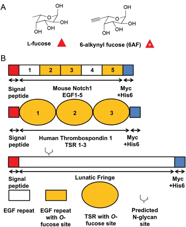
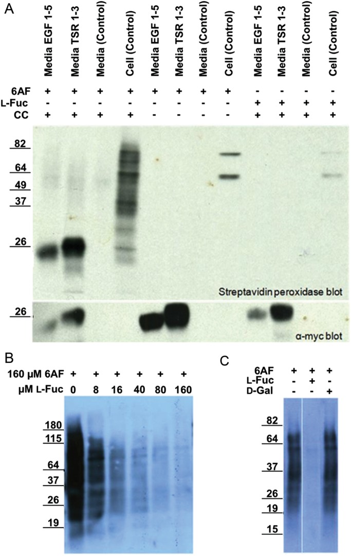
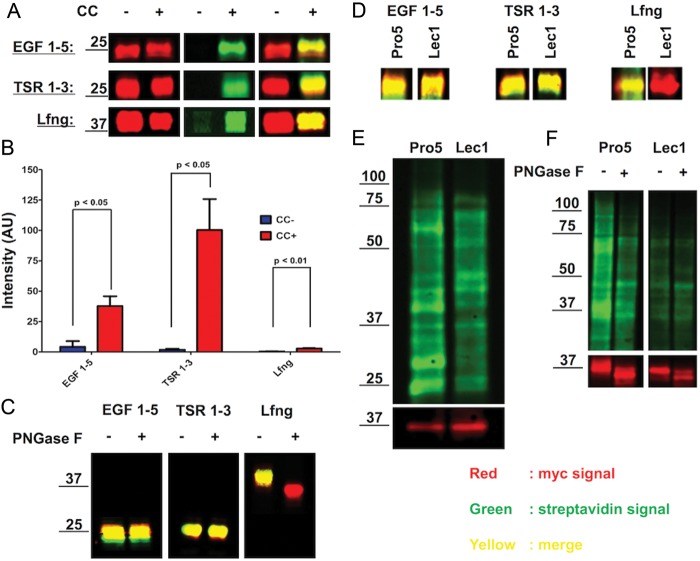
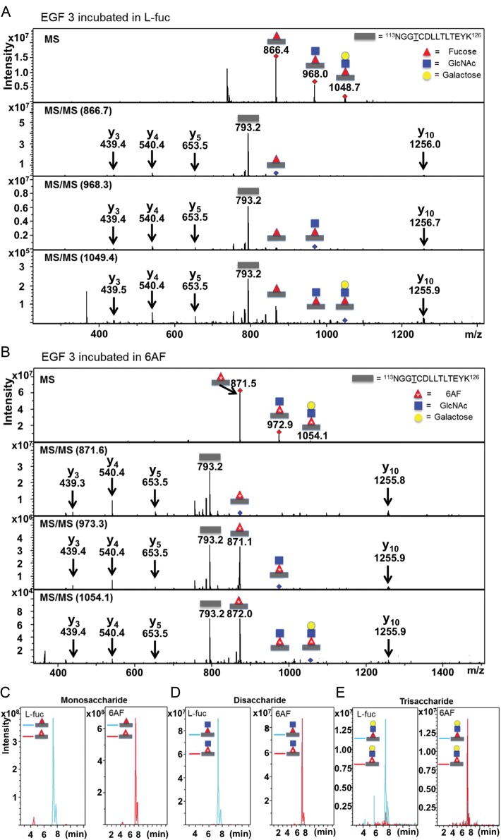
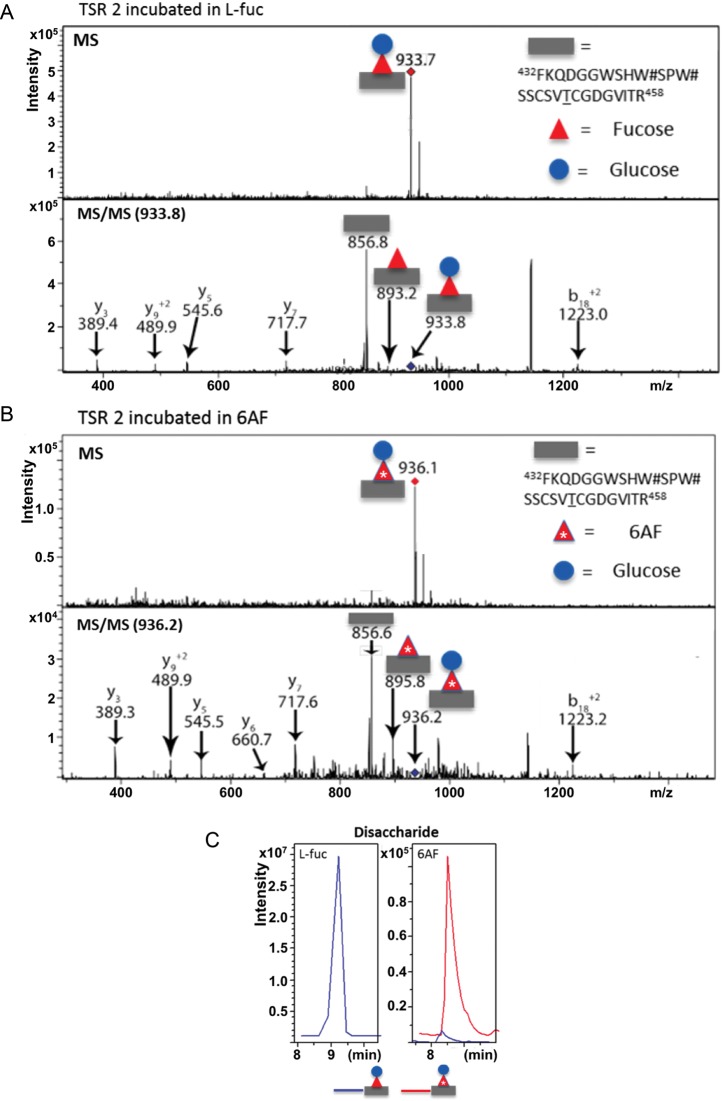
Protein O-fucosyltransferase 1 (Pofut1) and protein O-fucosyltransferase 2 (Pofut2) add O-linked fucose at distinct consensus sequences in properly folded epidermal growth factor (EGF)-like repeats and thrombospondin type-1 (TSR) repeats, respectively. Glycan chain elongation past O-fucose can occur to yield a tetrasaccharide on EGF repeats and a disaccharide on TSRs. Elimination of Pofut1 in mice causes embryonic lethality with Notch-like phenotypes demonstrating that O-fucosylation of Notch is essential for its function. Similarly, elimination of Pofut2 results in an early embryonic lethal phenotype in mice, although the molecular mechanism for the lethality is unknown. The recent development of sugar analogs has revolutionized the study of glycans by providing a convenient method for labeling and tracking glycosylation. In order to study O-fucosylation, we took advantage of the recently developed reporter, 6-alkynyl fucose. Using the Cu(I)-catalyzed azide-alkyne cycloaddition (CuAAC), or "click" reaction, azido-biotin allows tagging and detection of 6AF-modified proteins. Here we examine whether proteins containing EGF repeats or TSRs with O-fucose consensus sequences are specifically modified with 6AF in cell culture. Using mass spectrometry (MS), we demonstrate that 6AF is efficiently incorporated onto the appropriate consensus sequences on EGF repeats and TSRs. Furthermore, the elongation of the O-fucose monosaccharide on EGF repeats and TSRs is not hampered when 6AF is used. These results show that 6AF is efficiently utilized in a truly bioorthogonal manner by Pofut1, Pofut2 and the enzymes that elongate O-fucose, providing evidence that 6AF is a significant new tool in the study of protein O-fucosylation.


Figures

Fig. 1.
Fig. 1.
The structure of 6AF and domain map of the constructs used. (A) The structures of l-fucose and 6AF. (B) All expression constructs contained an N-terminal Igk signal peptide (red) and C-terminal Myc-His6 tags (blue) for detection by Western blot and purification. Mouse Notch 1 EGF 1–5 contains three O-fucosylated EGF repeats (indicated by shading) (Shao et al. 2003), and all the three TSRs in Human Thrombospondin 1 TSR 1–3 are O-fucosylated (Hofsteenge et al. 2001), while neither construct contains an N-glycosylation site. Lfng is not predicted to be O-fucosylated and contains one N-glycosylation site at Asn-167.
Fig. 2.
Fig. 2.
6AF is incorporated onto EGF repeats and TSRs in HEK293T cells. (A) HEK293T cells were either untransfected (Control) or transfected with plasmids encoding mN1 EGF1–5 or hT1 TSR1–3, and incubated with 200 µM peracetylated 6AF (6AF) or peracetylated l-fucose (Fuc) for three days. The media and the lysates were subjected to CuAAC with (+CC) or without (−CC) azido-biotin. The samples were then subjected to SDS–PAGE and Western blotting using Streptavidin-HRP and anti-myc. (B) Cell lysates were prepared from untransfected HEK293T cells incubated with 160 µM peracetylated 6AF and increasing amounts of l-Fucose, subjected to CuAAC with azido-biotin and analyzed as described above. (C) Cell lysates were prepared from untransfected HEK293T cells incubated with 200 µM peracetylated 6AF in the presence or absence of 200 µM l-fucose or d-galactose, subjected to CuAAC with azido-biotin and analyzed as described above.
Fig. 3.
Fig. 3.
6AF is incorporated into N-glycans found on Lfng and numerous proteins in crude extracts of CHO cells. (A) The Pro5 cells were transfected with plasmids encoding mN1 EGF1–5, hT1 TSR1–3 or Lfng (Figure 1) and grown in the presence of 200 µM peracetylated 6AF. The media samples were subjected to CuAAC with (+ Click Chemistry) or without (−CC) azido-biotin and analyzed as described in Materials and Methods. Protein was monitored using the anti-myc probe (red), and biotin using the streptavidin probe (green), while merging of the two channels results in yellow. (B) Bar graph illustrating the intensity (in arbitrary units (AU) of streptavidin signal in EGF 1–5, TSR 1–3 and Lfng samples, with (+CC) and without (−CC) performing the click chemistry reaction. The experiment was performed in triplicate and the streptavidin intensity values were normalized to anti-myc intensity values. Paired student t-test was performed to determine statistical significance. (C) The samples prepared as in A were subjected to PNGase F digestion as described in Materials and Methods. (D) The plasmids encoding mN1 EGF1–5, hT1 TSR1–3 and Lfng were transfected either into Pro5 or into Lec1 cells in the presence of 200 µM peracetylated 6AF, and the media samples were analyzed as in A. (E) The Pro5 and Lec1 cells transfected with Lfng were grown in the presence of 200 µM peracetylated 6AF and the cell lysates were biotinylated and analyzed as in A. Many proteins are labeled in both cell lines, but the signal is lost on several proteins in Lec1 cells. The bottom panel shows an anti-Myc blot for the Lfng-Myc-His6 to show that similar amounts of extract were loaded in each lane. (F) The Pro5 and Lec1 cells transfected with Lfng were grown in the presence of 200 µM peracetylated 6AF and the cell lysates were biotinylated, PNGase F digested and then analyzed as in A. The intensity of the streptavidin signal in Pro5 cell lysate is decreased after PNGase F digestion, closely resembling the Lec1 cell lysate sample. The bottom panel shows an anti-Myc blot for the transiently transfected Lfng-Myc-His6 to show that similar amounts of extract were loaded in each lane and to confirm that the PNGase F digestion was successful.
Fig. 4.
Fig. 4.
6AF is efficiently incorporated onto the O-fucosylation site in EGF3 of mouse Notch1 and elongated by Lfng. (A) The Pro5 cells were co-transfected with plasmids encoding mN1 EGF1–5 and Lfng grown in the presence of 200 µM peracetylated l-fucose. mN1 EGF1–5 was purified from the medium, digested with trypsin and subjected to nano-LC-MS/MS analysis as described in Materials and Methods. MS (top panel) and MS/MS (lower panels) spectra of a peptide from EGF3 containing the O-fucose consensus sequence (represented by a gray bar). The ions at m/z 866.4, 968.0 and 1048.7 correspond to the doubly charged forms of the mono-, di- and trisaccharide glycoforms of this peptide. The MS/MS spectra of each glycoform show loss of the modifying glycans to the unglycosylated form of the peptide (m/z 793.2), as well as several “b” and “y” fragmentation ions (indicated in red). (B) mN1 EGF1–5 was prepared as in A except that the cells were grown in 200 µM peracetylated 6AF. MS (top panel) and MS/MS (lower panels) spectra of the same peptide from EGF 3. The ions at m/z 871.5, 972.9 and 1054.1 correspond to the doubly charged forms of the mono-, di- and trisaccharide glycoforms of this peptide with 6AF. These masses closely match the expected increase of m/z of five compared with the l-fuc samples, as the peptides are in the 2+ charge state and as 6AF is 10 Da heavier than l-fucose. The MS/MS spectra confirm loss of the modifying glycans to the unglycosylated form of the peptide (m/z 793.2) and show several “b” and “y” fragmentation ions (in red). EIC for samples obtained from cells incubated in l-fucose (+l-Fuc) and for samples obtained from cells incubated in 6AF (+6AF), for the monosaccharide glycoform (C), the disaccharide glycoform (D) and the trisaccharide glycoform (E) of the peptide from EGF3. Gray rectangle, peptide; red triangle, fucose; red triangle with *, 6AF; blue square, GlcNAc; yellow circle, galactose.
Fig. 5.
Fig. 5.
6AF is efficiently incorporated onto TSR2 of Thromospondin1 and elongated by β3-glucosyltransferase. (A) The Pro5 cells were transfected with the plasmid encoding hT1 TSR1–3 and grown in the presence of 200 µM peracetylated fucose. hT1 TSR1–3 was purified from the medium, digested with trypsin and subjected to nano-LC-MS/MS analysis as described in Materials and Methods. MS (top panel) and MS/MS (bottom panel) spectra of a peptide from TSR 2 containing the O-fucose consensus sequence (represented by gray bar). The ion at m/z 933.7 corresponds to the quadruply charged form of this peptide modified with the O-fucose disaccharide. The MS/MS spectra indicate loss of the modifying glycans, first to the monosaccharide form (m/z 893.2) and then to the unglycosylated form of the peptide, (m/z 856.8). Several “b” and “y” fragmentation ions confirming the identity of the peptide are also indicated in black. (B) hT1 TSR1-3 was prepared as in A except that the cells were grown in the presence of 200 µM peracetylated 6AF. MS (top panel) and MS/MS (bottom panel) spectra of the same peptide from TSR 2 are shown. The ion at m/z 936.1 corresponds to the quadruply charged form of this peptide modified with the O-6AF disaccharide. The MS/MS spectra indicate sequential loss of glycans, first to the monosaccharide (m/z 895.8) and then to the unmodified form (m/z 856.6). These masses closely match the expected increase in m/z of 2.5, compared with the l-fucose labeled sample, as the peptides are in the 4+ charge state and as 6AF is 10 Da heavier than l-fucose. Several “b” and “y” ions are also indicated in black. (C) EIC for sample obtained from cells incubated in l-fucose (+l-Fuc) and for sample obtained from cells incubated in 6AF (+6AF), for the disaccharide glycoform of TSR 2. Gray rectangle, peptide; red triangle, fucose; red triangle with*, 6AF; blue circle, glucose.

Similar articles

Cited by

References

    1. Becker DJ, Lowe JB. Fucose: Biosynthesis and biological function in mammals. Glycobiology. 2003;13:41R–53R. doi:10.1093/glycob/cwg054. - DOI - PubMed
    1. Besanceney-Webler C, Jiang H, Wang W, Baughn AD, Wu P. Metabolic labeling of fucosylated glycoproteins in Bacteroidales species. Bioorg Med Chem Lett. 2011;21:4989–4992. doi:10.1016/j.bmcl.2011.05.038. - DOI - PMC - PubMed
    1. Bruckner K, Perez L, Clausen H, Cohen S. Glycosyltransferase activity of Fringe modulates Notch-Delta interactions. Nature. 2000;406:411–415. doi:10.1038/35019075. - DOI - PubMed
    1. Chen CI, Keusch JJ, Klein D, Hess D, Hofsteenge J, Gut H. Structure of human POFUT2: Insights into thrombospondin type 1 repeat fold and O-fucosylation. EMBO J. 2012 - PMC - PubMed
    1. Dehnert KW, Baskin JM, Laughlin ST, Beahm BJ, Naidu NN, Amacher SL, Bertozzi CR. Imaging the sialome during zebrafish development with copper-free click chemistry. Chembiochem. 2012;13:353–357. doi:10.1002/cbic.201100649. - DOI - PMC - PubMed

Publication types