. 2012 Oct 15;199(2):235-49.
doi: 10.1083/jcb.201112132. Epub 2012 Oct 8.

PARP1 promotes nucleotide excision repair through DDB2 stabilization and recruitment of ALC1

Affiliations

PARP1 promotes nucleotide excision repair through DDB2 stabilization and recruitment of ALC1

Alex Pines et al. J Cell Biol. .

Abstract

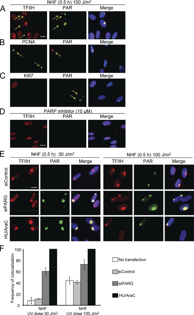
The WD40-repeat protein DDB2 is essential for efficient recognition and subsequent removal of ultraviolet (UV)-induced DNA lesions by nucleotide excision repair (NER). However, how DDB2 promotes NER in chromatin is poorly understood. Here, we identify poly(ADP-ribose) polymerase 1 (PARP1) as a novel DDB2-associated factor. We demonstrate that DDB2 facilitated poly(ADP-ribosyl)ation of UV-damaged chromatin through the activity of PARP1, resulting in the recruitment of the chromatin-remodeling enzyme ALC1. Depletion of ALC1 rendered cells sensitive to UV and impaired repair of UV-induced DNA lesions. Additionally, DDB2 itself was targeted by poly(ADP-ribosyl)ation, resulting in increased protein stability and a prolonged chromatin retention time. Our in vitro and in vivo data support a model in which poly(ADP-ribosyl)ation of DDB2 suppresses DDB2 ubiquitylation and outline a molecular mechanism for PARP1-mediated regulation of NER through DDB2 stabilization and recruitment of the chromatin remodeler ALC1.


Figures

Figure 1.
Figure 1.
PARP1 is a novel DDB2-associated factor. (A) SDS-PAGE electrophoresis and Coomassie staining of FLAG-DDB2 immunoprecipitates obtained from FLAG-DDB2–expressing MRC5 cells mock treated or irradiated with 20 J/m2 UV-C. Negative control (NC) indicates the eluate obtained from agarose beads incubated with MRC5 FLAG-DDB2 chromatin. The arrows in the zoom-in window indicate the position of the gel where DDB2 and the respective unique peptides were detected by MS (a unique peptide is defined as a peptide, irrespective of its length, that exists only in one protein of a proteome of interest). (B) Western blot of FLAG-DDB2 immunoprecipitates. Cells were mock treated or irradiated with 20 J/m2 UV-C and immunoblotted with DDB1- and DDB2-specific antibodies. Negative control indicates the eluate obtained from agarose beads incubated with chromatin from MRC5 FLAG-DDB2 cells. (C) Western blot of DDB2 and PARP1 immunoprecipitates (IP) from NHF cells mock treated or irradiated with 20 J/m2 UV-C, followed by 5-min incubation, and immunoblotted against DDB1, DDB2, or PARP1. (D) GFP-DDB2–PARP-1 binding assay. U2OS cells transfected with the indicated GFP constructs were lysed in denaturing buffer and subjected to immunoprecipitation with GFP-TRAP beads and then incubated with 100 ng purified recombinant PARP-1. The beads were then processed for immunoblotting. WB, Western blot.
Figure 2.
Figure 2.
PARylation of chromatin at sites of UV lesions. (A–D) NHF cells were locally UV irradiated (100 J/m2), fixed after the indicated time, and stained with an antibody recognizing PAR, TFIIH, PCNA, or Ki67. PAR colocalizes with the damage markers TFIIH and PCNA (A and B) including noncycling cells (Ki67-negative staining; C). Treatment with 10 µM of a specific PARPi resulted in a complete loss of PAR signal (D). Arrows indicate local damage sites. (E) NHF cells were transfected with the indicated siRNA or treated with HU/AraC. 48 h after transfection, the cells were locally UV exposed (30 or 100 J/m2), fixed after the indicated time, and stained with an antibody recognizing PAR or TFIIH. Bars, 20 µm. (F) The percentage of colocalization of PAR with TFIIH in NHF cells is plotted for the different siRNA transfections and HU/AraC treatment. The results are from three independent experiments in which about 100 cells per condition were analyzed. Error bars indicate SD. The data shown are from a single representative experiment out of three repeats.
Figure 3.
Figure 3.
Knockdown of PARG and DNA repair synthesis inhibition modulate UV-dependent PARylation. (A) XP-A cells expressing shControl or shDDB2 were transfected with the indicated siRNA or treated with HU/AraC. 48 h after transfection, the cells were locally exposed to 30 J/m2, fixed after the indicated time, and stained with an antibody recognizing PAR or TFIIH. The arrow indicates PAR chain synthesis at sites of local damage. (B) The percentage of colocalization of PAR with TFIIH in XP-A cells expressing shControl or shDDB2 is plotted for the different siRNA transfections and HU/AraC treatment. The results are from three independent experiments in which about 100 cells per condition were analyzed. (C) Scheme of the early stage of NER. (D) XP-E cells were transfected with indicated siRNA or treated with HU/AraC. The cells were locally UV exposed with 100 J/m2, fixed after the indicated time, and stained with an antibody recognizing PAR or TFIIH. Bars, 20 µm. (E) The percentage of colocalization of PAR with TFIIH in XP-E cells is plotted for the different siRNA transfections and HU/AraC treatment. The data shown are from a single representative experiment out of three repeats. (B and E) Error bars indicate SD.
Figure 4.
Figure 4.
DDB2 mediates PARylation. (A and B) NHF (A) and XP-E (B) cells were transfected with the indicated siRNA or treated with HU/AraC. 48 h after transfection, the cells were locally UV exposed with 30 or 100 J/m2, fixed after the indicated time, and stained with an antibody recognizing PAR or TFIIH. (C) XP-A cells expressing shControl or shDDB2 were transfected with the indicated siRNA or treated with HU/AraC. 48 h after transfection, the cells were locally UV exposed to 30 J/m2, fixed after the indicated time, and stained with an antibody recognizing PAR or TFIIH. (A and C) Arrows indicate PAR chain synthesis at sites of local damage. Bars, 20 µm. (D) The percentage of colocalization of PAR with TFIIH in NHF and XPE cells is plotted for the different siRNA transfections and HU/AraC treatment. (E) The percentage of colocalization of PAR with TFIIH in XP-A cells expressing shControl or shDDB2 is plotted for the different siRNA transfections and HU/AraC treatment. The data shown are from a single representative experiment out of three repeats. (D and E) The results are from three independent experiments in which about 100 cells per condition were analyzed. Error bars indicate SD.
Figure 5.
Figure 5.
PARylation affects the retention of DDB2 on UV-damaged chromatin. (A) NHF cells stably expressing GFP-DDB2 were transfected with the indicated siRNA or treated with 10 µM PARPi. 48 h after transfection, cells were UV irradiated using a 266-nm UV-C laser. To determine the dissociation kinetics of DDB2 from UV-damaged DNA, the undamaged nucleus was continuously bleached and the fluorescence decrease in the local damage was monitored. Relative fluorescence was normalized at 100% (before bleach at maximum level of accumulation). The half-time (t1/2) of a FLIP curve corresponds to the residence time of a protein molecule in the locally damaged area. Error bars indicate SEM. (B) VH10-tert cells stably expressing GFP-DDB2 were incubated in CO2-independent microscopy medium supplemented with 1% DMSO (mock treatment) or 10 µM PARP inhibitor dissolved in DMSO 3 h before FRAP analysis. Cells were mock treated or globally UV-C irradiated (10 J/m2) and transferred to the microscope chamber in microscopy medium. Cells were incubated on the microscope chamber at 37° for 10 min to allow repair proteins to accumulate at UV-induced DNA lesions after which the mobility of GFP-tagged NER factors was analyzed by strip-FRAP. The data were normalized to the prebleach intensity (set to 1) and bleach depth (set to 0). Three independent experiments were performed for each condition. (C) Western blot of normal fibroblasts transfected with the indicated siRNA or treated with 10 µM PARPi. Whole cell extracts of nonirradiated and UV-irradiated cells (30–100 J/m2) after the indicated time were probed with antibodies against DDB2, PAR, or H2B. Error bars indicate SD.
Figure 6.
Figure 6.
DDB2 is a target for PARP1-mediated PARylation. (A) The N terminus of DDB2 is targeted for PARylation. In vitro PARylation experiments using purified components reveal that both DDB2 and DDB1 are directly modified by PARP1. Human DDB2 lacking its first 40 N-terminal amino acids including 7 lysines (ΔUV-DDB), failed to undergo PARylation. The zebrafish orthologue of DDB2 (drDDB) lacking the first 93 N-terminal residues is also not PARylated in vitro. (B) 6His StrepII-tag DDB2 isolation using tandem purifications under denaturing conditions. NHF cells stably expressing 6His StrepII-tag DDB2 were irradiated with 100 J/m2 UV-C light in the presence or absence of 10 µM PARPi or mock irradiated and incubated for 30 min. The final Strep-Tactin column purifications were separated on SDS-PAGE gels, and proteins were visualized with antibodies against DDB2, PAR, or Ubiquitin. WB, Western blot.
Figure 7.
Figure 7.
DDB2-dependent recruitment of ALC1 to UV lesions. (A) NHF cells were locally UV irradiated (100 J/m2), fixed after the indicated time, and stained with an antibody recognizing ALC1 or TFIIH. ALC1 colocalizes with the damage marker TFIIH. (B) XP-A cells stably expressing GFP-ALC1 were infected with the indicated shRNA. The cells were UV damaged using UV-C (266 nm) laser irradiation. GFP fluorescence intensities at the site of UV damage were measured by real-time imaging until they reached a maximum. Assembly kinetic curves were derived from at least six cells for each protein. Error bars indicate SEM. (C) Clonal survival of UV-irradiated NHF cells expressing shControl or shALC1 and XPA cells. The percentage of surviving cells is plotted against the applied UV-C dose (J/m2). The results are from three independent experiments. Error bars indicate SD. Bars, 20 µm. (D) NHF cells expressing shControl or shALC1 RNAi or treated with 10 µM PARPi were irradiated with 10 J/m2 UV-C, fixed immediately, at 8 or 24 h after UV treatment, and stained with anti-CPD antibody (*, P < 0.05, analysis of variance). (E) NHF cells expressing shControl or shALC1 or treated with 10 µM PARPi were irradiated with 10 J/m2 UV-C, fixed immediately, at 1 or 2 h after UV treatment, and stained with an anti–6-4PP antibody. The total fluorescence intensity of the nucleus was quantified and divided by the surface area, resulting in a specific fluorescence intensity expressed in arbitrary units. Values are the result of three independent experiments (100 cells per time point). (D and E) Error bars indicate SD.
Figure 8.
Figure 8.
A model of DDB2- and PARP1-dependent regulation of NER. UV-DDB is the first NER factor to be recruited to UV damage as part of the Cullin-RING ubiquitin ligase (CRL4) complex CUL4A–RBX1. This complex binds to UV damage, and both DDB1 and DDB2 might be involved in binding of PARP1. In concert with PARP1, the CUL4A–RBX1 complex tightly regulates steady-state levels and retention time of DDB2 by opposing modifications (PARylation and ubiquitylation) of the same N-terminal region of DDB2. Additionally, PARP1-dependent PARylation of chromatin also effectuates recruitment of the Swi2/Snf2 chromatin remodeler ALC1 to UV-damaged DNA to locally modulate chromatin structure through nucleosome sliding, thereby stimulating the recruitment of XPC. The second distinct waves of PARylation and ALC1 recruitment require the generation of ssDNA gaps resulting from dual incision.

References

    1. Aboussekhra A., Biggerstaff M., Shivji M.K., Vilpo J.A., Moncollin V., Podust V.N., Protić M., Hübscher U., Egly J.M., Wood R.D. 1995. Mammalian DNA nucleotide excision repair reconstituted with purified protein components. Cell. 80:859–868 10.1016/0092-8674(95)90289-9 - DOI - PubMed
    1. Ahel D., Horejsí Z., Wiechens N., Polo S.E., Garcia-Wilson E., Ahel I., Flynn H., Skehel M., West S.C., Jackson S.P., et al. 2009. Poly(ADP-ribose)-dependent regulation of DNA repair by the chromatin remodeling enzyme ALC1. Science. 325:1240–1243 10.1126/science.1177321 - DOI - PMC - PubMed
    1. Bryant H.E., Petermann E., Schultz N., Jemth A.S., Loseva O., Issaeva N., Johansson F., Fernandez S., McGlynn P., Helleday T. 2009. PARP is activated at stalled forks to mediate Mre11-dependent replication restart and recombination. EMBO J. 28:2601–2615 10.1038/emboj.2009.206 - DOI - PMC - PubMed
    1. Cleaver J.E., Bodell W.J., Morgan W.F., Zelle B. 1983. Differences in the regulation by poly(ADP-ribose) of repair of DNA damage from alkylating agents and ultraviolet light according to cell type. J. Biol. Chem. 258:9059–9068 - PubMed
    1. Cleaver J.E., Lam E.T., Revet I. 2009. Disorders of nucleotide excision repair: the genetic and molecular basis of heterogeneity. Nat. Rev. Genet. 10:756–768 10.1038/nrg2663 - DOI - PubMed

Publication types