Review
. 2012 Oct;1269(1):110-6.
doi: 10.1111/j.1749-6632.2012.06651.x.

Treatment of neurological injury with thymosin β4

Affiliations
Review

Treatment of neurological injury with thymosin β4

Daniel C Morris et al. Ann N Y Acad Sci. 2012 Oct.

Abstract

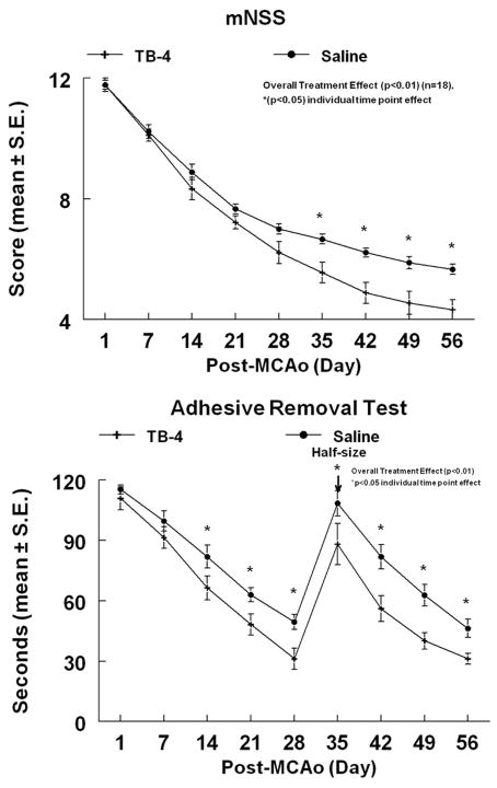
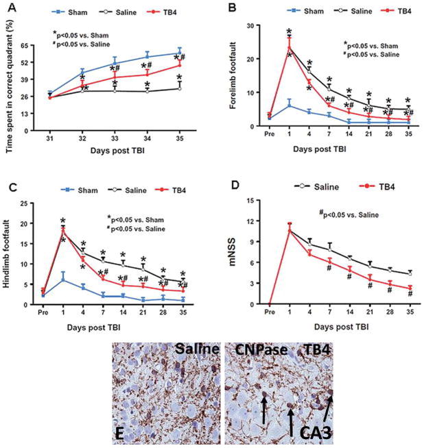
Neurorestorative therapy targets multiple types of parenchymal cells in the intact tissue of injured brain tissue to increase neurogenesis, angiogenesis, oligodendrogenesis, and axonal remodeling during recovery from neurological injury. In our laboratory, we tested thymosin β4 (Tβ4) as a neurorestorative agent to treat models of neurological injury. This review discusses our results demonstrating that Tβ4 improves neurological functional outcome in a rat model of embolic stroke, a mouse model of multiple sclerosis, and a rat model of traumatic brain injury. Tβ4 is a pleiotropic peptide exhibiting many actions in several different types of tissues. One mechanism associated with improvement of neurological improvement from Tβ4 treatment is oligodendrogenesis involving the differentiation of oligodendrocyte progenitor cells to mature myelin-secreting oligodendrocytes. Moreover, our preclinical data provide a basis for movement of Tβ4 into clinical trials for treatment of these devastating neurological diseases and injuries.


Conflict of interest statement

Conflicts of interest

The authors declare no conflicts of interest.

Figures

Figure 1
Figure 1
Embolic stroke rat model treated with Tβ4. The mNSS of embolic stroke rats treated with Tβ4 demonstrated a significant overall (treatment effect) improvement of neurological function (P < 0.01). The adhesive removal test of embolic stroke rats treated with Tβ4 also demonstrated a significant overall (treatment effect) improvement (P < 0.01). Significant effect (P < 0.05) at individual time points are indicated. Adhesive-backed paper dots were reduced in size by one-half at day 35 (arrow) to increase sensitivity. Reprinted from Ref. with permission from Elsevier.
Figure 2
Figure 2
Embolic stroke rat model treated with Tβ4. The staining by Bielshowsky and Luxol fast blue (A) shows the myelin and axons in the white matter bundles of the striatum of saline and Tβ4-treated rats (see arrows). There is a significantly increased density of Bielshowsky and Luxol fast blue staining in the Tβ4-treated rats compared to the demyelination of the saline control. LV = lateral ventricle and IC = ischemic core. NG-2 staining (B) is significantly increased in the ipsilateral SVZ and striatum adjacent to the ischemic core of Tβ4-treated rats when compared to saline control (see arrows). CNPase (C) is significantly increased in the striatum of Tβ4-treated rats when compared to saline control (see arrows). P < 0.05 for A, B and C. Reprinted from Ref. with permission from Elsevier.
Figure 3
Figure 3
EAE mouse model of multiple sclerosis treated with Tβ4. The neurological response of EAE mice treated with or without Tβ4. The significant therapeutic Tβ4 effects were detected as early as day 11 after EAE onset. Nearly 50% relative functional recovery was observed in the Tβ4 treated group, compared to the saline controls with P < 0.01 for either the median score or the cumulative score up to 30 days. NG-2 cells (B) and CNPase cells (C) were significantly increased at 30 days after EAE onset in the Tβ4 treatment group compared to that in the saline group (p<0.05). Reprinted from Ref. with permission from Elsevier.
Figure 4
Figure 4
Rat model of traumatic brain injury treated with Tβ4. Tβ4 treatment improves spatial learning performance measured by the Morris water maze test at days 33–35 compared with the saline group (A). Tβ4 treatment significantly reduces forelimb foot faults at days 7–35 compared with the saline-treated group (B). Tβ4 treatment significantly reduces hindlimb foot faults at days 7–35 compared with the saline-treated group (C). Line graph showing the functional improvement detected on the mNSS (D). Treatment with Tβ4 significantly lowers mNSS at days 7–35 compared with the saline group. Pre = preinjury level. Treatment with Tβ4 significantly increases CNPase cells in the CA3 region of the hippocampus (E) (P < 0.05). Reprinted from Ref. with permission from JNS Publishing Group.

References

    1. Sharma JC, Fletcher S, Vassallo M. Strokes in the elderly - higher acute and 3-month mortality - an explanation. Cerebrovasc Dis. 1999;9:2–9. - PubMed
    1. Reitz C, Brayne C, Mayeux R. Epidemiology of Alzheimer disease. Nat Rev Neurol. 2011;7:137–152. - PMC - PubMed
    1. Fuchs VR. Three “inconvenient truths” about health care. N Engl J Med. 2008;359:1749–1751. - PubMed
    1. Zhang ZG, Chopp M. Neurorestorative therapies for stroke: underlying mechanisms and translation to the clinic. Lancet Neurol. 2009;8:491–500. - PMC - PubMed
    1. Hacke W, et al. Thrombolysis with alteplase 3 to 4.5 hours after acute ischemic stroke. N Engl J Med. 2008;359:1317–1329. - PubMed

Publication types