. 2013 Mar;19(5-6):707-15.
doi: 10.1089/ten.TEA.2012.0112. Epub 2012 Nov 19.

Osteogenic differentiation of human dental pulp stromal cells on 45S5 Bioglass® based scaffolds in vitro and in vivo

Affiliations

Osteogenic differentiation of human dental pulp stromal cells on 45S5 Bioglass® based scaffolds in vitro and in vivo

Reem El-Gendy et al. Tissue Eng Part A. 2013 Mar.

Abstract

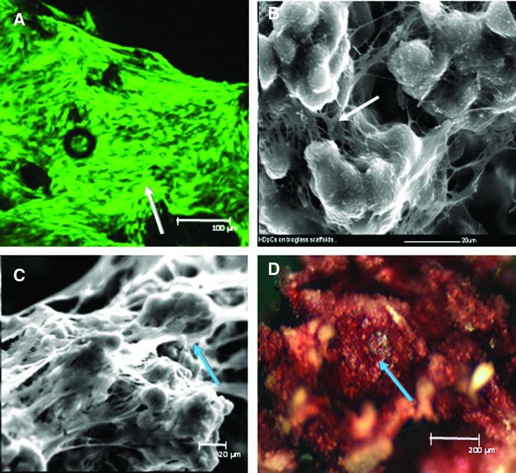
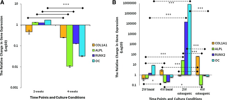
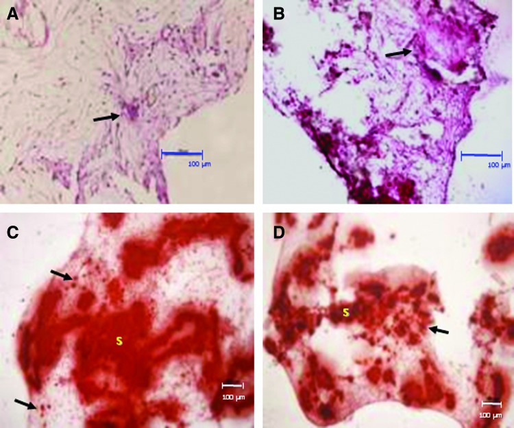
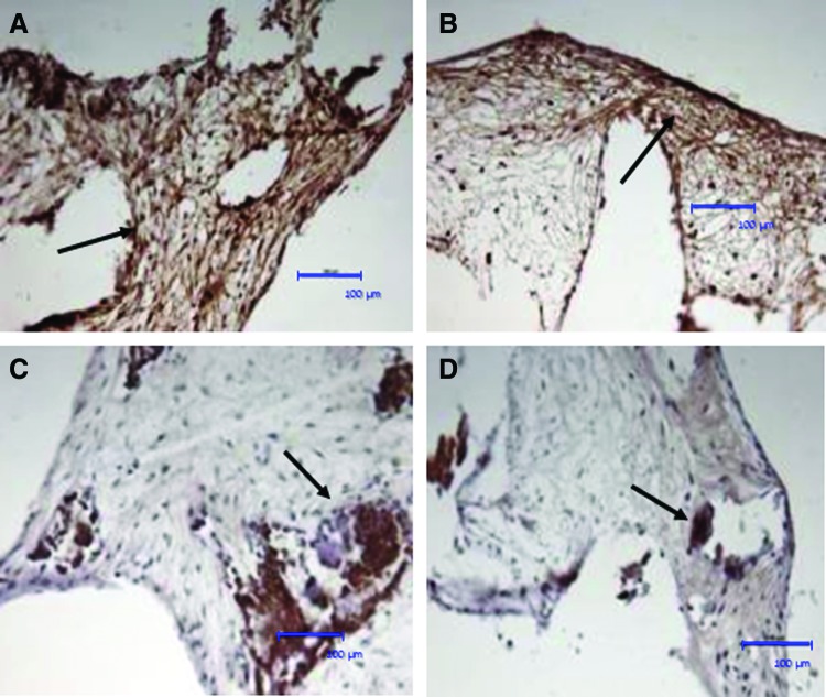
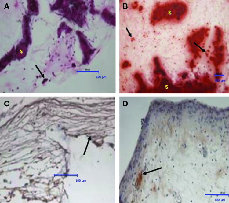
The increasing clinical demand for bone substitutes has driven significant progress in cell-based therapies for bone tissue engineering. The underpinning goals for success are to identify the most appropriate cell source and to provide three-dimensional (3D) scaffolds that support cell growth and enhance osteogenic potential. In this study, human dental pulp stromal cells (HDPSCs) were cultured under basal or osteogenic conditions either in monolayers or on 3D Bioglass® scaffolds in vitro for 2 or 4 weeks. Cell-scaffold constructs were also implanted intraperitoneally in nude mice for 8 weeks. Osteogenic potential was assessed using quantitative real-time polymerase chain reaction and histological/immunohistochemical assays. In monolayer culture, osteoinductive conditions enhanced HDPSC expression of osteogenic gene markers (COL1A1, RUNX2, OC, and/or OCN) compared with basal conditions while culture of HDPSCs on 3D scaffolds promoted osteogenic gene expression compared with monolayer culture under both basal and osteogenic conditions. These results were confirmed using histological and immunohistochemical analyses. In vivo implantation of the HDPSC 3D Bioglass constructs showed evidence of sporadic woven bone-like spicules and calcified tissue. In conclusion, this study has demonstrated the potential of using a combination of HDPSCs with 3D 45S5 Bioglass scaffolds to promote bone-like tissue formation in vitro and in vivo, offering a promising approach for clinical bone repair and regeneration.


Figures

FIG. 1.
FIG. 1.
Human dental pulp stromal cell (HDPSC) viability and growth on three-dimensional (3D) 45S5 Bioglass® scaffolds. (A) Confocal laser scanning microscope image showing HDPSC viability (arrow–CMFDA) on 45S5 Bioglass scaffolds after 2 weeks of in vitro culture under basal condition. (B, C) scanning electron microscope (SEM) images showing HDPSC spreading and cell bridge formation (arrows) on 45S5 Bioglass scaffolds after in vitro culture under basal conditions for 2 weeks (B) and 4 weeks (C). (D) Alkaline phosphatase (ALP) staining (arrow) indicative of osteogenic differentiation of HDPSC–45S5 Bioglass scaffolds after 4 weeks of culture in basal medium. Color images available online at www.liebertpub.com/tea
FIG. 2.
FIG. 2.
Relative changes in osteogenic marker gene expression for HDPSCs cultured on 45S5 Bioglass scaffolds in vitro. (A) The effect of osteogenic culture conditions on the expression of osteogenic marker genes by HDPSCs on 3D Bioglass scaffolds after 2 and 4 weeks (n=3). The data for osteogenic conditions were normalized to corresponding controls cultured under basal conditions using the ΔΔct method. (B) The effect of 3D Bioglass scaffolds on osteogenic marker gene expression by HDPSCs cultured under basal or osteogenic condition on 3D Bioglass scaffolds for 2 and 4 weeks (n=3). The data for 3D culture were normalized to corresponding controls in monolayers using the ΔΔct method. The data are presented as log10 of the mean 2−ΔΔct±SD (*p<0.05, ***p<0.001). Color images available online at www.liebertpub.com/tea
FIG. 3.
FIG. 3.
Hematoxylin and eosin (H&E) and Alizarin red staining of HDPSCs cultured on 3D Bioglass scaffolds in vitro. (A, B) H&E staining following 6 weeks of culture under basal conditions (A) and osteogenic conditions (B). Arrows point to regions of extracellular matrix condensation/woven bone-like spicules. (C, D) Alizarin red staining for calcium deposits in the cell–scaffold constructs cultured in vitro for 6 weeks under basal conditions (C) and osteogenic conditions (D). Despite the fact that the calcium-rich scaffolds themselves (“S”) are intensely stained with the Alizarin red, staining can also be seen in the “tissue” surrounding the scaffold (arrows). Scale bars=100 μm. Color images available online at www.liebertpub.com/tea
FIG. 4.
FIG. 4.
Immunohistochemical staining of HDPSC–scaffold constructs cultured in vitro. After 6 weeks of culture in basal condition in vitro, positive immunohistochemical staining was seen for collagen type I (A, B); arrows indicate collagen-I-positive fibers/bundles running parallel to the construct surface. The constructs were also positive for osteocalcin (C, D). All sections were counterstained with Harris hematoxylin. Scale bars=100 μm. Color images available online at www.liebertpub.com/tea
FIG. 5.
FIG. 5.
H&E and Alizarin red staining of HDPSC–3D Bioglass scaffold constructs following intraperitoneal implantation in vivo in diffusion chambers. After 8 weeks in vivo implantation, H&E staining (A) indicated extracellular matrix condensation/woven bone-like spicule formation (arrows). Alizarin red staining (B) for calcium deposits in HDPSC–scaffold constructs showed that despite the fact that the calcium-rich scaffolds themselves (“S”) were intensely stained with the Alizarin red, additional staining was also seen in the “tissue” surrounding the scaffold (arrows). (C) Immunohistochemical staining for collagen type I shows parallel collagen-I-positive fiber/bundle formation (arrows). (D) Immunohistochemical staining for osteocalcin was positive in all samples (sections counterstained with Harris hematoxylin). Scale bars=100 μm. Color images available online at www.liebertpub.com/tea

References

    1. Arrington E.D. Smith W.J. Chambers H.G. Bucknell A.L. Davino N.A. Complications of iliac crest bone graft harvesting. Clin Orthop Relat Res. 1996;329:300. - PubMed
    1. Laurencin C. Khan Y. El-Amin S.F. Bone graft substitutes. Expert Rev Med Devices. 2006;3:49. - PubMed
    1. Gu Y.D. Cheng D.S. Zhang G.M. Chen X.M. Xu J.G. Yang X.B. Long-term results of toe transfer: retrospective analysis. J Reconstr Microsurg. 1997;13:405. - PubMed
    1. Yang X.B. Oreffo R. Bone tissue engineering. In: Hong-wen Deng Y.-z.L., editor; Guo C.-Y., editor. Current topics in bone biology. 1st. Singapore: World Scientific Publishing Ltd; 2005. p. 435.
    1. Kroon F.H. Perren S.M. van den Hoof A. Behavior of cortical bone grafts under different types of fixation. Int J Oral Surg. 1977;6:131. - PubMed

Publication types

MeSH terms

LinkOut - more resources