Review
. 2012 Nov;250(1):82-101.
doi: 10.1111/imr.12006.

Diversity-oriented approaches for interrogating T-cell receptor repertoire, ligand recognition, and function

Affiliations
Review

Diversity-oriented approaches for interrogating T-cell receptor repertoire, ligand recognition, and function

Michael E Birnbaum et al. Immunol Rev. 2012 Nov.

Abstract

Molecular diversity lies at the heart of adaptive immunity. T-cell receptors and peptide-major histocompatibility complex molecules utilize and rely upon an enormous degree of diversity at the levels of genetics, chemistry, and structure to engage one another and carry out their functions. This high level of diversity complicates the systematic study of important aspects of T-cell biology, but recent technical advances have allowed for the ability to study diversity in a comprehensive manner. In this review, we assess insights gained into T-cell receptor function and biology from our increasingly precise ability to assess the T-cell repertoire as a whole or to perturb individual receptors with engineered reagents. We conclude with a perspective on a new class of high-affinity, non-stimulatory peptide ligands we have recently discovered using diversity-oriented techniques that challenges notions for how we think about T-cell receptor signaling.


Figures

Fig. 1
Fig. 1. Sources of diversity in pMHC-TCR interactions
Left: Overall binding topology of representative pMHC-TCR complex. Right: close-up of pMHC-TCR binding interface. TCR CDR1 and CDR2 (boxed in cyan) are encoded in the germline and primarily bind MHC (green). The TCR CDR3 interface (boxed in magenta) is uniquely derived for each receptor and primarily binds the peptide (yellow). The table (below) lists areas of diversity. Overall theoretical TCR diversity is 1015 unique receptors. The potential peptidome displayed on MHC is a comparable number.
Fig. 2
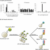
Fig. 2. Synthetic versus cell-based methods for discovery of new TCR peptide ligands
(A) Split-pool libraries use mixtures of peptides to activate a T-cell clone of interest. The response of a set amino acid at a given position can lead to information about residues that are favored or disfavored. Schematic data is provided for three peptide positions (P1-P3). In this data, Asp and Glu are favored for P1 (left), Pro is disfavored for P2 (middle), and Lys is strongly favored for P3 (right). This information can be combined to find an optimized peptide ligand and even to deorphanize a TCR. (B) Cell-based display systems can be used to generate libraries of diverse peptides. Library members are then selected for peptide binding to MHC or for pMHC binding to TCR. This approach can optimize peptide affinity for MHC (top), higher TCR affinity (middle), or be used to identify new peptides of interest (bottom).
Fig. 3
Fig. 3. Diversity of structural features exhibited by yeast display-derived peptides recognized by 42F3 TCR
(A) Overall binding topology for p5E8, p4B10, P3A1, and QL9 shows a marked difference for p3A1. (B) 42F3 CDR3 loops (red and blue) adopt different topologies to bind to peptides (yellow) with markedly different chemistries. (C) Peptide-CDR3 interaction map showing van der Waals contacts (black) and hydrogen bonds (red). (D) Electron density maps of peptides from Ld-42F3 complex crystal structures.
Fig. 4
Fig. 4. Potential mechanisms for high affinity, non-stimulatory pMHC ligands
(A) 42F3 TCR binding footprint for agonist (QL9, black) and nonagonist (p3A1, red) peptides. While there is a large deviation in binding footprint, the overall TCR binding polarity is maintained. (B) Schematic of binding/function differences between agonist and high affinity nonagonist. (C) Potential models for agonist (yellow) vs. nonagonist (red) peptides. Efficient pMHC binding may not be possible in the context of a T cell (left), binding may not lead to productive signaling (center), or TCR oligomerization may be disrupted (right).

References

    1. Davis MM, Bjorkman PJ. T-cell antigen receptor genes and T-cell recognition. Nature. 1988;334:395–402. - PubMed
    1. Rudolph MG, Stanfield RL, Wilson IA. How TCRs bind MHCs, peptides, and coreceptors. Annu Rev Immunol. 2006;24:419–466. - PubMed
    1. Irvine DJ, Purbhoo MA, Krogsgaard M, Davis MM. Direct observation of ligand recognition by T cells. Nature. 2002;419:845–849. - PubMed
    1. Juang J, Ebert PJ, Feng D, Garcia KC, Krogsgaard M, Davis MM. Peptide-MHC heterodimers show that thymic positive selection requires a more restricted set of self-peptides than negative selection. J Exp Med. 2010;207:1223–1234. - PMC - PubMed
    1. Hochweller K, Wabnitz GH, Samstag Y, Suffner J, Hammerling GJ, Garbi N. Dendritic cells control T cell tonic signaling required for responsiveness to foreign antigen. Proc Natl Acad Sci USA. 2010;107:5931–5936. - PMC - PubMed

Publication types

MeSH terms